Bleeding Disorders
One lecture, six patients: the two arms of hemostasis, four bedside questions, the first panel, and the common disorders with their first-line care.
These slides are an outline only. Read the notes under each slide below with them, and chapters 20 to 25 of the Hematology Handbook. Then work through Case 53 in the Internal Medicine Casebook and the Hematology Question Bank. This lecture replaces the two-part version, Introduction to Bleeding Disorders and Disorders of Hemostasis.
Bleeding Disorders
These slides are an outline only. Read the notes under each slide on this page with them, and chapters 20 to 25 of the Hematology Handbook. Then work through Case 53 in the Internal Medicine Casebook and the Hematology Question Bank.
This lecture is about the patient who bleeds. It replaces the two earlier lectures, Introduction to Bleeding Disorders and Disorders of Hemostasis, with one lecture built around six patients.
The plan has four parts. First, how bleeding normally stops, and the two arms of hemostasis. Second, four questions to ask at the bedside. Third, the first panel of blood tests. Fourth, the disorders themselves, one patient at a time, each with its first-line care.
What is left out on purpose: how the laboratory works up an unexplained long clotting time (sample problems, mixing studies and specialist factor assays). That is taught in a later lecture. Here you need to recognise the pattern and know when to refer.
Why hemostasis matters
Start with the idea that runs through the whole lecture. Blood has two jobs that pull against each other. It must stay liquid everywhere, all the time, or the tissues starve. And it must turn solid within minutes at one place, the place where a vessel has broken, and nowhere else.
Hemostasis is the system that keeps that balance. When it tips one way, the patient bleeds. When it tips the other way, the patient clots where they should not, which is thrombosis.
This lecture is about the first failure: the patient who bleeds. Thrombosis has its own teaching, in handbook chapter 26 and again in Med-2.
Four patients
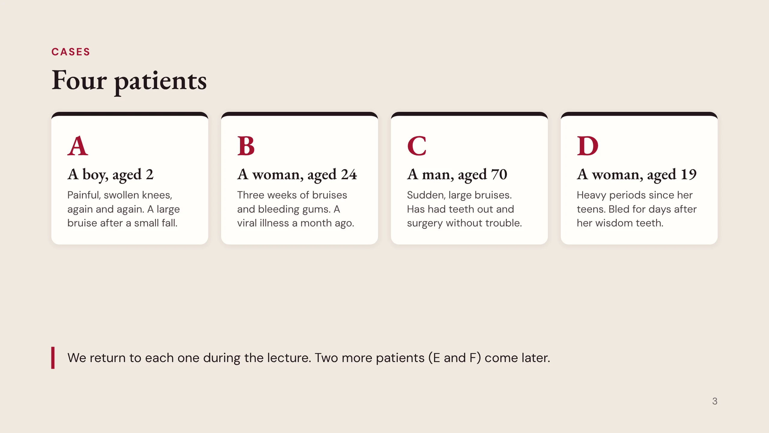
Four people come to clinic. Each one says the same thing: I bleed.
Patient A is a boy of 2. His knees swell and hurt, again and again, and he had a large bruise after a small fall.
Patient B is a woman of 24. For three weeks she has bruised easily and her gums bleed. She had a viral illness a month ago.
Patient C is a man of 70 with sudden large bruises. He has had teeth out and surgery in the past without any trouble.
Patient D is a woman of 19. Her periods have been heavy since her teens, and she bled for days after her wisdom teeth were removed.
Before going on, guess. Which of them was born with the problem, and which acquired it? Which has a problem with platelets, and which with the clotting factors? Keep your guesses. We meet each patient again during the lecture, and two more, E and F, join them on the ward.
By the end, you can
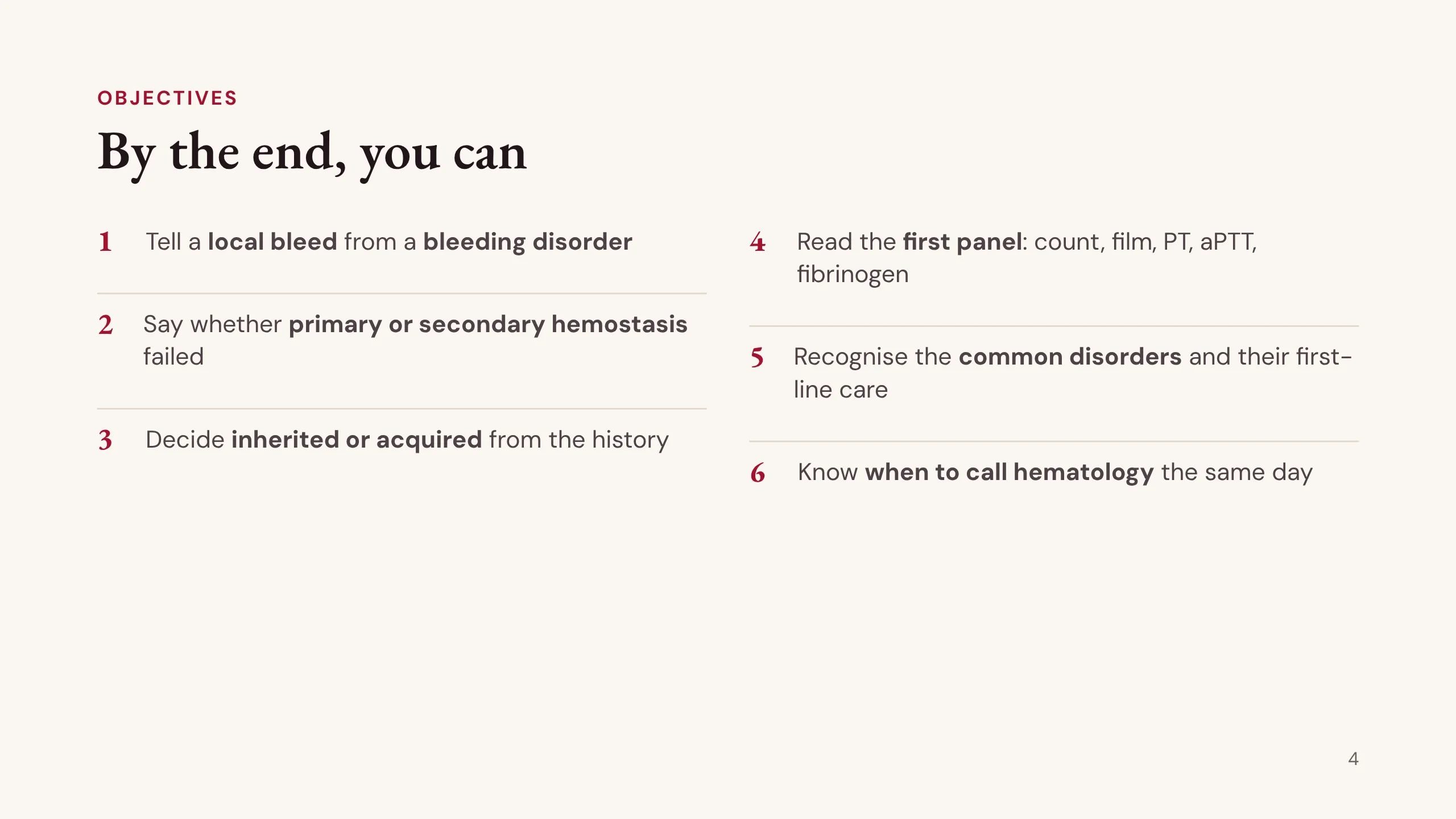
By the end of this lecture you should be able to do six things.
1. Decide from the history whether the bleeding comes from a local lesion or from a bleeding disorder.
2. Say whether primary hemostasis (the platelet plug) or secondary hemostasis (the fibrin clot) has failed, from the pattern of bleeding alone.
3. Decide whether the problem is inherited or acquired, from past challenges, the family, and drugs and illness.
4. Read the first panel: the platelet count, the blood film, the prothrombin time (PT), the activated partial thromboplastin time (aPTT) and the fibrinogen.
5. Recognise the common disorders, immune thrombocytopenia, von Willebrand disease, hemophilia, acquired hemophilia and disseminated intravascular coagulation, and name their first-line care.
6. Know which patients need hematology the same day.
The first four are the four bedside questions. The last two are recognition and safe escalation.
Part 1: How bleeding stops
Part 1 is the physiology, but only as much as the clinic needs. Every step you meet here will have a test that measures it and a disease in which it fails.
Two arms of hemostasis
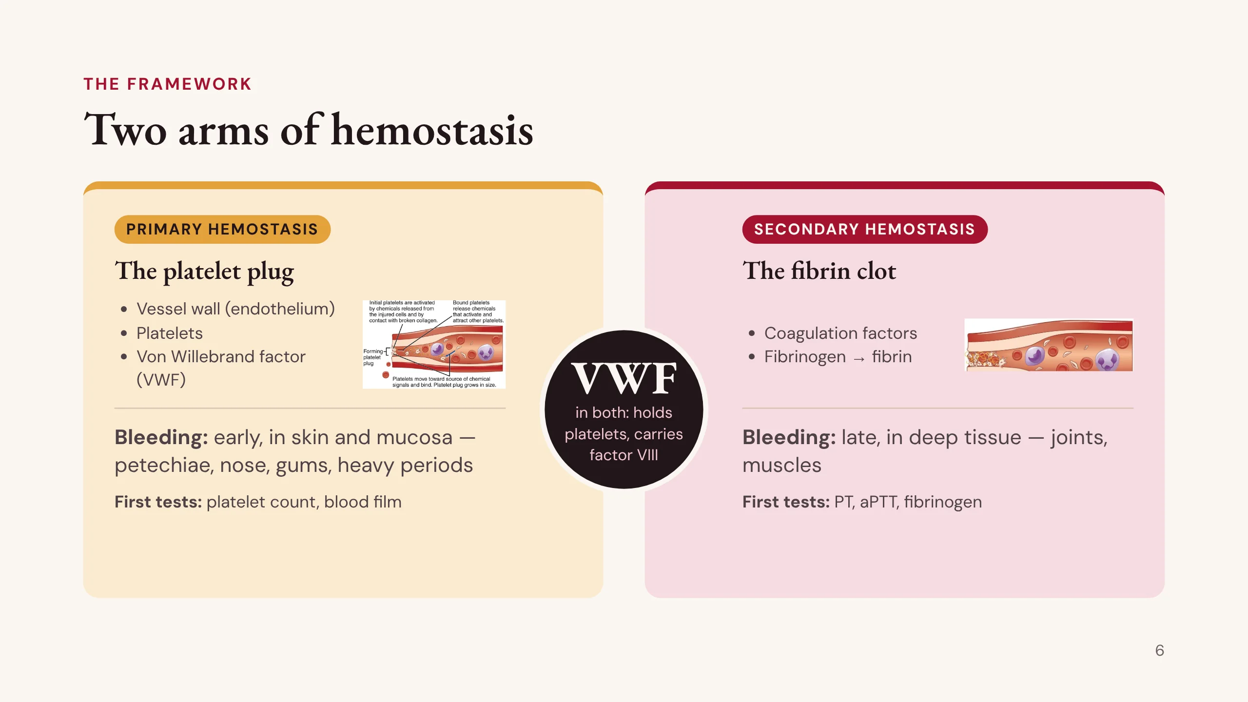
This is the framework for the whole lecture. Hemostasis has two arms.
Primary hemostasis makes the platelet plug. It needs three things: the vessel wall (the endothelium), the platelets, and von Willebrand factor (VWF), the protein that glues platelets to the injured wall. When primary hemostasis fails, bleeding starts early, straight after the injury, and it is in the skin and mucosa: petechiae, bruises, nosebleeds, bleeding gums, heavy periods. The first tests are the platelet count and the blood film.
Secondary hemostasis makes the fibrin clot that holds the plug in place. It needs the coagulation factors, which end by turning fibrinogen into fibrin. When secondary hemostasis fails, the plug forms and the first bleeding stops, but the plug is not reinforced. Bleeding starts again later, and it is deep: into joints and muscles. The first tests are the PT, the aPTT and the fibrinogen.
Notice the circle in the middle. Von Willebrand factor belongs to both arms. It holds platelets to the wound, which is primary hemostasis, and it carries and protects factor VIII, which is secondary hemostasis. That is why severe von Willebrand disease can bleed like hemophilia. Keep this slide in mind: almost every slide that follows is tagged with the arm it belongs to.
The two small pictures are from OpenStax Anatomy and Physiology, under a CC BY 3.0 licence.
From injury to clot
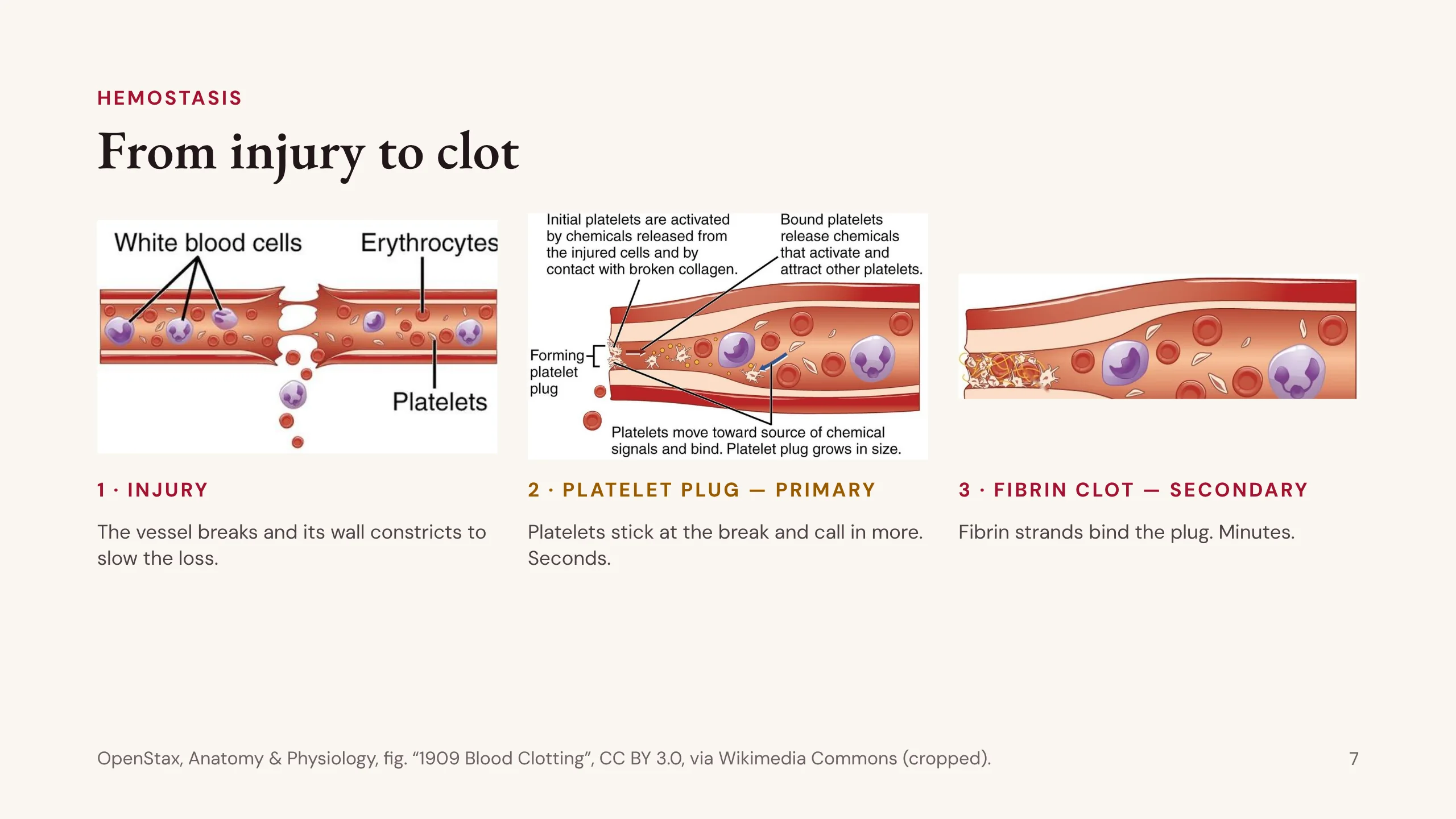
Here is the sequence when a vessel is cut.
1. Injury. The vessel breaks and blood leaks out. Straight away the smooth muscle in the wall contracts, which narrows the vessel and slows the loss.
2. The platelet plug. Platelets stick to the collagen exposed at the break. They change shape and release signals that call in more platelets. Within seconds there is a soft plug. This is primary hemostasis.
3. The fibrin clot. At the same time the clotting factors make thrombin, and thrombin turns fibrinogen into fibrin. Fibrin strands bind the plug and trap red cells. This takes minutes. This is secondary hemostasis.
Later, when the vessel has healed, the clot is dissolved.
The illustration is from OpenStax Anatomy and Physiology, under a CC BY 3.0 licence, cropped from the original figure.
Four verbs: START, MAKE, STOP, CLEAN
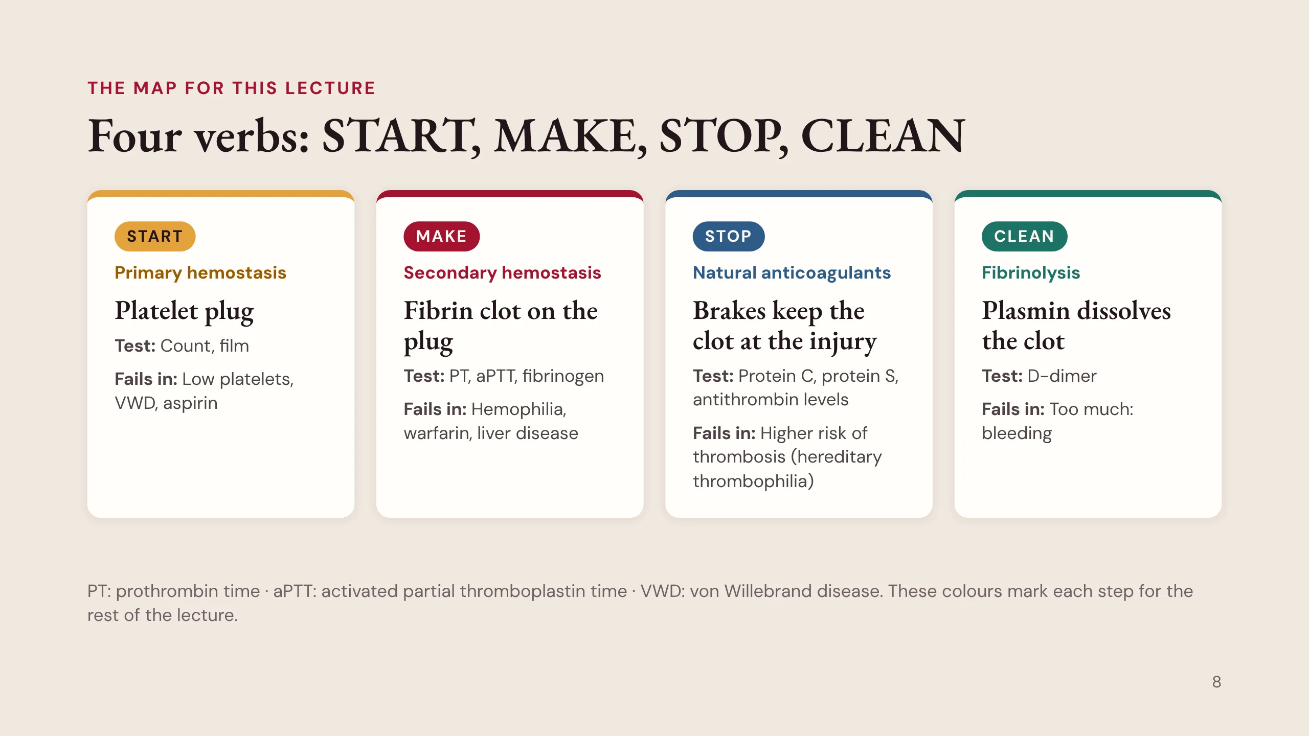
Four verbs carry the subject: START, MAKE, STOP and CLEAN. The colours are used for the rest of the lecture.
START is primary hemostasis, the platelet plug. It forms within seconds. We test it with the platelet count and the film. It fails with a low count, in von Willebrand disease (VWD) and with aspirin.
MAKE is secondary hemostasis, the fibrin clot on the plug. We test it with the PT, the aPTT and the fibrinogen. It fails in hemophilia, with warfarin and in liver disease.
STOP is the natural anticoagulants: protein C, protein S and antithrombin, with tissue factor pathway inhibitor. At rest they keep blood liquid; after an injury they keep the clot at the site. They are tested by measuring protein C, protein S and antithrombin levels. When one is lacking, the risk is not bleeding but thrombosis: a hereditary thrombophilia.
CLEAN is fibrinolysis. Plasmin dissolves the clot once the vessel heals, and D-dimer is one of its products. Too much fibrinolysis can itself cause bleeding.
Each verb has a test and a disease. That is the point of learning them.
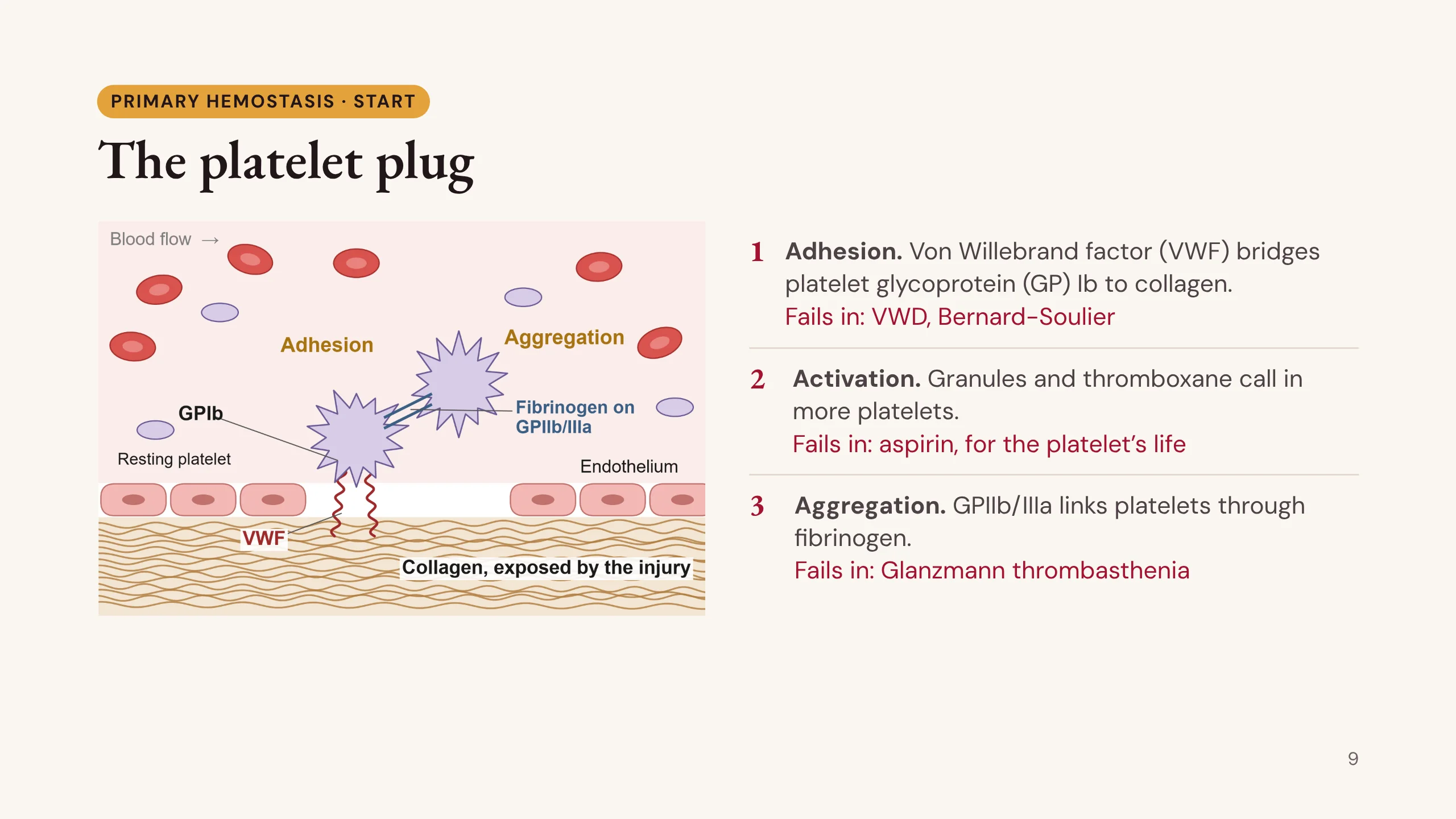
The platelet plug
Primary hemostasis in three steps, each with its disease.
Step 1, adhesion. The injury exposes collagen. Von Willebrand factor binds the collagen and catches platelets by their glycoprotein Ib (GPIb) receptor. If VWF is low or faulty, that is von Willebrand disease. If GPIb is missing, that is Bernard-Soulier syndrome, where the platelets are large and the count is a little low.
Step 2, activation. The platelet changes shape, empties its granules and makes thromboxane. These signals call in more platelets. Aspirin blocks the enzyme that makes thromboxane (cyclooxygenase), and the block lasts for the life of the platelet, about 7 to 10 days. That is why aspirin is stopped well before surgery.
Step 3, aggregation. Glycoprotein IIb/IIIa links one platelet to the next through fibrinogen. If it is missing, that is Glanzmann thrombasthenia. The count and platelet size are normal, but the platelets do not stick together, and the patient bleeds badly.
The drawing was made for the lecture.
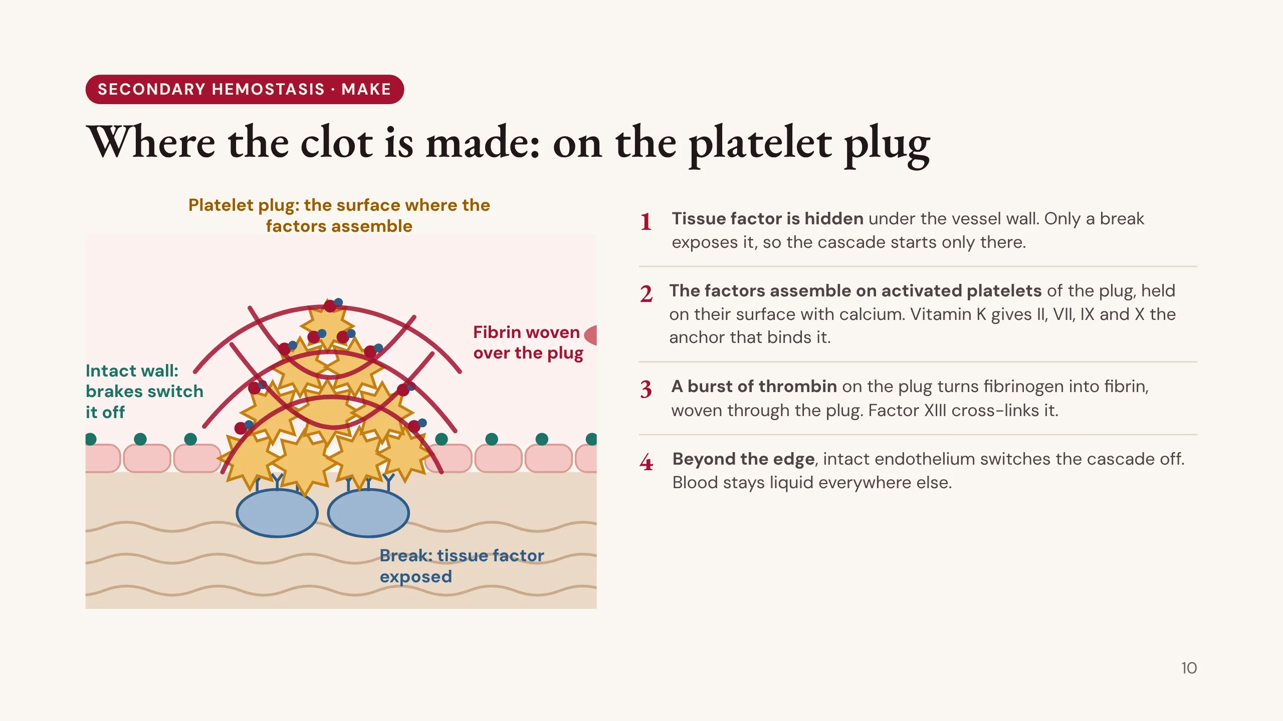
Where the clot is made: on the platelet plug
Secondary hemostasis, the coagulation cascade, is often drawn as a chain of factors floating in the blood. That picture misleads. The cascade runs only at the site of the injury, on the surface of the platelet plug that primary hemostasis has already built.
1. Tissue factor (TF) is the starter, and it is hidden. It sits on cells under the vessel wall, out of contact with the blood. Only a break in the wall exposes it, so the cascade can start only at the break.
2. The factors need a surface to work on, and the activated platelets of the plug provide it. Activated platelets turn a negatively charged phospholipid layer outward. With calcium as the bridge, the factor complexes (VIIIa with IXa, and Va with Xa) assemble on that surface, where they work thousands of times faster than in free plasma. Vitamin K is what gives factors II, VII, IX and X the anchor (the Gla domain) that binds this surface; without vitamin K, as with warfarin, they cannot attach.
3. On the plug, the complexes make a burst of thrombin. Thrombin turns fibrinogen into fibrin, which is woven through and over the plug, and factor XIII cross-links it into a firm clot. The soft platelet plug becomes a stable clot.
4. Beyond the edge of the injury, the intact endothelium switches the cascade off: thrombomodulin turns thrombin into an activator of protein C, and tissue factor pathway inhibitor and antithrombin mop up what escapes. So blood stays liquid everywhere else.
Two consequences follow. The two arms work in sequence: plug first, then the clot on the plug. And a very low platelet count weakens the clot as well as the plug, because the cascade has less surface to run on. The drawing was made for this lecture.
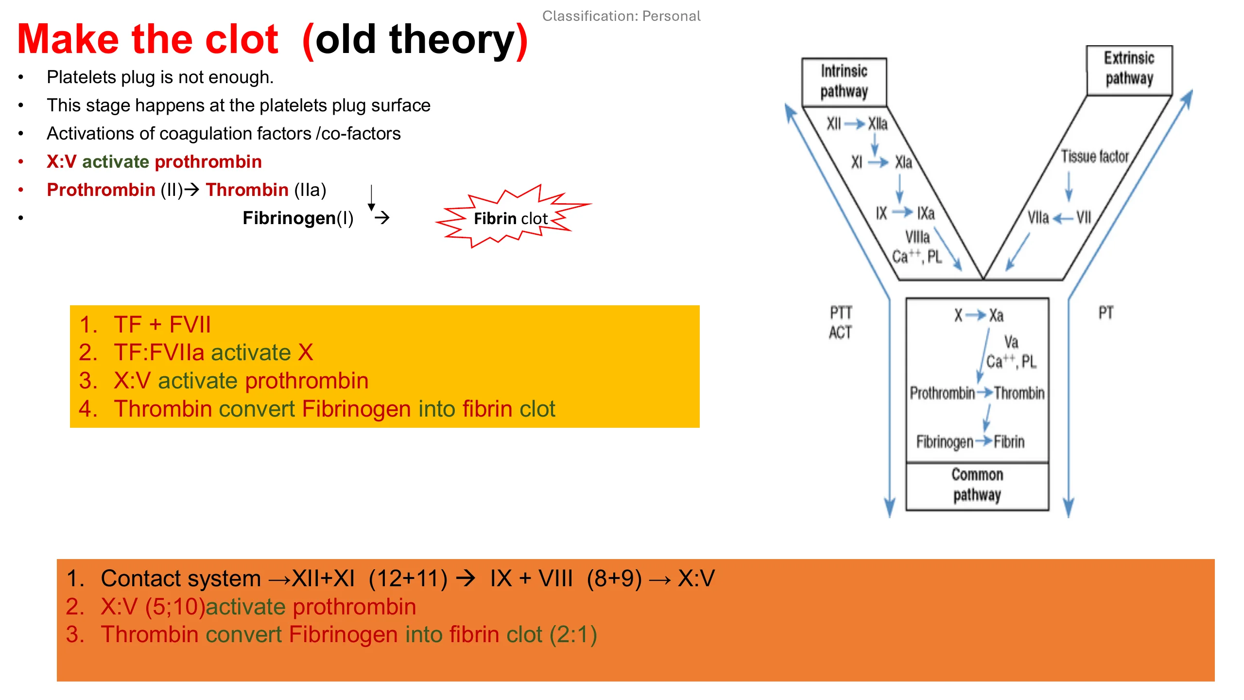
Make the clot: the extrinsic and intrinsic pathways
This is the classic picture of the cascade, from the earlier version of this lecture, and it is still the clearest way to learn the names.
The platelet plug is not enough on its own. The clot is made on the surface of the plug: the coagulation factors and their cofactors are activated there, factor X with factor V activates prothrombin, prothrombin (II) becomes thrombin (IIa), and thrombin turns fibrinogen (I) into a fibrin clot.
The extrinsic pathway, in the yellow box: tissue factor (TF) meets factor VII; TF with VIIa activates X; X with V activates prothrombin; thrombin converts fibrinogen into fibrin.
The intrinsic pathway, in the orange box: the contact system, XII then XI (12 then 11), activates IX with VIII (9 with 8), which activates X with V (10 with 5). From there it is the same common pathway: prothrombin to thrombin, fibrinogen to fibrin.
The diagram shows the two arms meeting at X. The PTT (aPTT) tests the intrinsic arm and the common pathway; the PT tests the extrinsic arm and the common pathway. That is how the screening tests are read later in the lecture.
The next slide adds the newer view: in the body these two are not separate pathways but one loop.
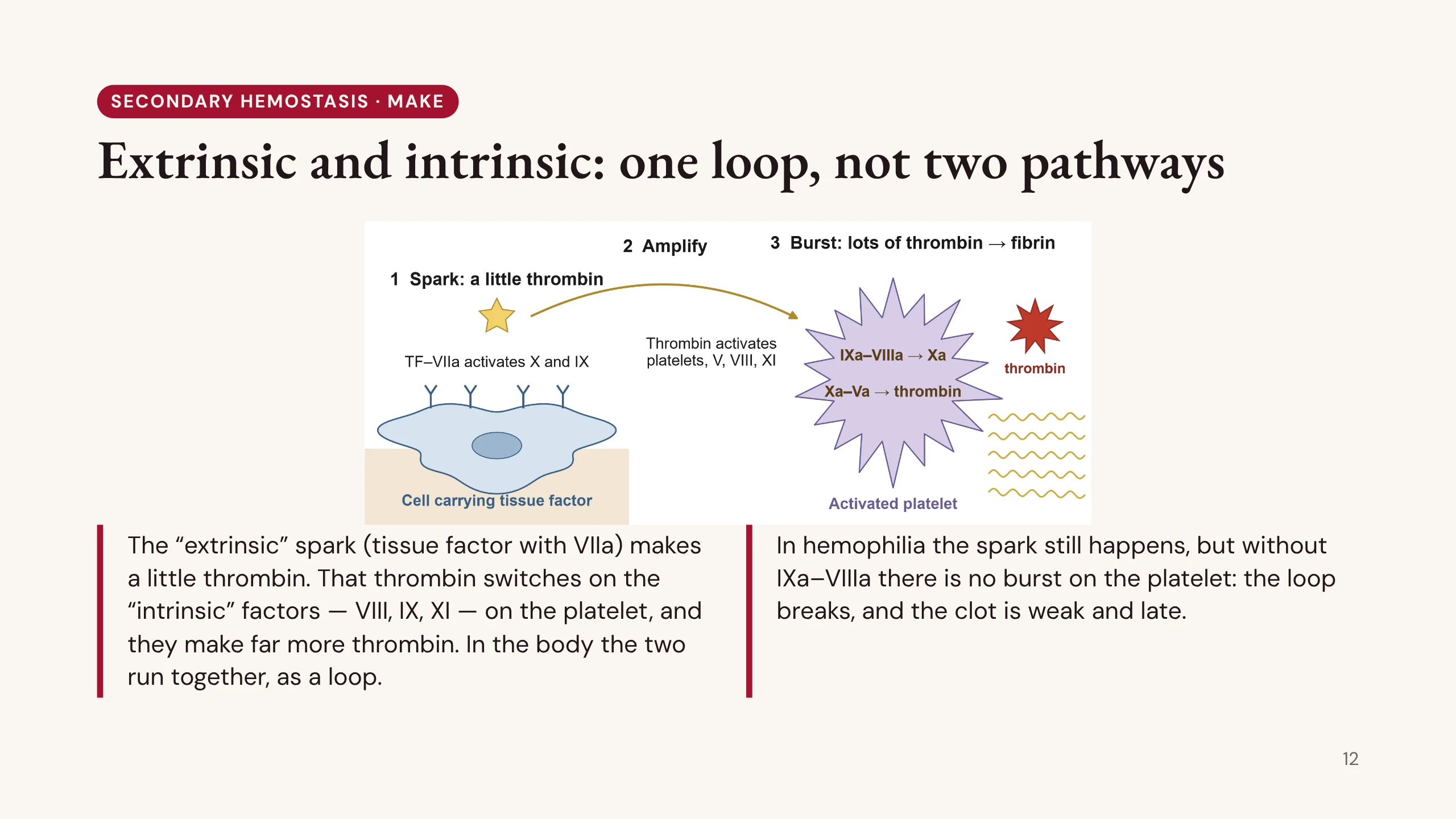
Extrinsic and intrinsic: one loop, not two pathways
You will meet the words extrinsic pathway and intrinsic pathway in every textbook and on every coagulation report. They come from the test tube: the PT starts the reaction with tissue factor added from outside the plasma (extrinsic), and the aPTT starts it with factors already inside the plasma (intrinsic). In the body they are not two separate pathways. They run together, as one loop, on the cells at the injury.
1. The spark. On a cell carrying tissue factor at the injury, tissue factor with factor VIIa, the “extrinsic” step, activates factors X and IX. Xa with Va makes a little thrombin, not enough for a good clot.
2. Amplify. That little thrombin activates platelets, and switches on factors V, VIII and XI, the “intrinsic” factors.
3. The burst. On the activated platelet, IXa with VIIIa makes large amounts of Xa, and Xa with Va makes a burst of thrombin. That thrombin activates still more V, VIII and XI, so the reaction feeds itself: a loop. Thrombin turns fibrinogen into fibrin, and factor XIII cross-links it.
This answers a puzzle. A boy with hemophilia has normal factor VII and tissue factor, so his “extrinsic pathway” works and his PT is normal. Yet he bleeds badly into his joints. Without factor VIII or IX there is no IXa–VIIIa on the platelet, so the loop breaks and there is no burst. The spark alone is not enough: the clot is weak and late.
It also explains factor XII. The aPTT needs it in the tube, but it plays no part in the loop in the body, so people who lack it have a long aPTT and do not bleed. This is the cell-based model of Hoffman and Monroe (2001). The drawing was made for the lecture.
Stop: the natural anticoagulants
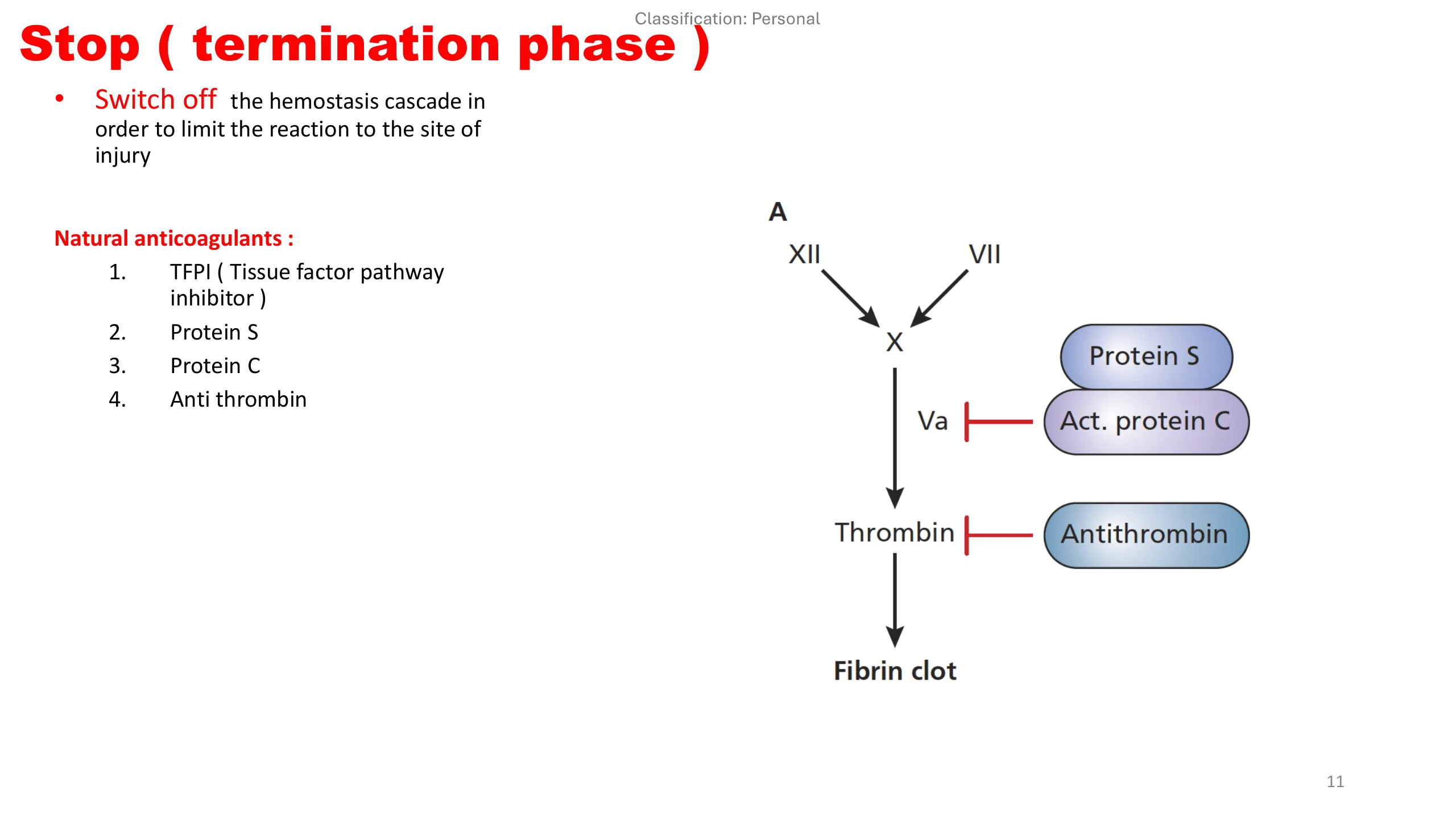
Stop, the termination phase, from the earlier version of this lecture. Once the clot has started, the hemostasis cascade must be switched off, so that the reaction stays at the site of injury and blood stays liquid everywhere else.
Four natural anticoagulants do this. 1. Tissue factor pathway inhibitor (TFPI) switches off the spark, tissue factor with VIIa. 2 and 3. Protein C, once activated, with protein S as its helper, cuts factor Va (and VIIIa). Thrombin switches protein C on when it binds thrombomodulin on intact endothelium. 4. Antithrombin blocks thrombin (and Xa); heparin works by boosting it.
The diagram shows where each acts: activated protein C and protein S on Va, antithrombin on thrombin.
These are tested by measuring protein C, protein S and antithrombin levels. Their failure does not cause bleeding. A deficiency of any of them raises the risk of thrombosis: a hereditary thrombophilia. Factor V Leiden, in which Va resists activated protein C, does the same. Thrombosis is taught in handbook chapter 26.
Clean: fibrinolysis
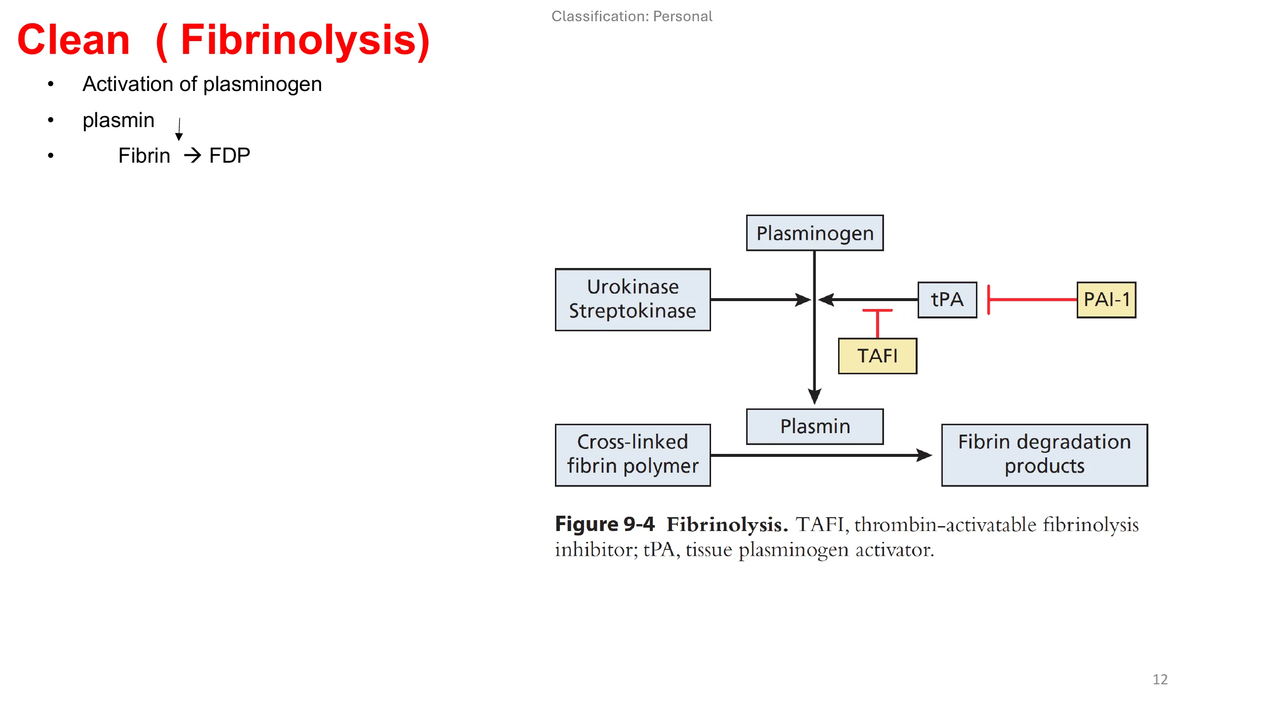
Clean, fibrinolysis, from the earlier version of this lecture. When the vessel has healed, the clot must be removed.
Plasminogen is activated to plasmin. The main activator in the body is tissue plasminogen activator (tPA), released by the endothelium; urokinase and streptokinase do the same job and have been used as clot-dissolving drugs. Plasmin then cuts cross-linked fibrin into fibrin degradation products (FDP), one of which is the D-dimer.
There are brakes on this step too. Plasminogen activator inhibitor-1 (PAI-1) blocks tPA, and thrombin-activatable fibrinolysis inhibitor (TAFI) slows the breakdown of the clot.
D-dimer is sensitive but not specific: surgery, infection, pregnancy, cancer and age all raise it. And too much fibrinolysis can itself cause bleeding, which is why tranexamic acid, a drug that blocks it, helps so many bleeders.
Part 2: Four questions at the bedside
Part 2 uses the physiology at the bedside. A bleeding patient answers four questions, in order. Is it a bleeding disorder at all? Did primary or secondary hemostasis fail? Was the patient born with it, or did they acquire it? And what does the first panel show? The first three come from the history and examination.
Local bleed, or a bleeding disorder?
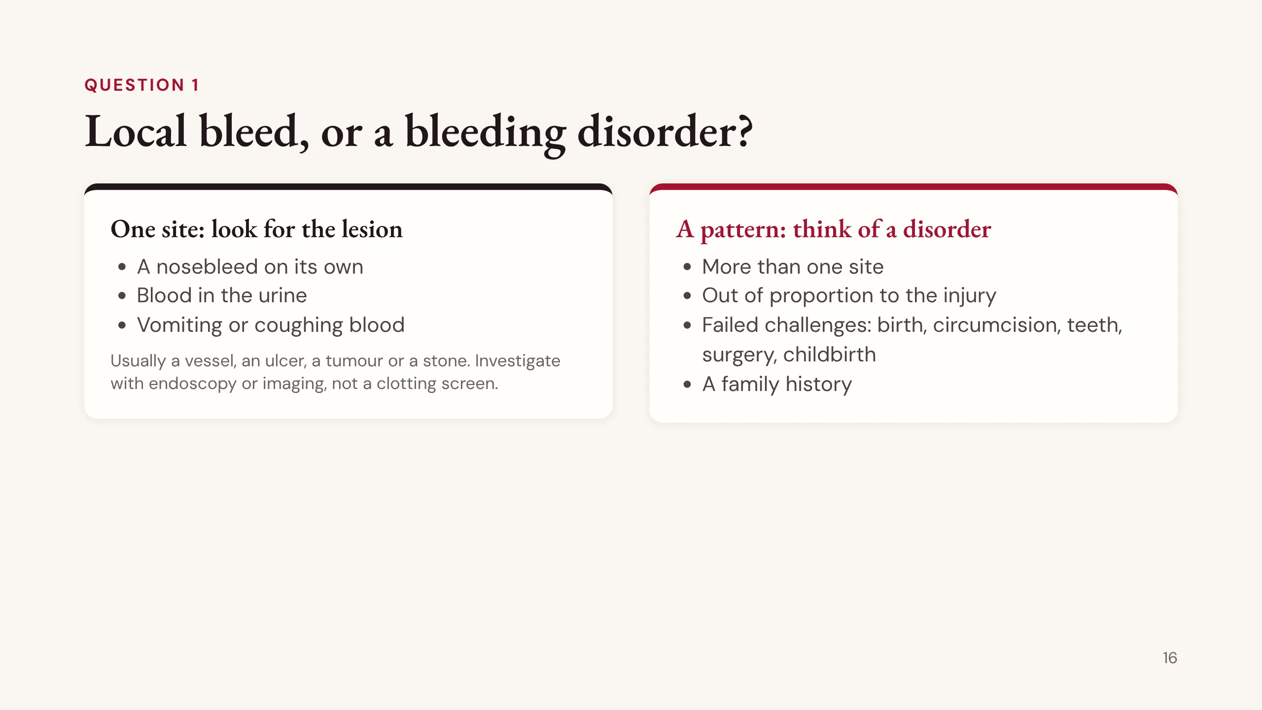
Question 1: is this a bleeding disorder at all?
Most people who bleed do not have one. Bleeding from one site on its own, such as a nosebleed, blood in the urine, or vomiting or coughing blood, usually comes from a local lesion: a vessel, an ulcer, a tumour or a stone. Look for it with endoscopy or imaging. A clotting screen will not find it.
A bleeding disorder shows itself as a pattern. More than one site. Bleeding out of proportion to the injury. Bleeding after the natural tests of life: birth, circumcision, a tooth extraction, surgery, childbirth. A family history.
One caution. A patient with a disorder can still present first with one site, so the rule tells you where to look first, not where to stop.
Primary or secondary: which arm failed?
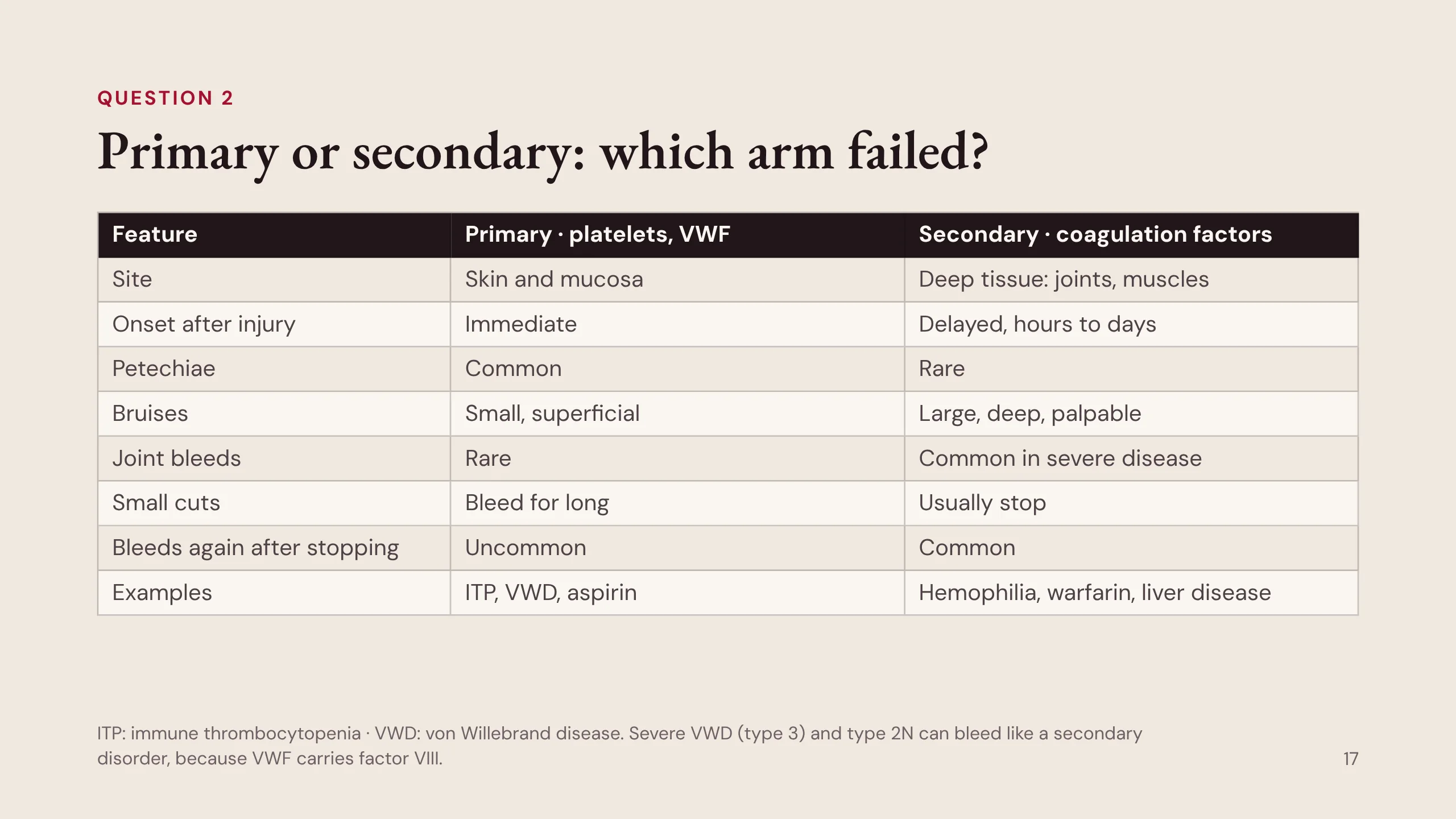
Question 2: which arm failed? This table is the most useful clinical split in the subject.
Primary hemostasis, the platelets and von Willebrand factor. Bleeding is in the skin and mucosa, and it starts immediately, because the plug is the first thing that should have stopped it. Petechiae are common. Bruises are small and superficial. Joint bleeds are rare. Small cuts bleed for a long time. Once stopped, bleeding rarely starts again. Examples: immune thrombocytopenia (ITP), von Willebrand disease, aspirin.
Secondary hemostasis, the coagulation factors. Bleeding is deep, into joints and muscles, and it is delayed by hours or days, because the plug forms first. Petechiae are rare. Bruises are large, deep and palpable. Joint bleeds are common in severe disease. Small cuts usually stop. But bleeding often starts again after it seemed to stop. Examples: hemophilia, warfarin, liver disease.
One line to remember: petechiae are a platelet sign. People with hemophilia do not get petechiae.
The exception is severe von Willebrand disease (type 3) and type 2N, which can bleed like a secondary disorder because VWF carries factor VIII.
Skin signs: primary or secondary?
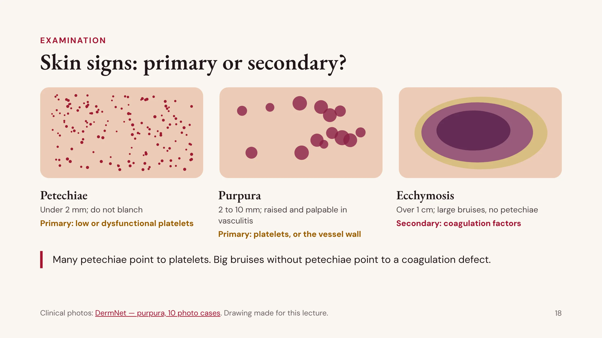
On examination, the size of the skin bleeding points to the arm.
Petechiae are pinpoint spots, under 2 mm, that do not blanch when pressed. They come from capillaries that leak because there are too few platelets, or platelets that do not work. They point to primary hemostasis.
Purpura are larger, 2 to 10 mm. Flat purpura usually also mean platelets. Raised purpura that you can feel (palpable purpura) mean inflamed vessel walls, a vasculitis, not a bleeding disorder.
An ecchymosis is a bruise over 1 cm. Large, deep bruises without petechiae point to a coagulation defect, secondary hemostasis.
The drawing was made for this lecture. For real photographs, work through the DermNet set of ten purpura cases: DermNet — different causes of purpura.
Inherited, or acquired?
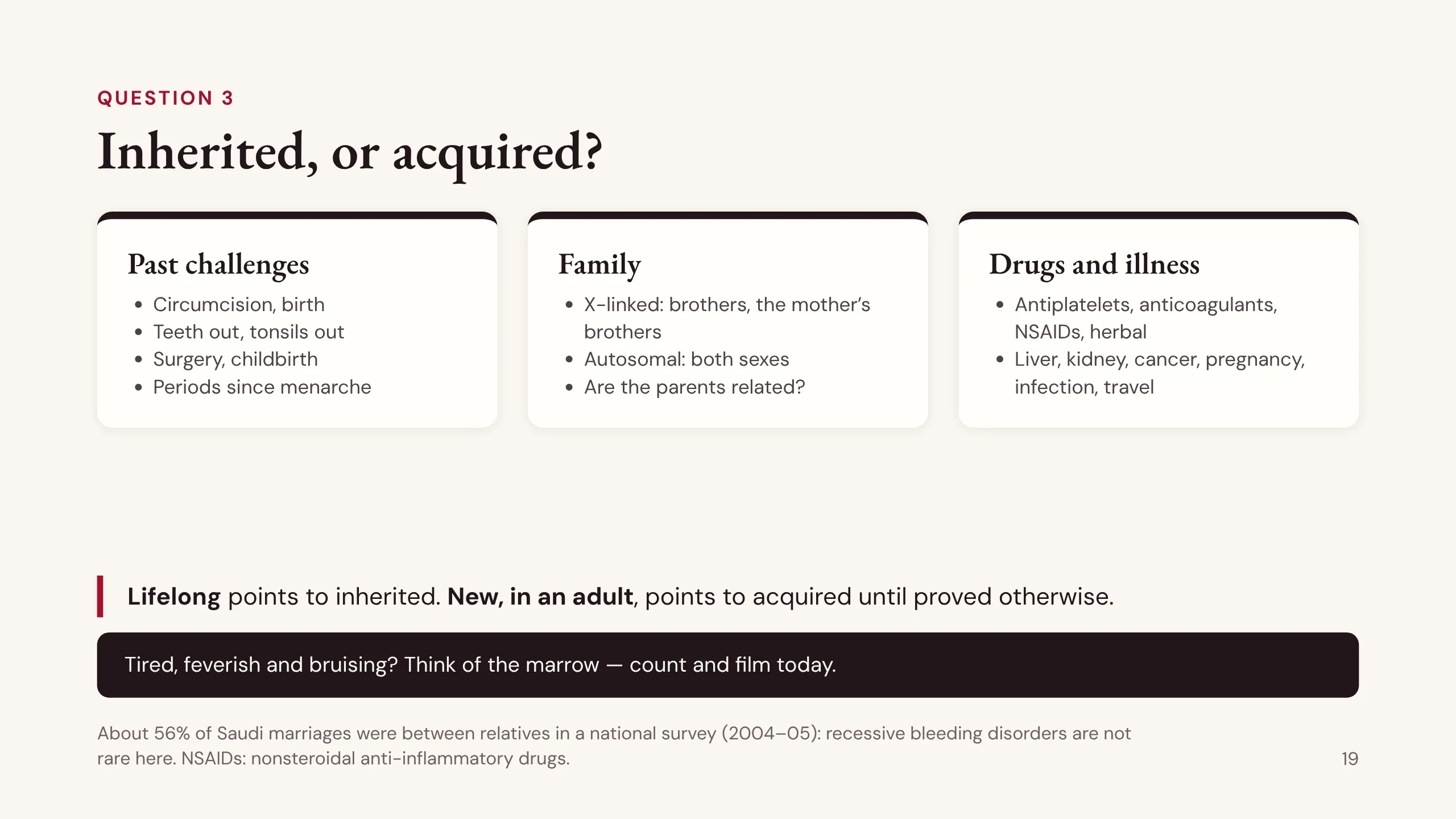
Question 3: was the patient born with it, or did they acquire it?
Past challenges are natural tests: circumcision, birth, a tooth or tonsils out, surgery, childbirth, periods since menarche. Someone who passed all of these and now bleeds has an acquired problem. Someone who bled at each was probably born with it.
The family. Hemophilia is X-linked, so ask about brothers and the mother’s brothers. Von Willebrand disease and most rare factor deficiencies are autosomal, so both sexes are affected. Ask whether the parents are related. A national Saudi survey in 2004 to 2005 found about 56% of marriages were between relatives, so recessive disorders such as Glanzmann thrombasthenia are not rare here.
Drugs and illness. In adults most bleeding is acquired, and antiplatelet drugs and anticoagulants are the commonest cause of all. Ask about nonsteroidal anti-inflammatory drugs (NSAIDs) and herbal products by name, because patients do not count them as medicines. Ask about liver disease, kidney failure, cancer, pregnancy, infection and travel.
The rule: lifelong points to inherited; new in an adult points to acquired until proved otherwise.
And the warning. A patient who is tired, feverish and bruising may have all three cell lines low: a marrow problem, perhaps leukemia. Get the count and film today.
“How are your periods?” “Fine.”
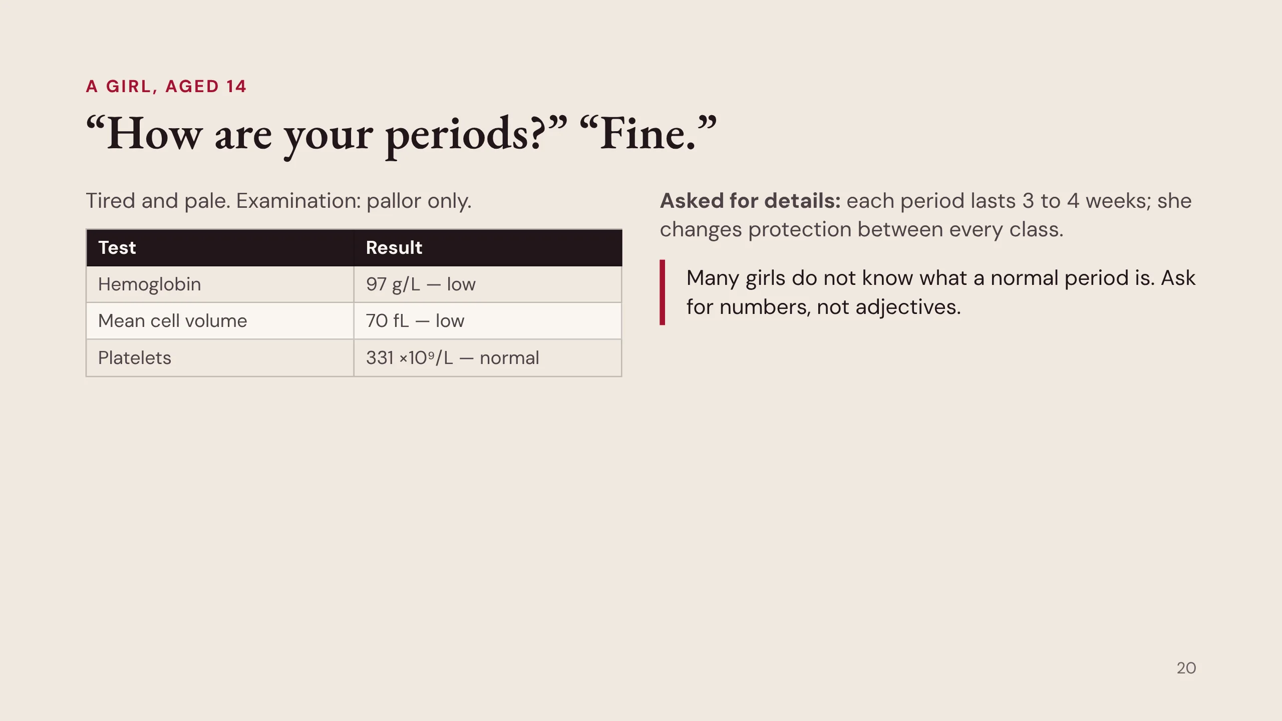
A 14-year-old girl sees her doctor because she is tired. She is pale. Her hemoglobin is 97 g/L and her red cells are small: this is iron deficiency. Her platelets are normal.
Where did the iron go? In a girl of this age the usual answer is menstrual loss. So the doctor asks about her periods, and she says they are fine.
“Fine” should open the question, not close it. Asked for details, she says each period lasts three to four weeks, and she changes her pad between every class at school. That is heavy bleeding by any standard, and it explains her anemia.
The lesson: many girls have nothing to compare their periods with, and their mothers and sisters may bleed the same way because the same disorder runs in the family. Ask for numbers, not adjectives: how many days, how often products are changed, clots, changing at night, soaking through clothes. (Case courtesy of @acweyand.)
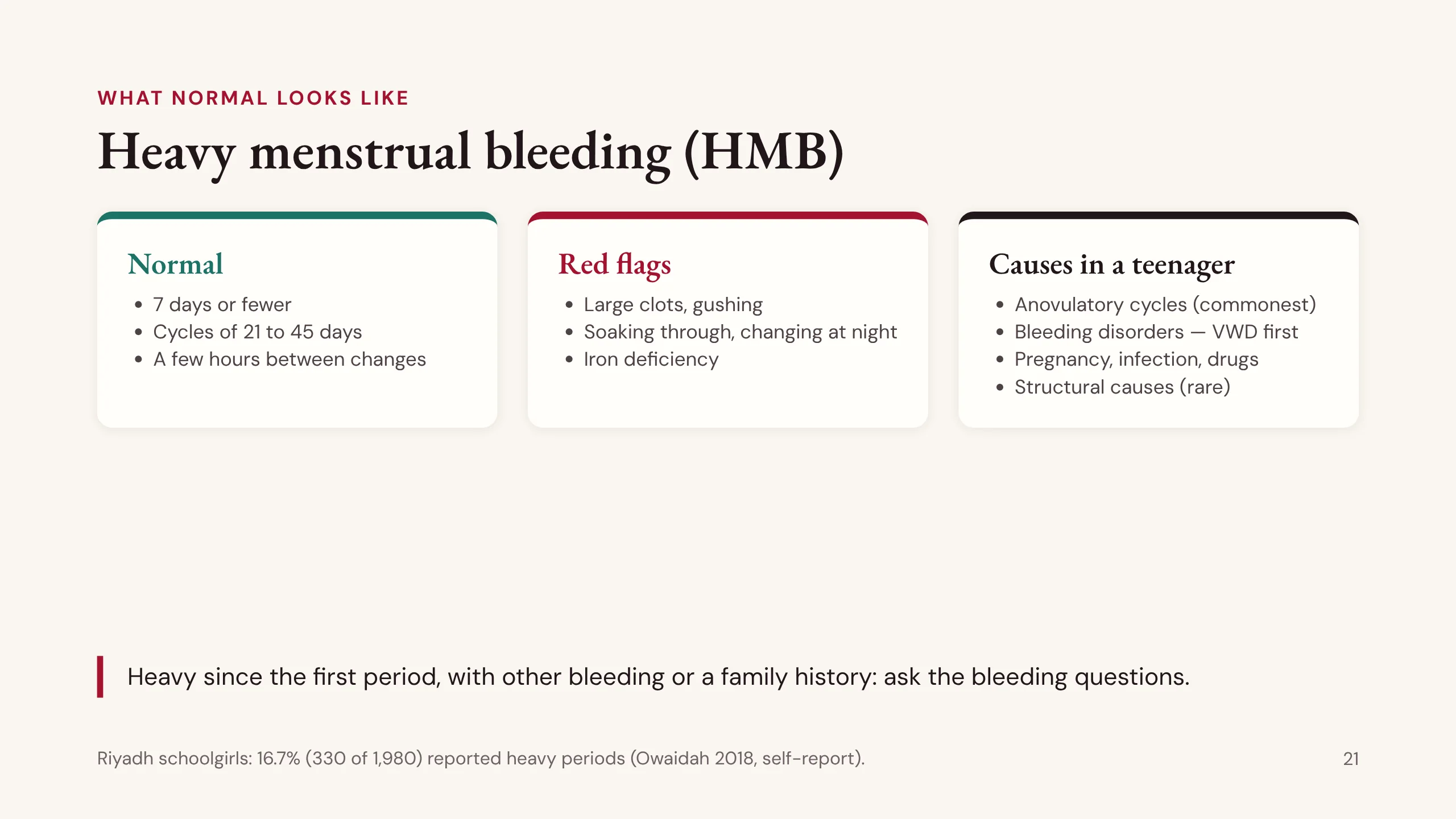
Heavy menstrual bleeding (HMB)
To call a period heavy you need a standard. A normal period lasts 7 days or fewer, cycles come 21 to 45 days apart, and a few hours pass between changes of protection.
The red flags: large clots, a gushing feeling, soaking through clothes, changing at night, and iron deficiency.
Heavy periods are common. A Riyadh school survey of 1,980 girls found 16.7% reported them (Owaidah 2018, self-report). So a bleeding disorder that shows itself this way is easy to lose among many girls who bleed heavily for other reasons.
In a teenager the commonest cause is anovulatory cycles, because the hormonal axis is still immature. Bleeding disorders come next, and von Willebrand disease above all. Fibroids and polyps, which are common in older women, are rare at this age.
The practical rule: heavy bleeding since the first period, in a girl with other bleeding or a family history, must not be put down to anovulation until you have asked the bleeding questions.
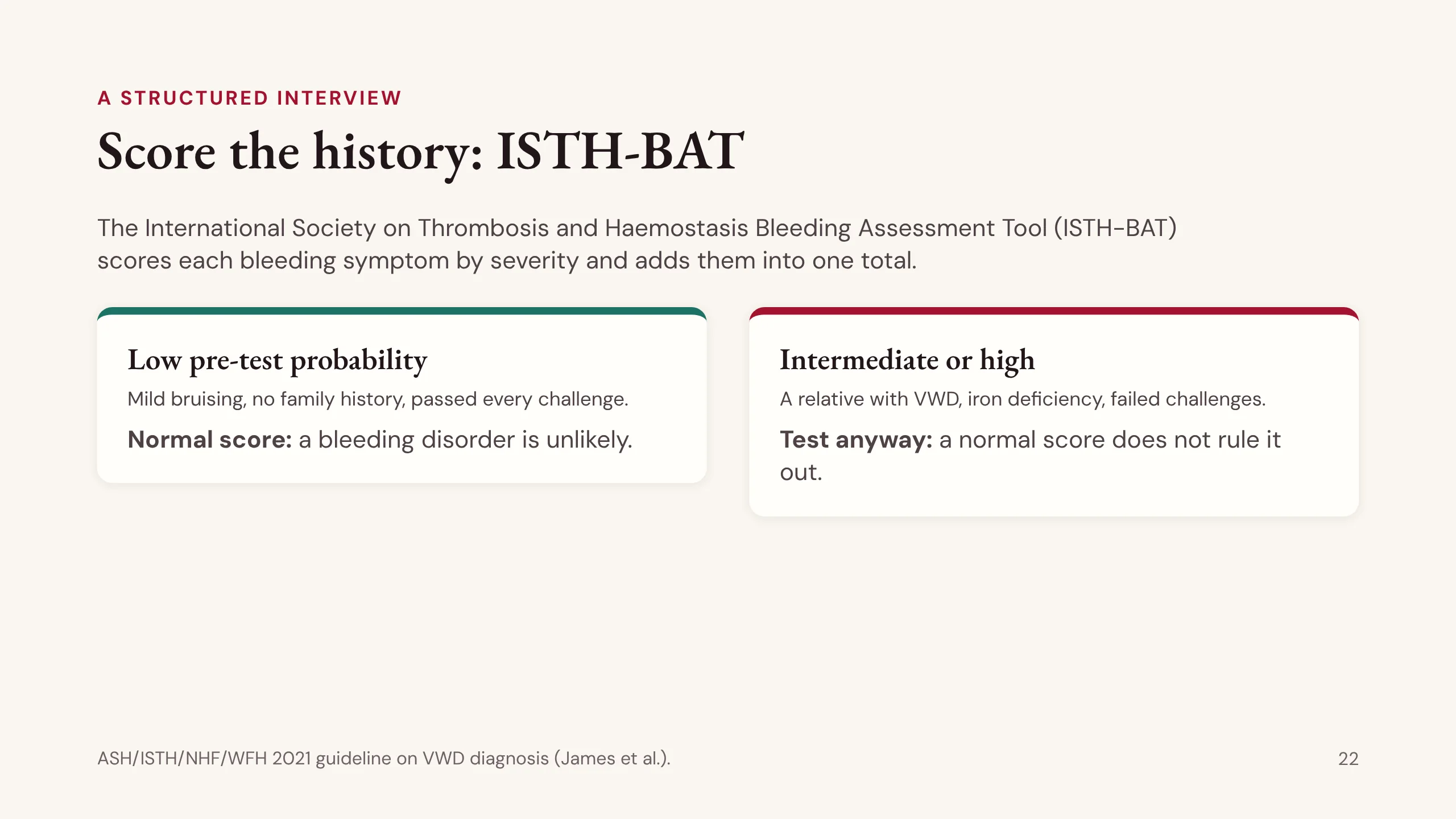
Score the history: ISTH-BAT
A bleeding history from memory is vague. The International Society on Thrombosis and Haemostasis Bleeding Assessment Tool (ISTH-BAT) makes it structured. It asks about each bleeding symptom, scores how severe it was (for example, whether it needed a doctor, a transfusion or an operation) and adds the scores into one total.
Know its limit. The 2021 guideline on the diagnosis of von Willebrand disease (ASH, ISTH, NHF and WFH; James and colleagues) says the score is useful when the chance of a disorder is low to begin with: mild bruising, no family history, every challenge passed. There a normal score is reassuring.
When the chance is intermediate or high, such as a relative with VWD, heavy periods that have caused iron deficiency, or failed challenges, go straight to blood tests. A normal score in that patient does not rule a disorder out.
Patient D has lifelong heavy periods, bruising, and bleeding after her wisdom teeth. Her probability is high. She needs testing whatever the score says.
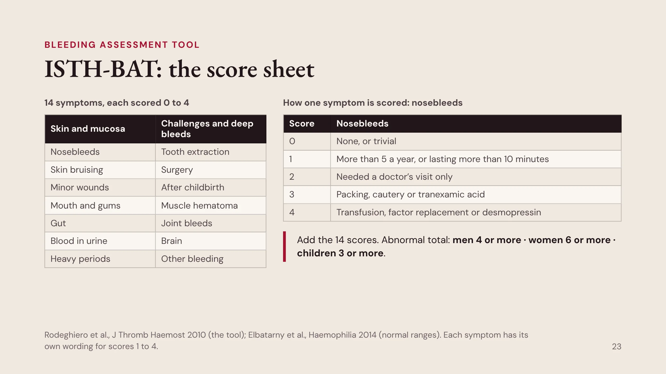
ISTH-BAT: the score sheet
This is what the ISTH-BAT actually scores. It asks about 14 kinds of bleeding. On the left are the bleeds of skin and mucosa, the primary-hemostasis pattern: nosebleeds, skin bruising, minor wounds, the mouth and gums, the gut, blood in the urine and heavy periods. On the right are the challenges and the deep bleeds: tooth extraction, surgery, childbirth, muscle hematoma, joint bleeds, bleeding into the brain, and any other bleeding.
Each symptom is scored from 0 to 4, and the scale climbs the same way for all of them: from nothing, to bleeding that needed a doctor, to bleeding that needed treatment, to bleeding that needed transfusion, factor replacement, desmopressin or surgery. Nosebleeds are the worked example. 0: none or trivial. 1: more than five a year, or lasting more than ten minutes. 2: needed a doctor’s visit only. 3: needed packing, cautery or tranexamic acid. 4: needed a transfusion, factor replacement or desmopressin. Each symptom has its own wording, but the steps are the same.
The 14 scores are added. A total of 4 or more is abnormal in adult men, 6 or more in adult women (higher because of periods and childbirth), and 3 or more in children (Rodeghiero and colleagues 2010; normal ranges from Elbatarny and colleagues 2014).
And remember the previous slide: a normal total is reassuring only when the pre-test probability is low. With a convincing history or a family history, test anyway.
Part 3: The first panel
Part 3 is question 4: the first panel. At this stage you need to read the pattern and know when to refer. How the laboratory then pins down an unexplained long result is taught in a later lecture.
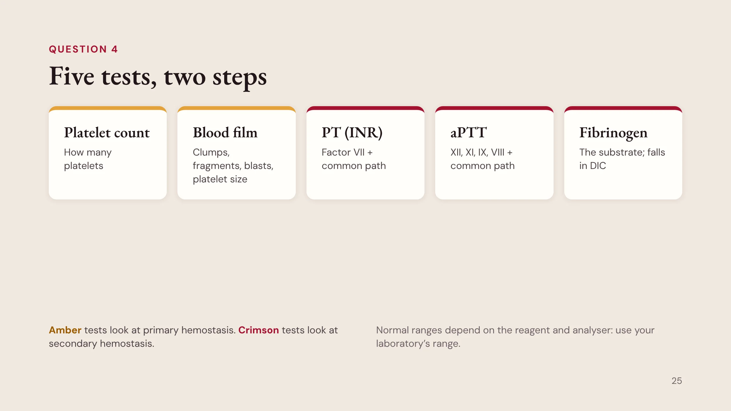
Five tests, two steps
The first panel is five tests, and they map onto the two arms.
The platelet count and the blood film look at primary hemostasis. The film is part of the panel, not an extra. Never interpret a low count without it: it shows clumps, fragmented red cells, blasts and platelet size.
The PT, the aPTT and the fibrinogen look at secondary hemostasis. The PT is reported as the INR for patients on warfarin. Fibrinogen is the substrate of the last step; it falls in disseminated intravascular coagulation (DIC).
Normal ranges depend on the reagent and the analyser, so always use the range on your own laboratory’s report.
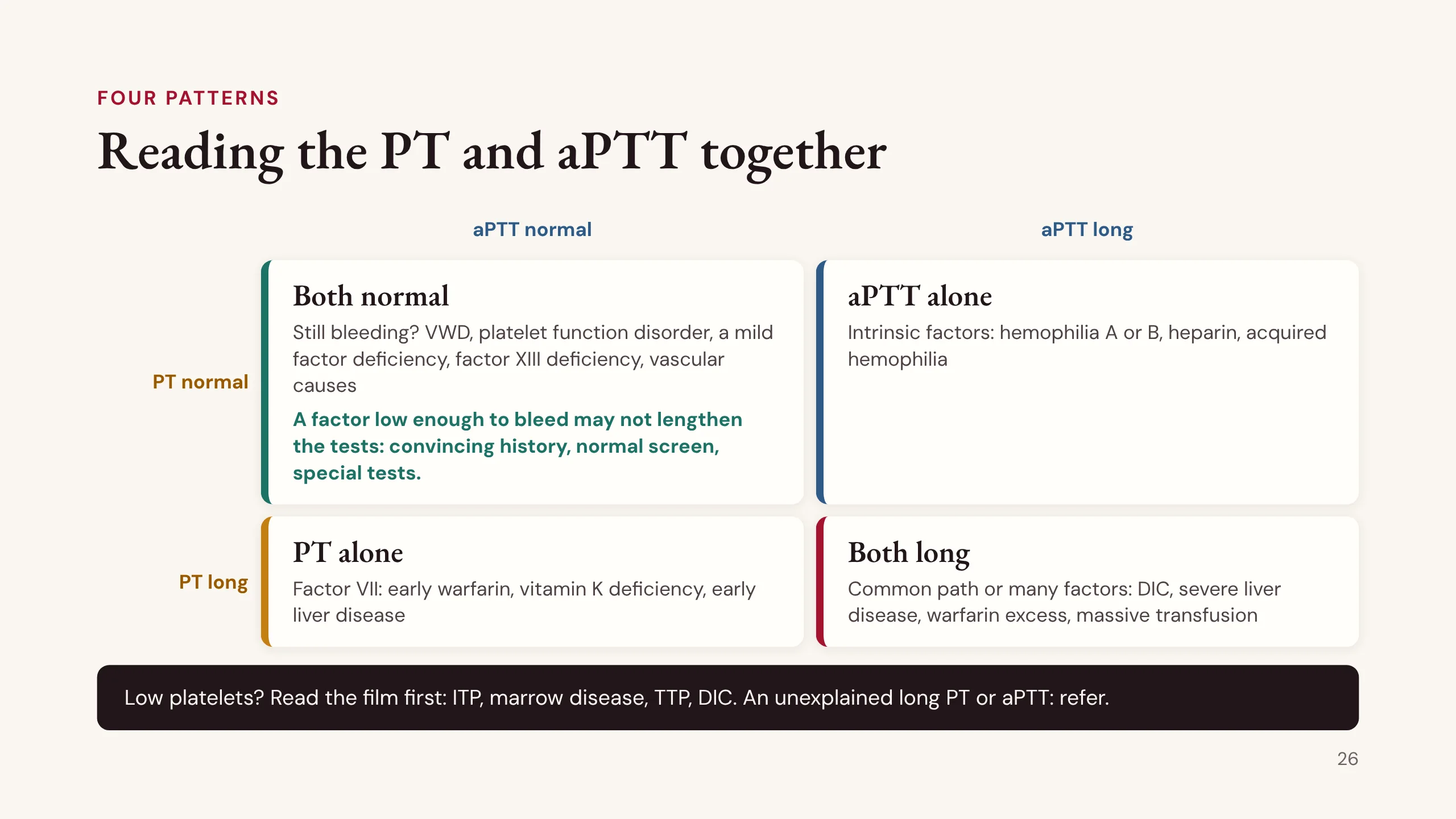
Reading the PT and aPTT together
This grid is the key slide of Part 3. Read the PT and the aPTT together.
Both normal. If the patient still bleeds, think of what the screen misses: von Willebrand disease, a platelet function disorder, a mild factor deficiency, factor XIII deficiency and causes in the vessel wall.
The mild factor deficiency needs a word. The PT and aPTT only lengthen when a factor falls quite low, often below about 30 to 40% of normal, depending on the reagent. A patient with factor VIII, IX or XI at a level just above that has a normal screen, yet may bleed badly after a tooth extraction or an operation. So when the history is convincing and the screen is normal, the screen has not answered the question: specific tests (von Willebrand studies, factor levels, platelet function) are needed. The next slide is about this box.
aPTT long alone. The intrinsic factors: hemophilia A or B, heparin, and acquired hemophilia, an antibody against factor VIII.
PT long alone. Factor VII, which falls first when vitamin K factors are not being made: early warfarin, vitamin K deficiency, early liver disease.
Both long. The common path, or many factors at once: DIC, severe liver disease, warfarin excess, massive transfusion.
And if the platelets are low, read the film first: immune thrombocytopenia (ITP), marrow disease, thrombotic thrombocytopenic purpura (TTP), DIC.
If a long PT or aPTT has no explanation in the drug chart or the history, refer. The laboratory work-up that follows is taught later.
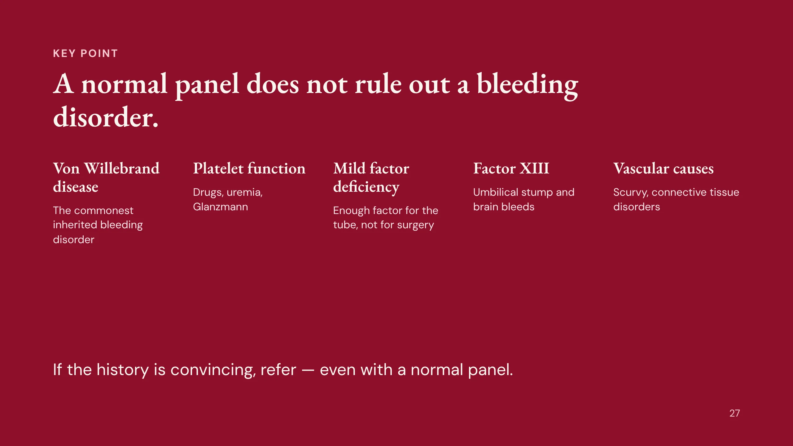
A normal panel does not rule out a bleeding disorder
This is the most important single fact in Part 3. A normal count, PT and aPTT do not exclude a bleeding disorder.
Von Willebrand disease is the commonest inherited bleeding disorder, and in mild disease factor VIII is high enough to keep the aPTT normal.
Platelet function disorders: the count is normal but the platelets do not work. Drugs most often, uremia, and rarely Glanzmann thrombasthenia.
Mild factor deficiencies: there is enough factor to clot in the tube in normal time, but not enough to stop bleeding after a challenge such as surgery or a dental extraction. Mild hemophilia A or B and factor XI deficiency are the usual examples; specific factor assays find them.
Factor XIII deficiency: factor XIII cross-links fibrin after the clot forms, and the clotting tests stop the clock when the clot first appears, so they are normal. It classically bleeds from the umbilical stump and into the brain.
Vascular causes: the problem is in the vessel wall, not the blood, as in scurvy or connective tissue disorders.
So if the history is convincing, refer, even with a normal panel. Patient D proves the point.
Part 4: The disorders, step by step
Part 4 takes the disorders one patient at a time. Primary hemostasis first (the platelets and von Willebrand factor), then secondary hemostasis (hemophilia), then the acquired problems of the ward. Each patient runs the same four questions, and each disorder ends with its first-line care.
Patient B: a woman, aged 24
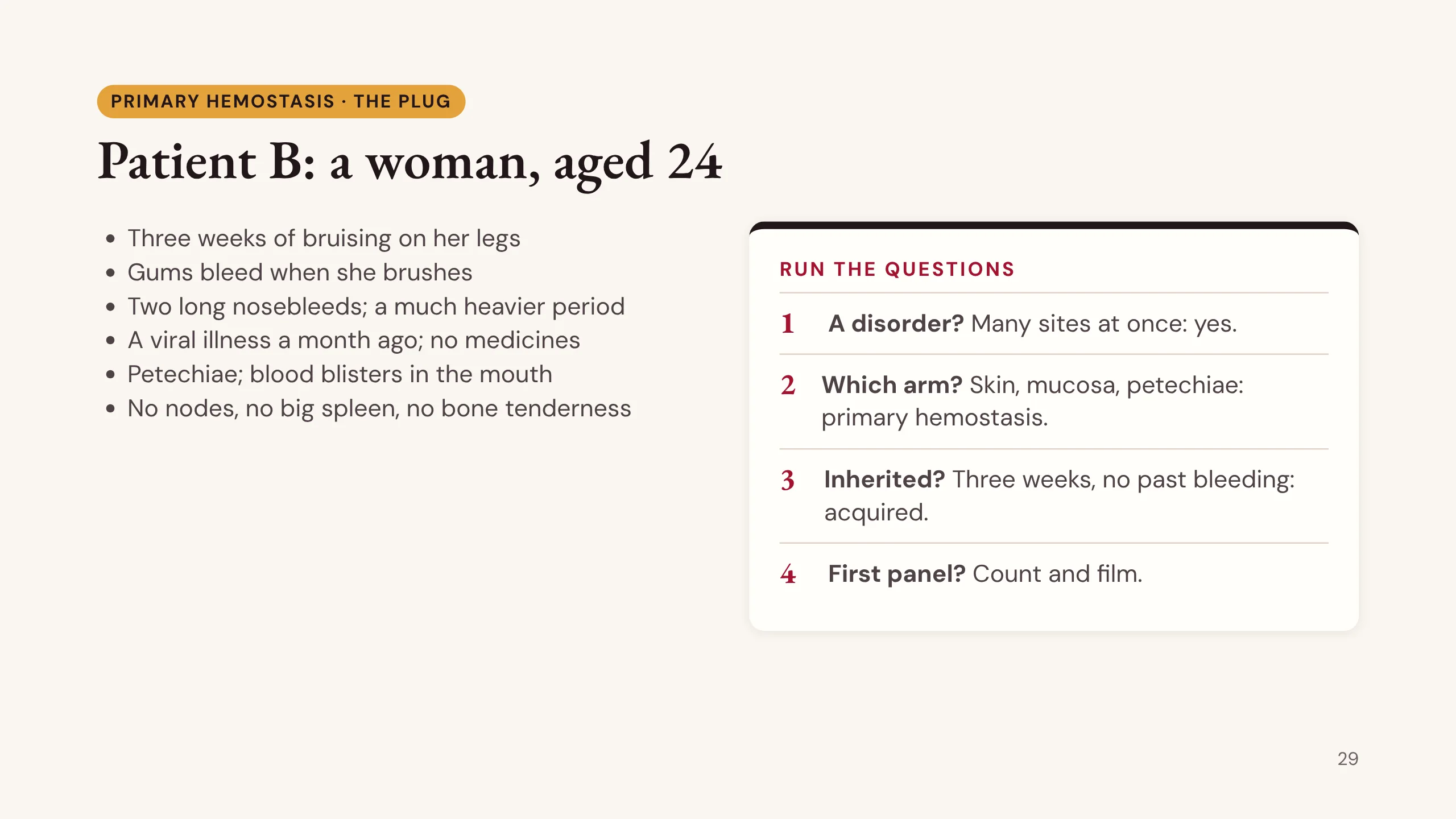
Patient B, from Case 53 in the casebook. A woman of 24. For three weeks she has bruised easily on her legs and her gums bleed when she brushes. She has had two long nosebleeds and a much heavier period. She had a viral illness a month ago and takes no medicines.
On examination: petechiae on the shins and forearms, and blood blisters inside the mouth. No enlarged lymph nodes, no enlarged spleen, no bone tenderness.
Run the questions. A disorder? Yes, many sites at once. Which arm? Skin and mucosa with petechiae: primary hemostasis. Inherited? Three weeks of new symptoms with no past bleeding: acquired. First panel? For a primary pattern, start with the count and the film.
The blood blisters in the mouth are called wet purpura. Keep them in mind; they change the urgency.
Patient B: the panel and the film
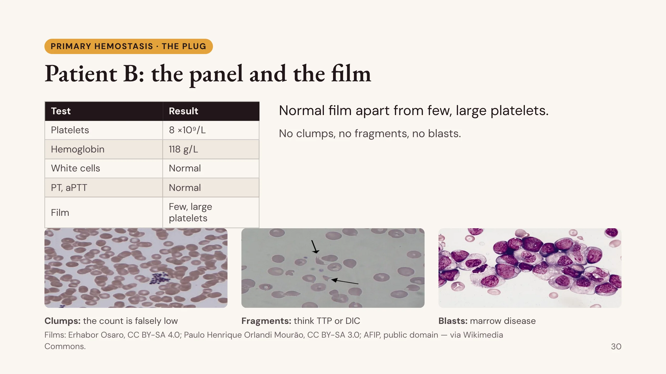
Her first panel. Platelets 8 ×10⁹/L. Hemoglobin 118 g/L. White cells normal. PT and aPTT normal. Low platelets, everything else close to normal.
Before you act on a low count, look at the film. Hers shows few platelets, some large, and nothing else.
The film could have shown other things, and each changes the problem. Clumps: the platelets stuck together in the tube and the machine counted each clump as one cell, so the count is false; repeat it in a different tube. Fragmented red cells (schistocytes): something is shearing red cells; think of TTP or DIC, both emergencies. Blasts: marrow disease, such as acute leukemia.
A low count has three causes: the marrow makes too few, the body destroys too many, or a big spleen holds them back.
The three photographs are from Wikimedia Commons: platelet clumping by Erhabor Osaro (CC BY-SA 4.0), schistocytes by Paulo Henrique Orlandi Mourão (CC BY-SA 3.0), and blasts from the Armed Forces Institute of Pathology (public domain).
Why the count falls in ITP
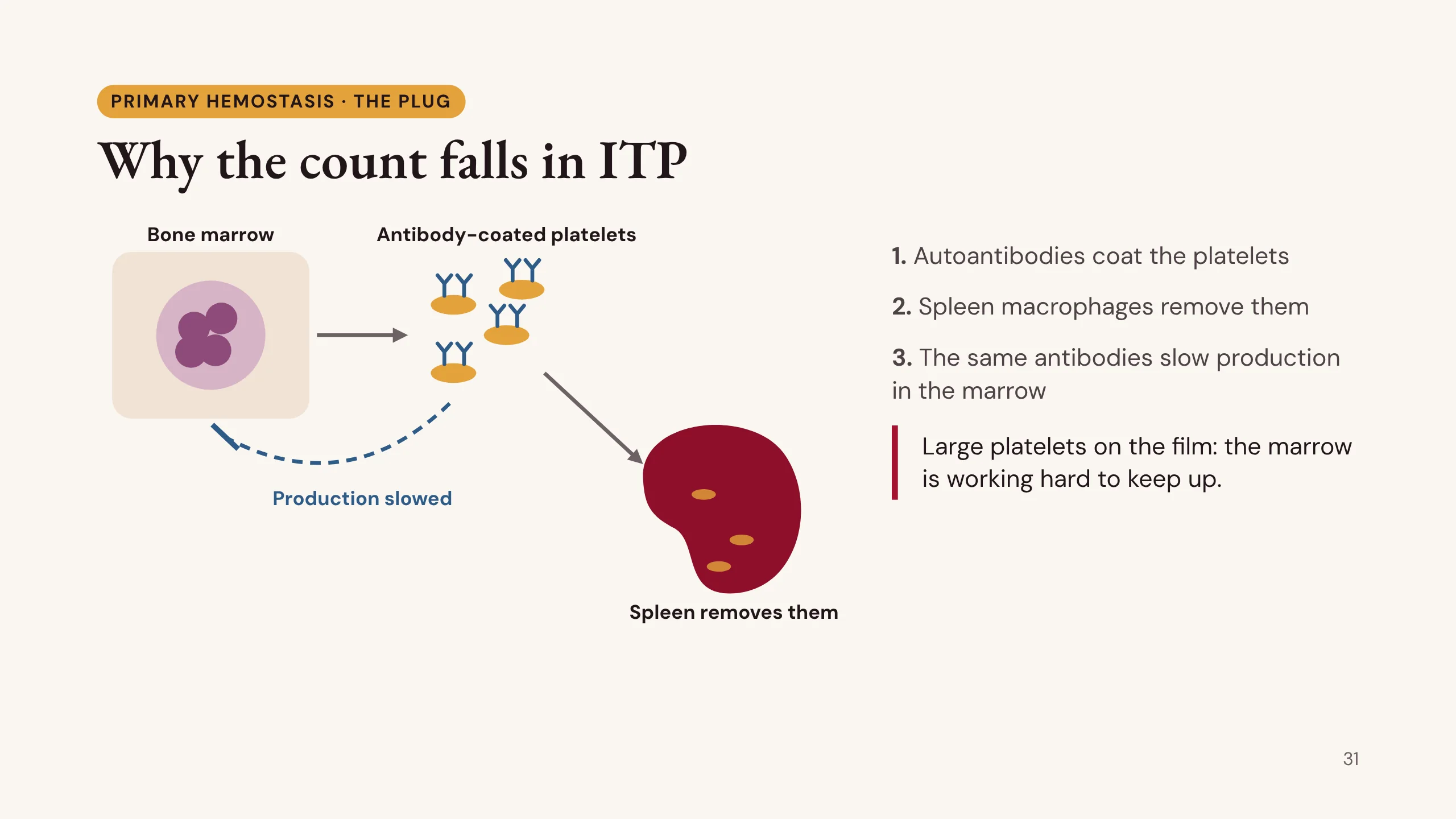
Why does the count fall in immune thrombocytopenia?
1. The patient makes autoantibodies against their own platelets, often after a viral illness, especially in children.
2. Macrophages in the spleen recognise the antibody-coated platelets and remove them, so platelets survive only a short time.
3. The same antibodies also act on the megakaryocytes in the marrow and slow platelet production. So ITP is both too much destruction and too little production.
The marrow responds by releasing young, large platelets. That is why large platelets on the film fit ITP. The drawing was made for this lecture.
Immune thrombocytopenia (ITP)
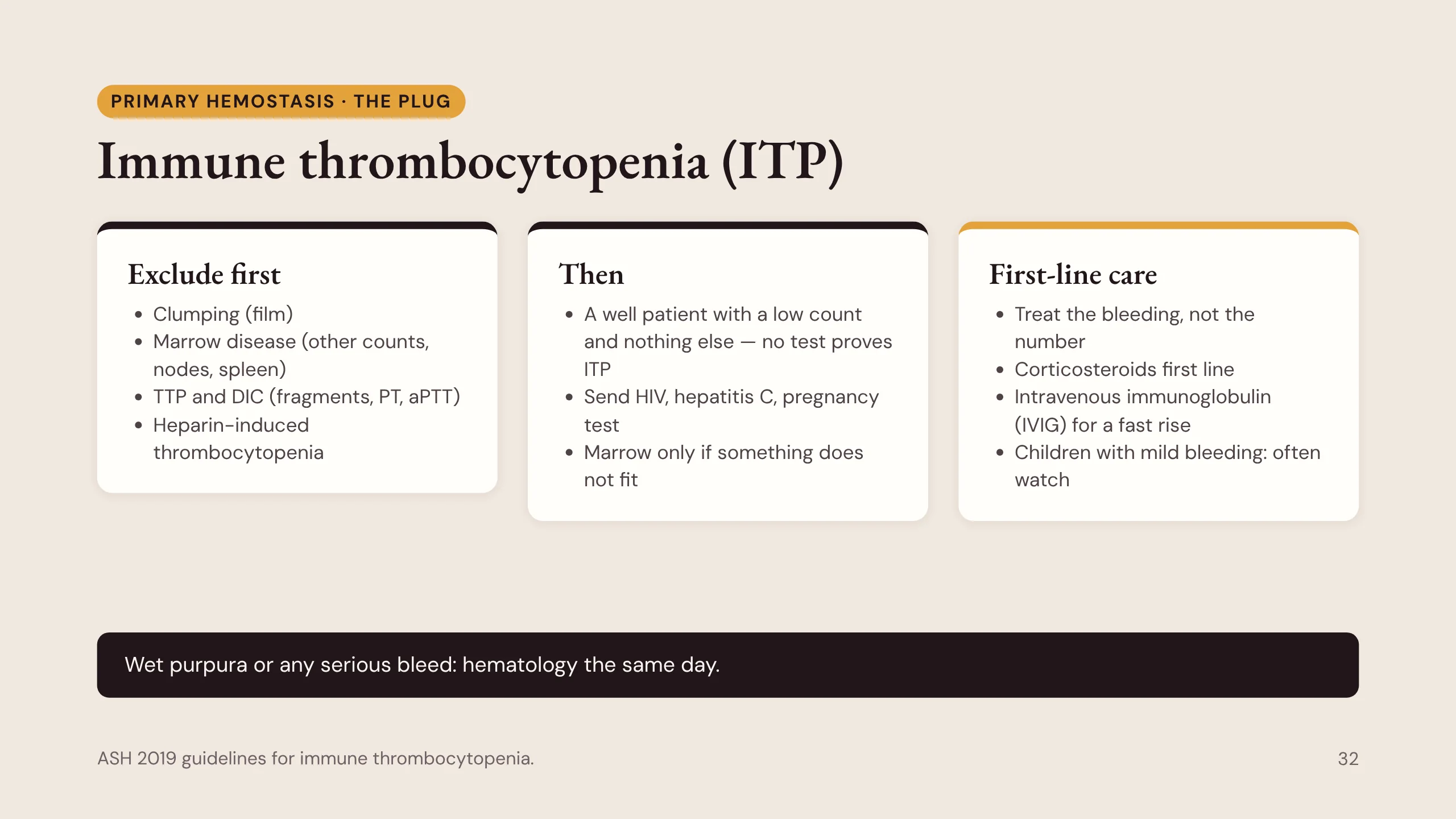
ITP is what is left once everything else is excluded. No test proves it.
Exclude first. Clumping: her film shows none. Marrow disease: no nodes, no big spleen, no bone pain, and the other counts are near normal. TTP and DIC: no fragments, and normal PT and aPTT. Heparin-induced thrombocytopenia: no heparin.
Then. ITP can follow another illness, so send HIV and hepatitis C serology and a pregnancy test. A bone marrow test is not needed in a typical young patient; do it only when something does not fit, such as older age, fever or weight loss, other low counts, an abnormal film, or no response to treatment.
First-line care. Treat the bleeding, not the number. Corticosteroids are the first-line treatment. Intravenous immunoglobulin (IVIG) is used when a fast rise is needed, such as serious bleeding or before surgery. Children with mild bleeding are often watched without treatment. Platelet transfusion is kept for life-threatening bleeding.
Wet purpura, retinal bleeding or any serious bleed means hematology the same day. (ASH 2019 guidelines for immune thrombocytopenia.)
When platelets are present but do not work
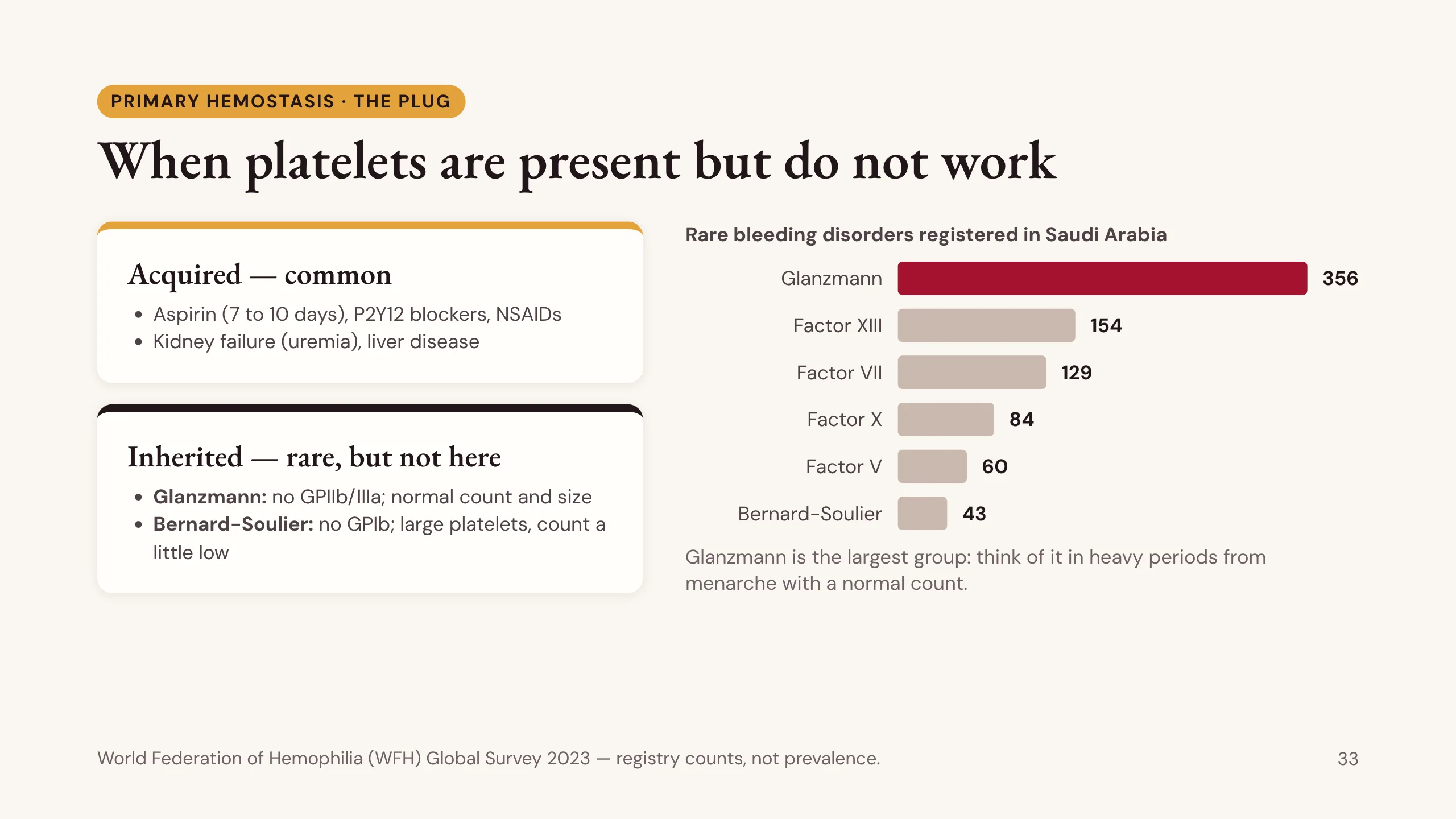
Sometimes the platelets are there in normal numbers but do not work. The bleeding is the primary type, and the count and screen are normal, so it is easy to miss.
Acquired causes are by far the commonest: aspirin (its effect lasts 7 to 10 days), the P2Y12 blockers such as clopidogrel, NSAIDs, kidney failure and liver disease.
Inherited causes are rare in most places, but not here. Glanzmann thrombasthenia: no GPIIb/IIIa, a normal count and normal-sized platelets that do not aggregate, and severe bleeding. Bernard-Soulier syndrome: no GPIb, large platelets and a count that is a little low.
The chart is from the World Federation of Hemophilia (WFH) Global Survey 2023. Glanzmann is the largest group of rare bleeding disorders registered in Saudi Arabia, with 356 patients, ahead of factor XIII (154) and factor VII (129). These are registry counts, not prevalence. Glanzmann is recessive, so it is commoner where parents are related.
Think of it in a girl with heavy periods from menarche, a normal count, a normal screen and normal von Willebrand studies, and refer her for platelet function testing.
Patient D: a woman, aged 19
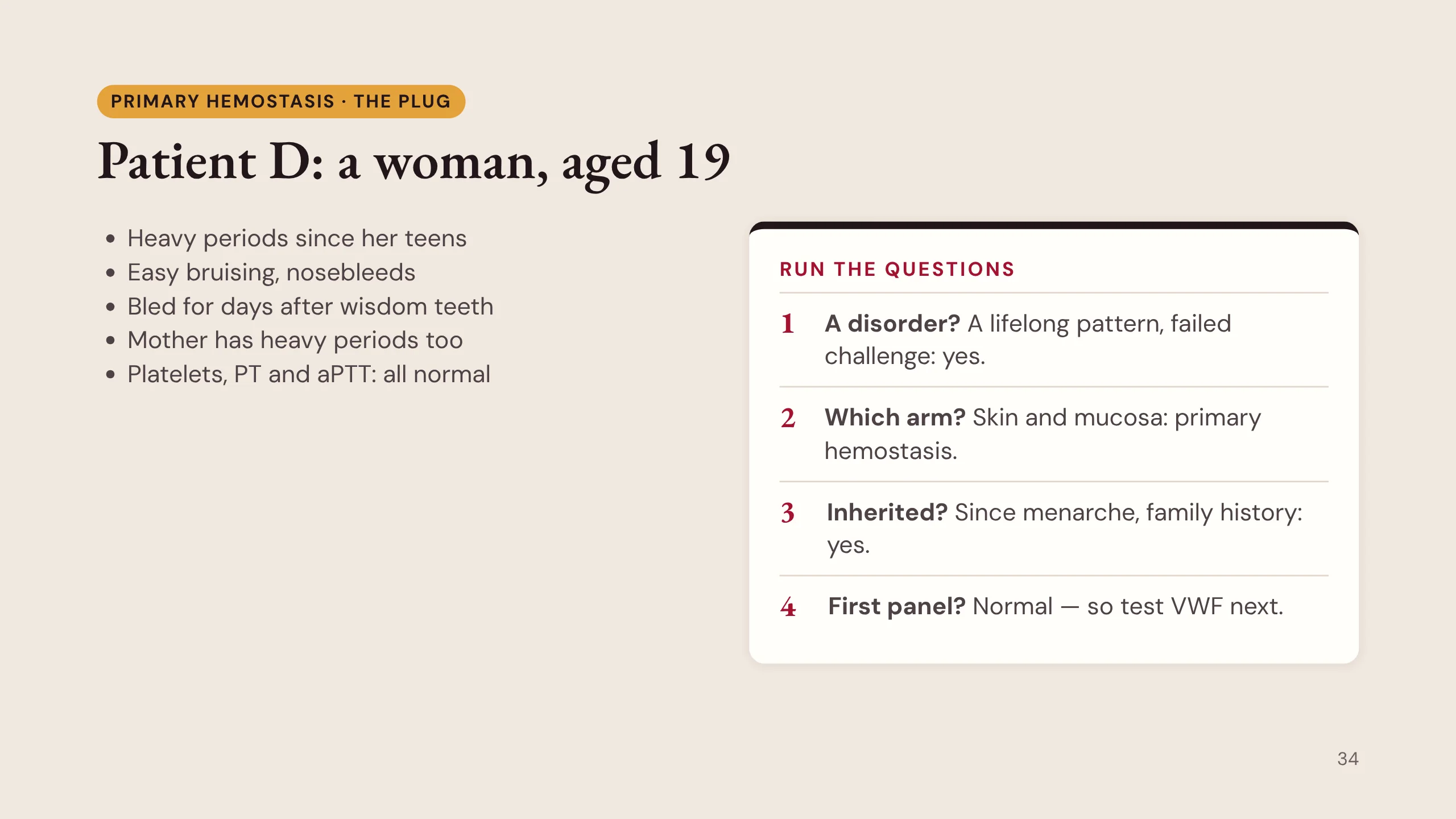
Patient D, a woman of 19. Heavy periods since her teens, easy bruising, nosebleeds, and days of bleeding after her wisdom teeth were removed. Her mother also has heavy periods. Her platelet count, PT and aPTT are all normal.
Many students, and many doctors, would look at three normal results and tell her she has no bleeding disorder. That would be wrong.
Run the questions. A disorder? A lifelong pattern and a failed challenge: yes. Which arm? Skin and mucosa: primary hemostasis. Inherited? Since menarche, with a family history: yes. First panel? Normal, which is exactly the box on the grid that sends you to von Willebrand studies.
Von Willebrand factor has two jobs
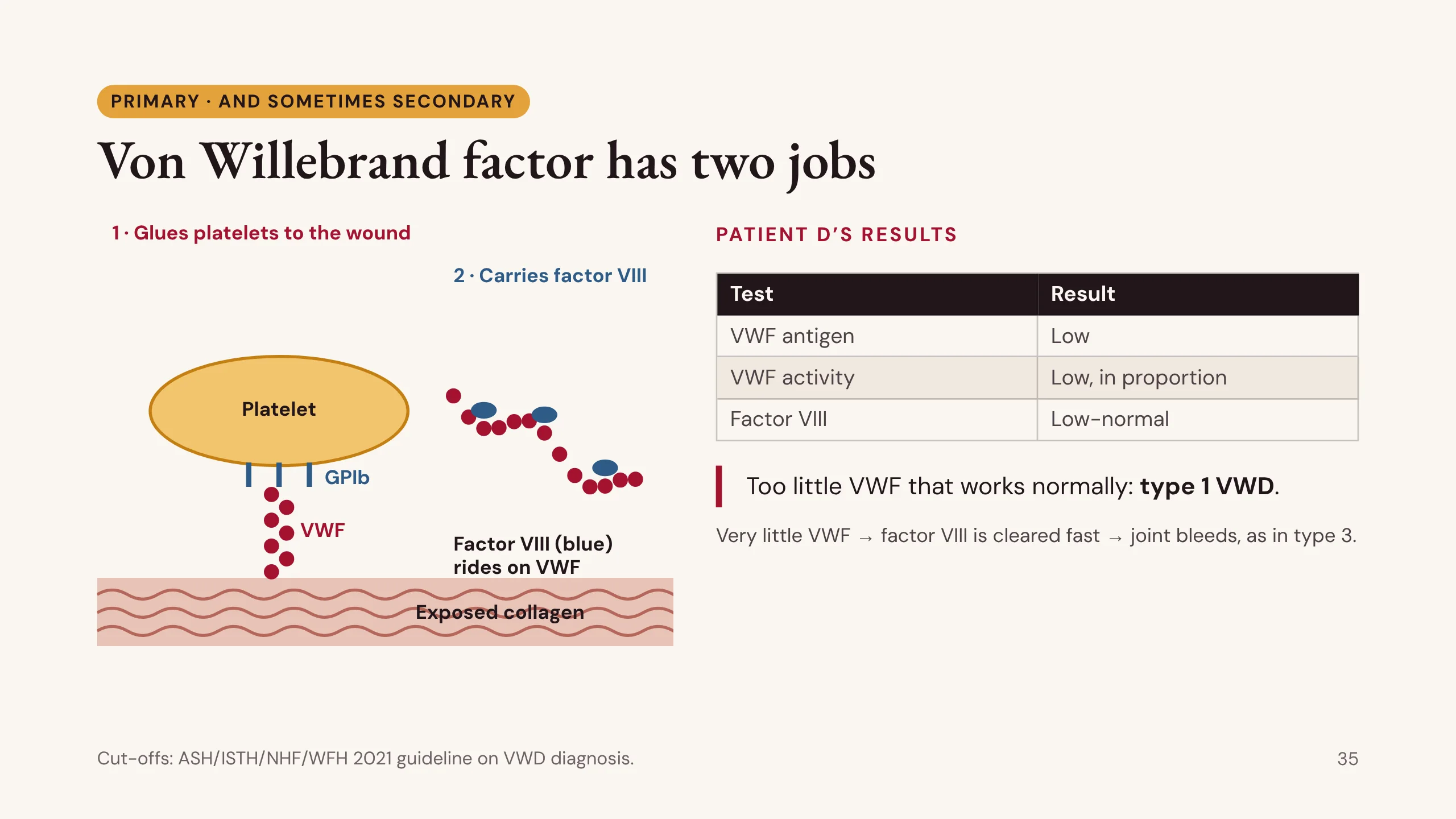
Von Willebrand factor has two jobs, and they explain the whole disease.
Job one: it glues platelets to the wound. One end binds exposed collagen, the other binds the platelet’s GPIb receptor. When it is short, platelets do not stick, and the bleeding is the primary type: nosebleeds, bruising, heavy periods.
Job two: it carries factor VIII in the blood and protects it from being cleared. When VWF is very low, factor VIII falls too, and the patient bleeds into joints like a person with hemophilia. That is type 3.
Patient D’s results. VWF antigen, the amount, is low. VWF activity, how well it binds platelets, is low in proportion. Factor VIII is low-normal. Too little VWF that works normally: type 1 von Willebrand disease.
The cut-offs come from the 2021 international guideline: a VWF level below 0.30 IU/mL is VWD whatever the history, and below 0.50 IU/mL it is VWD in a patient with abnormal bleeding. Levels are lower in blood group O. And because VWD is usually autosomal, her family should be tested. The drawing was made for this lecture.
Von Willebrand disease: three types
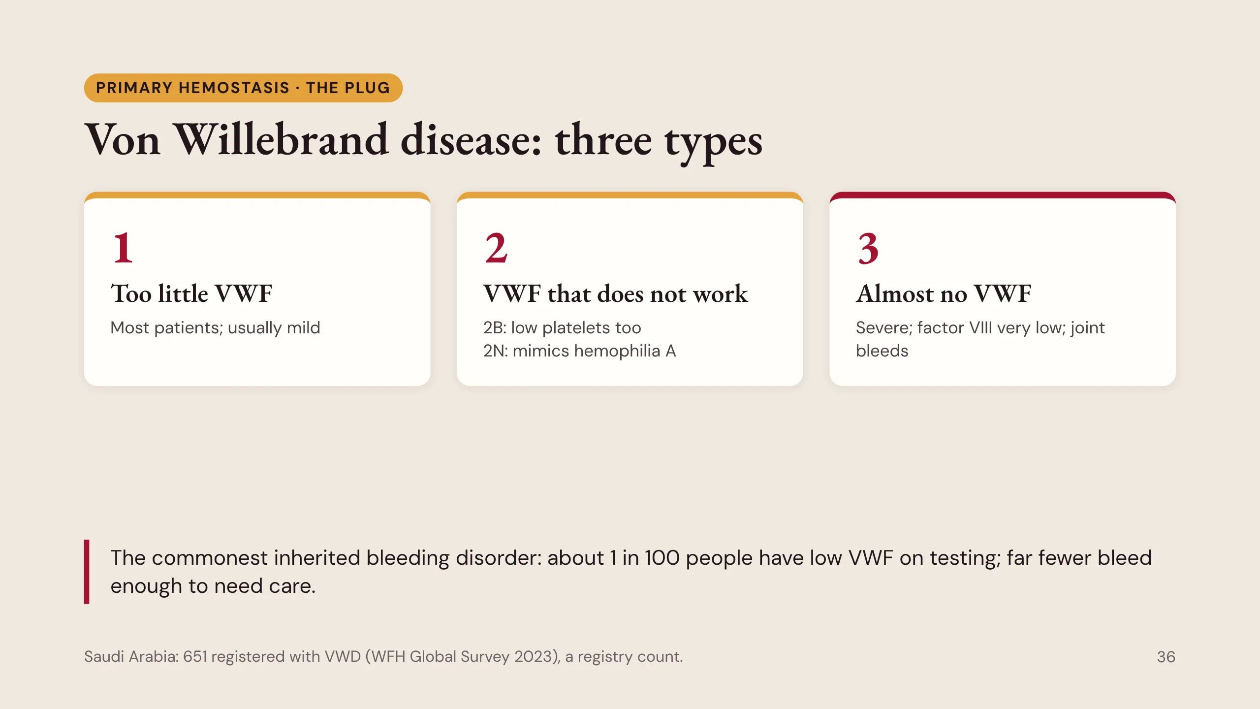
At this level, learn the types as clinical pictures.
Type 1: too little of a normal protein. Most patients, and usually mild. Patient D.
Type 2: the protein is there but does not work. Two subtypes are worth knowing. 2B binds platelets too well, even in the circulation, so platelets are cleared and the count is low. 2N cannot carry factor VIII, so factor VIII is low and the patient looks like mild hemophilia A, but it is autosomal and affects women. Test VWF before labelling a woman with hemophilia.
Type 3: almost no VWF. Severe, with very low factor VIII and joint bleeds.
How common? About 1 in 100 people have low VWF on testing, but far fewer bleed enough to need care. The WFH 2023 survey lists 651 people registered with VWD in Saudi Arabia, a registry count, not a prevalence.
Von Willebrand disease: first-line care
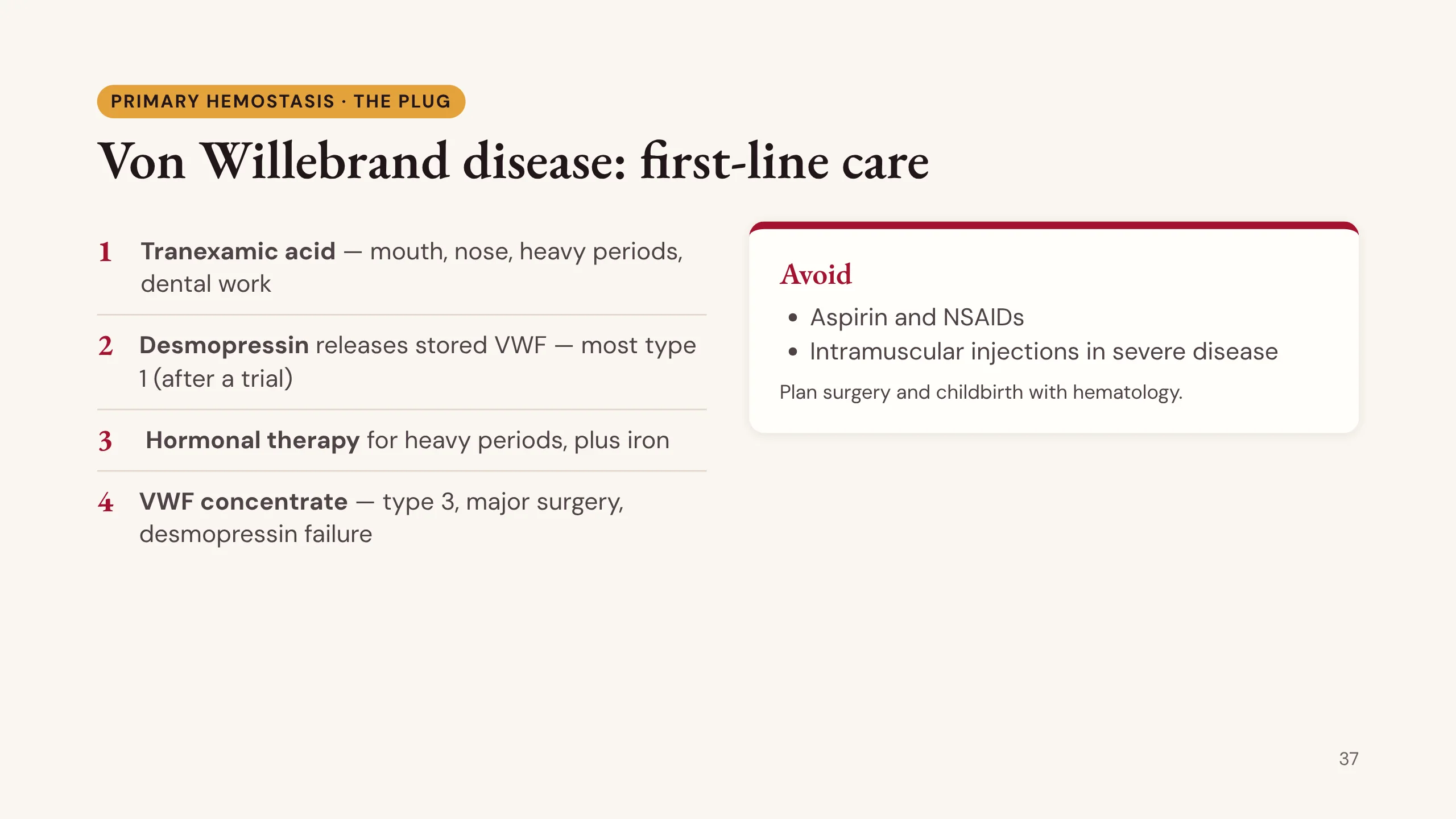
First-line care for von Willebrand disease.
1. Tranexamic acid, which blocks fibrinolysis. It helps bleeding from the mouth and nose, heavy periods and dental work.
2. Desmopressin, which releases the patient’s own stored VWF and factor VIII. It works in most type 1 patients, after a test dose shows a response. It is not used in type 2B (it lowers the platelet count further) and does not work in type 3.
3. Hormonal therapy for heavy periods, planned with gynecology, plus iron to treat the deficiency.
4. VWF-containing concentrate for type 3, for major surgery, and when desmopressin fails.
Avoid aspirin and NSAIDs, and intramuscular injections in severe disease. Plan surgery and childbirth with hematology. For patient D: tranexamic acid and hormonal therapy for her periods, iron, and a desmopressin trial.
Patient A: a boy, aged 2
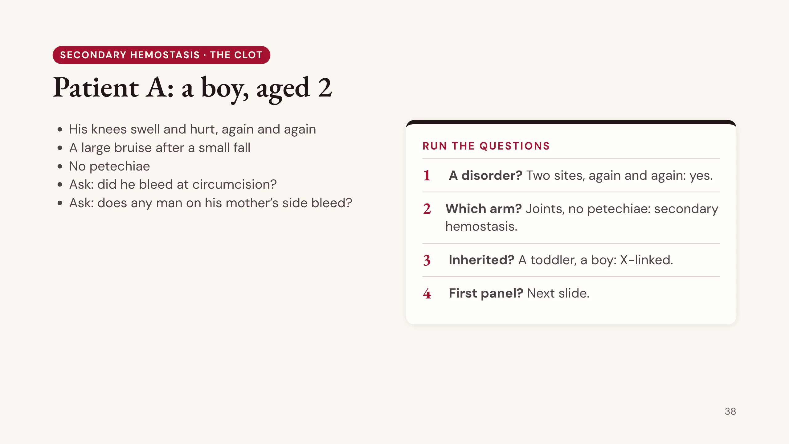
Patient A, from handbook chapter 25. A boy of 2. His knees swell and hurt, again and again, and he had a large bruise after a small fall. There are no petechiae.
Ask two questions: did he bleed at circumcision? Does any man on his mother’s side bleed?
Run the questions. A disorder? Two sites, again and again: yes. Which arm? Bleeding into joints, with no petechiae: secondary hemostasis. Inherited? A toddler, and a boy: think X-linked. First panel? Predict it before the next slide: which test will be long?
Patient A: the panel
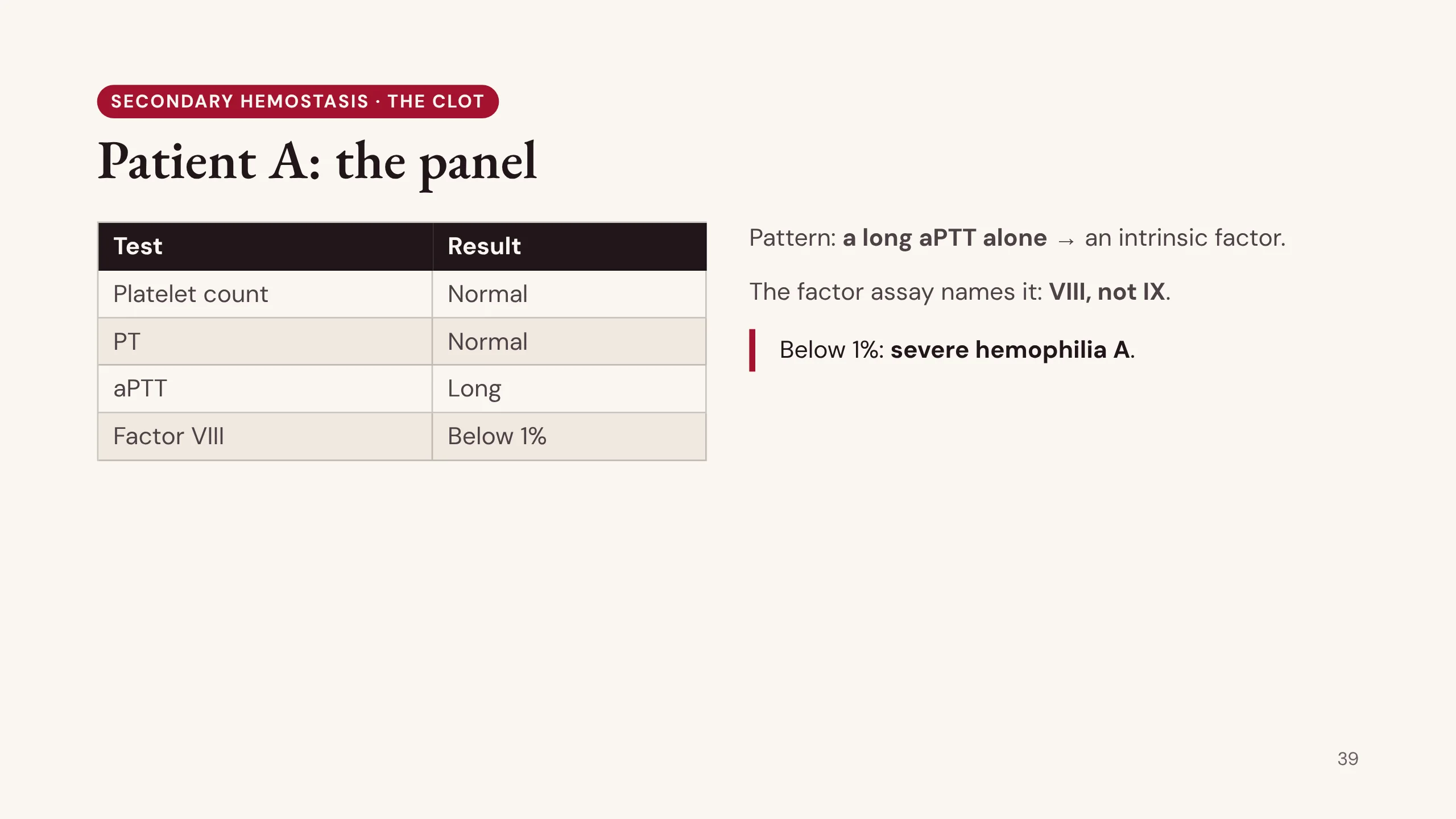
His panel. Platelet count normal. PT normal. aPTT long. That is the aPTT-alone box on the grid: an intrinsic factor.
The factor assay names it. Factor VIII is below 1%. This is severe hemophilia A.
Hemophilia A (factor VIII) and B (factor IX) look the same at the bedside. Only the assay tells them apart.
Hemophilia: the factor level sets the pattern
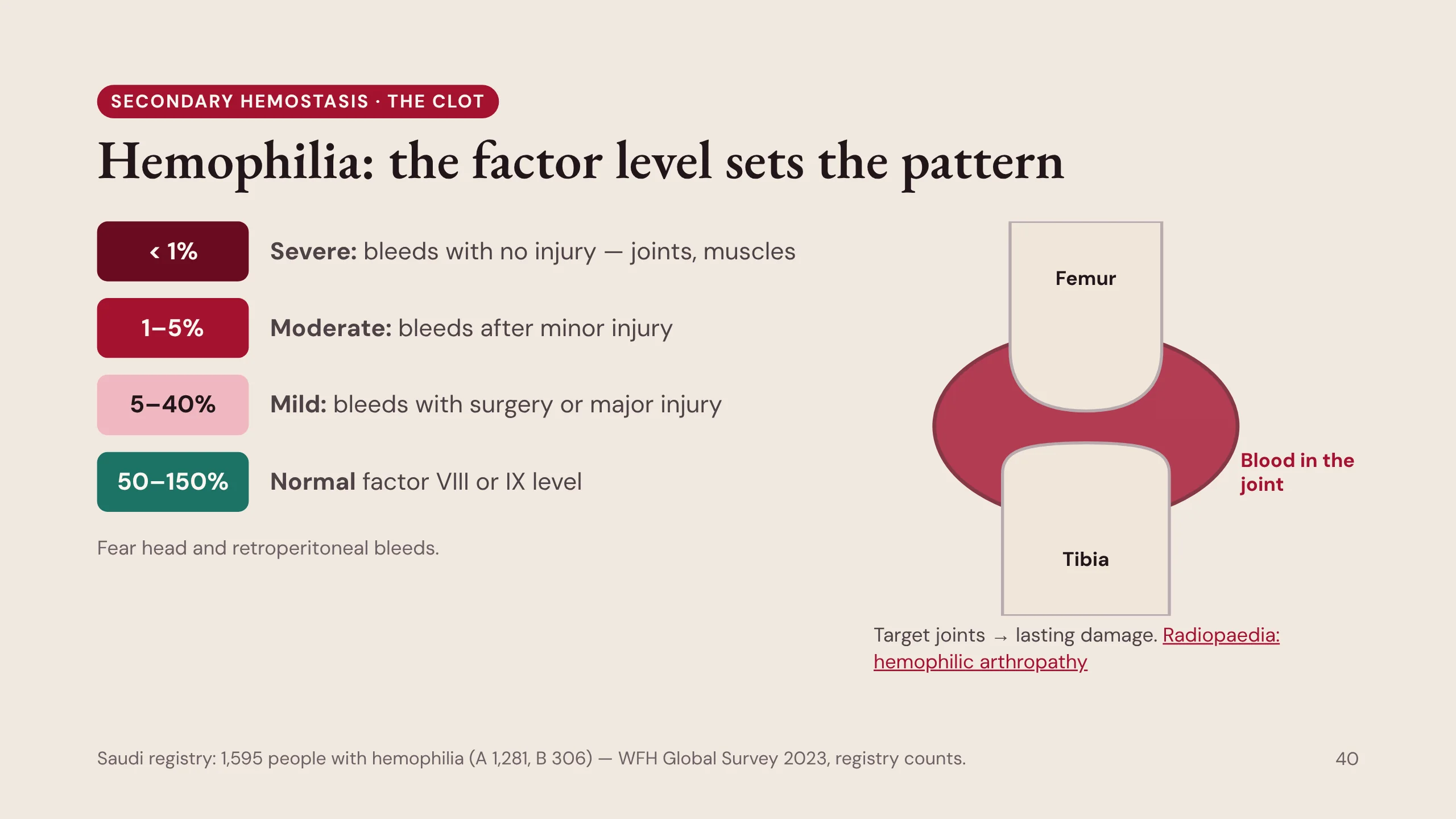
In hemophilia, the factor level sets the pattern.
Below 1% is severe: bleeding into joints and muscles with no injury at all. 1 to 5% is moderate: bleeding after minor injury. 5 to 40% is mild: bleeding mainly with surgery or major injury, so a mild patient may not be found until the first operation. A normal level is about 50 to 150%.
The hallmark is joint bleeding. Blood in the joint inflames the lining (the synovium), the joint bleeds again more easily, and over years it becomes a damaged target joint: knees, elbows and ankles. The bleeds to fear are inside the head and behind the abdomen (retroperitoneal).
The Saudi registry for 2023 counts 1,595 people with hemophilia: 1,281 with A and 306 with B (WFH Global Survey 2023, registry counts).
The knee drawing was made for this lecture. For imaging of a damaged joint, see Radiopaedia — haemophilic arthropathy.
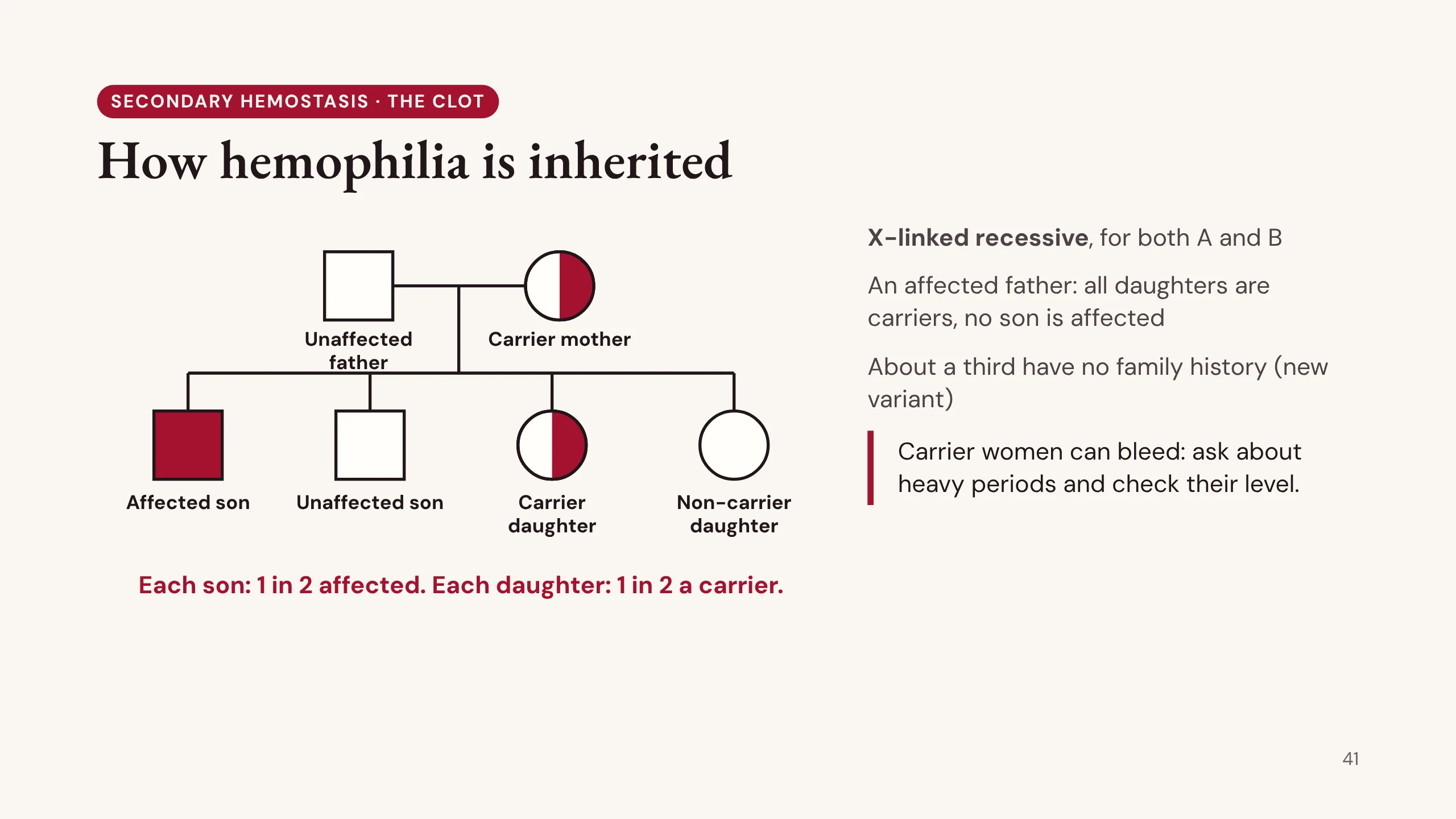
How hemophilia is inherited
Hemophilia A and B are both X-linked recessive.
Follow the tree. The mother carries the gene on one X chromosome. Each son receives one of her two X chromosomes, so each son has a 1 in 2 chance of being affected. Each daughter has a 1 in 2 chance of being a carrier.
An affected father passes his X to every daughter and his Y to every son. So all his daughters are carriers, and none of his sons is affected.
About a third of patients have no family history: the variant is new.
Carrier women can bleed, if their factor level is low. Ask about heavy periods and bleeding after childbirth, and check their level. Do not dismiss a carrier who bleeds.
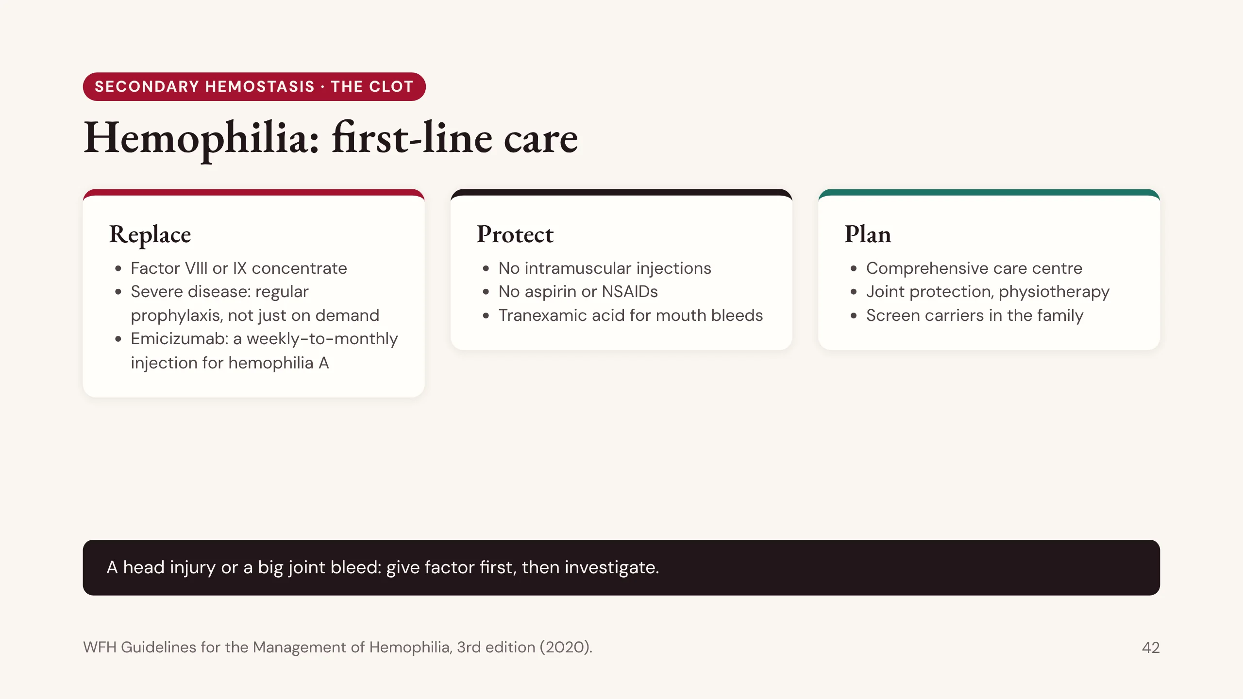
Hemophilia: first-line care
First-line care in hemophilia, in three words.
Replace. Give the missing factor as a concentrate: factor VIII for A, factor IX for B. In severe disease the standard is regular prophylaxis to prevent bleeds, not just treatment after they happen. Emicizumab, an antibody given by injection under the skin weekly to monthly, does the job of factor VIII and is used for prophylaxis in hemophilia A.
Protect. No intramuscular injections and no aspirin or NSAIDs, because both cause bleeding. Tranexamic acid helps mouth and dental bleeding.
Plan. Care belongs in a comprehensive hemophilia centre, with physiotherapy and joint protection, and screening of female relatives who may be carriers.
In an emergency, such as a head injury or a big joint bleed, give factor first and investigate second. Some patients develop inhibitors, antibodies against the infused factor, which the hemophilia team manages. (WFH Guidelines for the Management of Hemophilia, 3rd edition, 2020.)
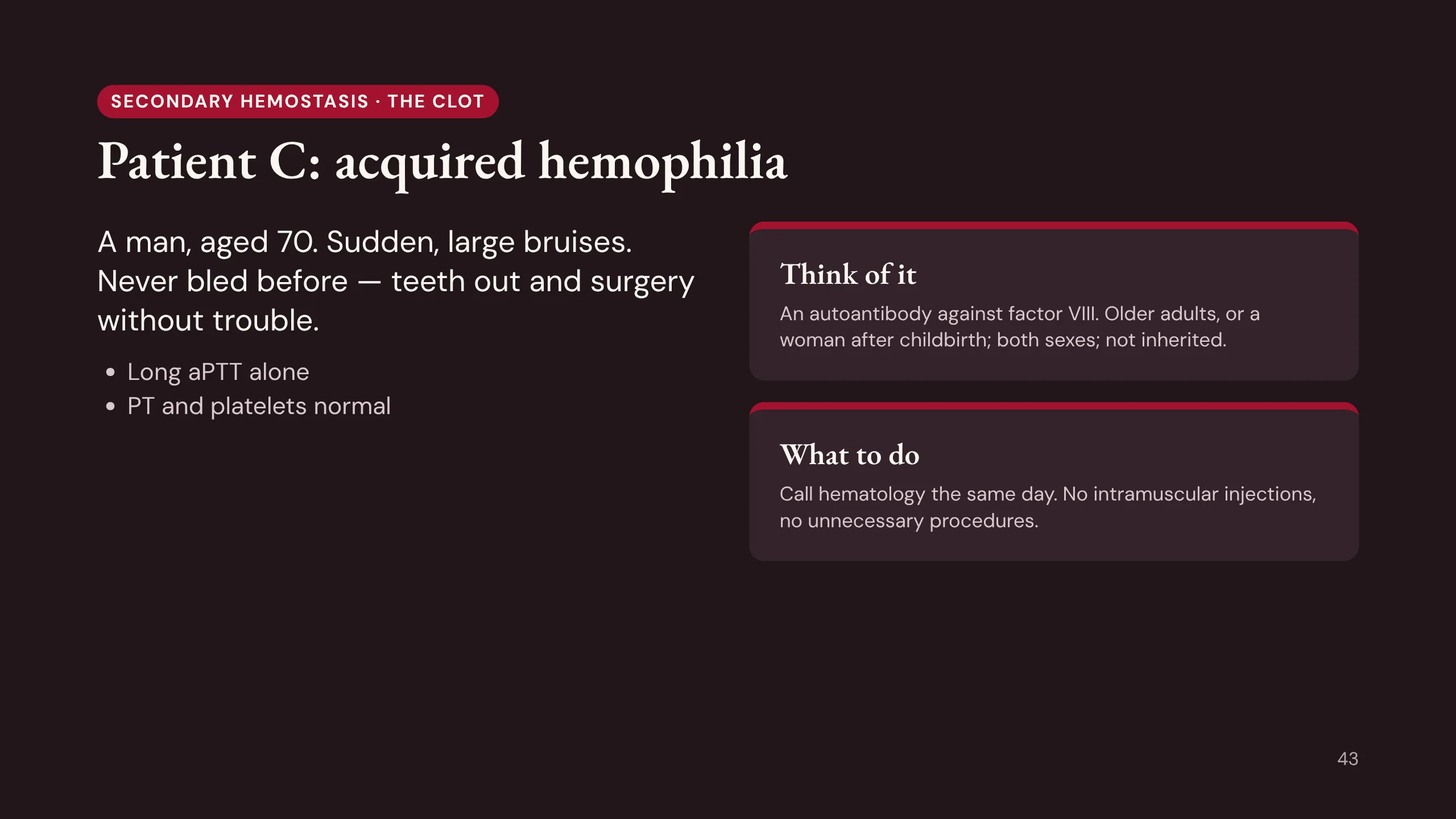
Patient C: acquired hemophilia
Patient C, the man of 70 with sudden large bruises. He has had teeth out and surgery without trouble, so whatever this is, it is new. His aPTT is long; his PT and platelets are normal.
First check the drug chart, because drugs are the commonest acquired cause. If nothing explains it, think of acquired hemophilia.
Acquired hemophilia is not the inherited disease. The patient makes an autoantibody against their own factor VIII. It appears in people who never bled before, usually older adults or women after childbirth, sometimes with autoimmune disease or cancer. It affects both sexes. The bleeding is often into skin and muscle, sudden and severe.
The panel looks like hemophilia, a long aPTT alone. The history is what differs. Recognise it, avoid intramuscular injections and unnecessary procedures, and call hematology the same day. It is rare, but it kills when missed.
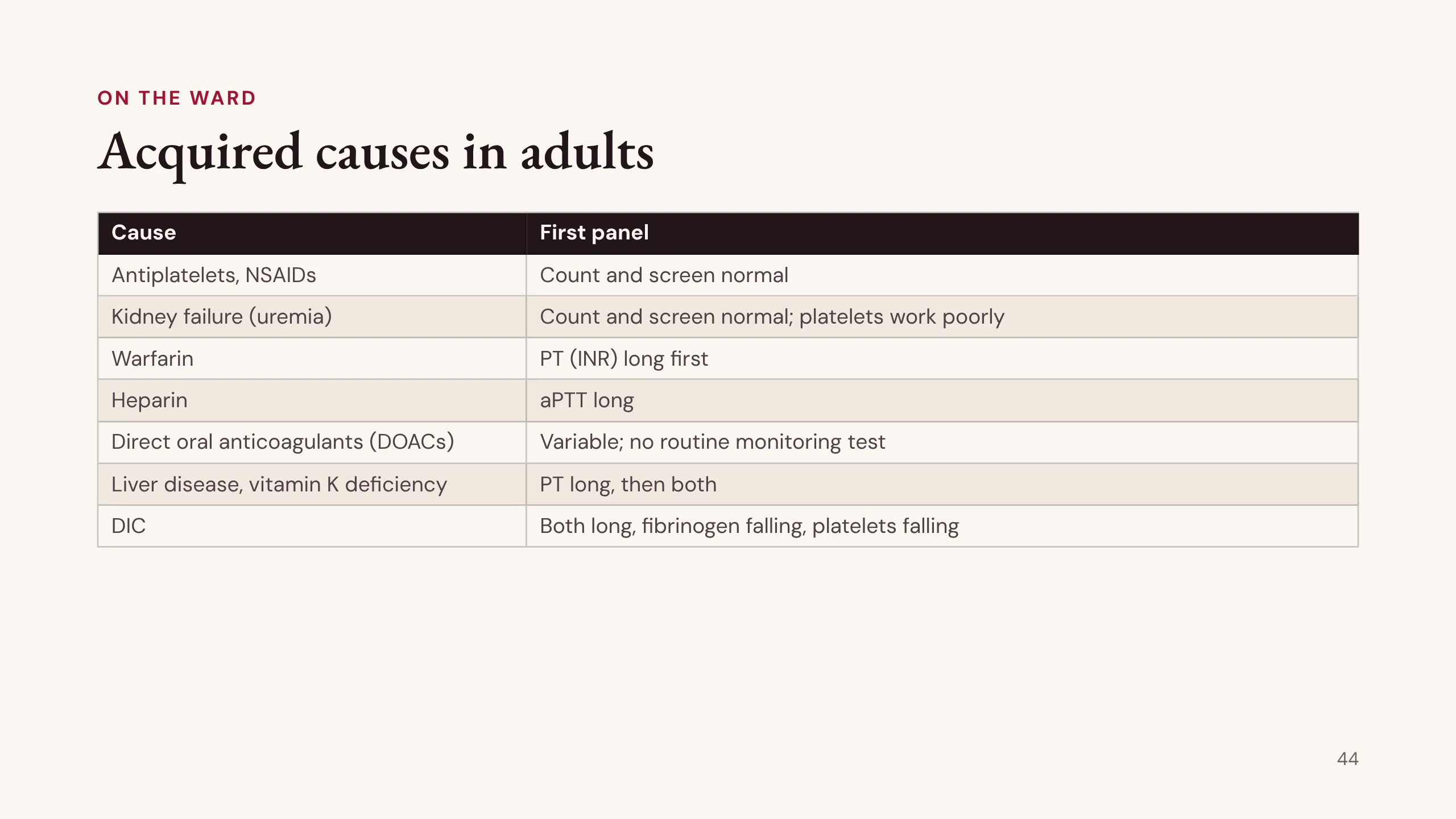
Acquired causes in adults
In an adult who starts to bleed, think of the acquired causes first. They are far more common than inherited ones.
Antiplatelet drugs and NSAIDs: the commonest cause of all. The count and screen are normal, because the platelets are there but do not work. Kidney failure does the same: uremia makes platelets work poorly.
Warfarin: the PT (INR) rises first. Heparin: the aPTT rises. The direct oral anticoagulants (DOACs) have no routine monitoring test, so the drug history finds them, not the screen.
Liver disease and vitamin K deficiency: the PT rises first, then both.
DIC: both times long, fibrinogen falling, platelets falling. Patient E is next.
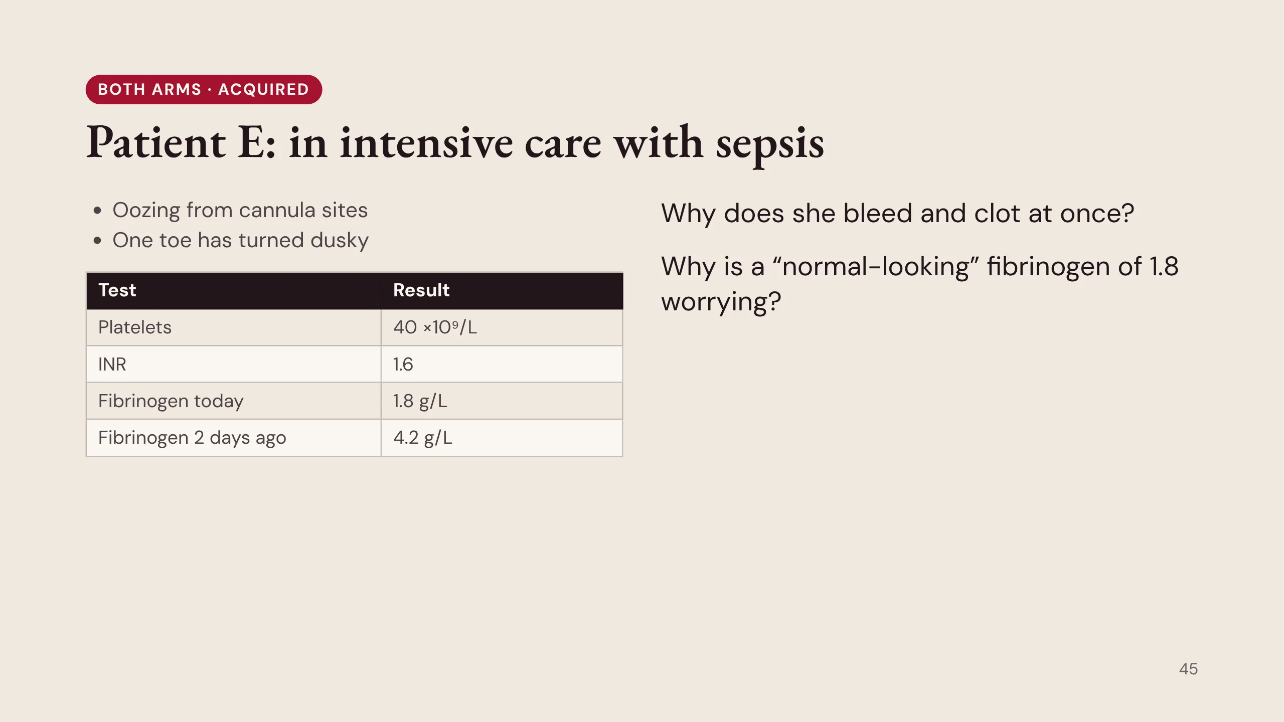
Patient E: in intensive care with sepsis
Patient E is the kind of bleeding you will meet on every night on call. A patient in intensive care with sepsis starts to ooze from the cannula sites, and one toe has turned dusky. Platelets 40 ×10⁹/L. INR 1.6. Fibrinogen 1.8 g/L; two days ago it was 4.2.
Two questions. First, how can the same patient bleed from the lines and clot in a toe at once? It sounds like two diseases. It is one.
Second, why is a fibrinogen of 1.8 worrying, when it is close to normal? Because fibrinogen rises in inflammation. A septic patient should have a high fibrinogen. A fall from 4.2 to 1.8 in two days means it is being used up. The trend tells you more than any single value.
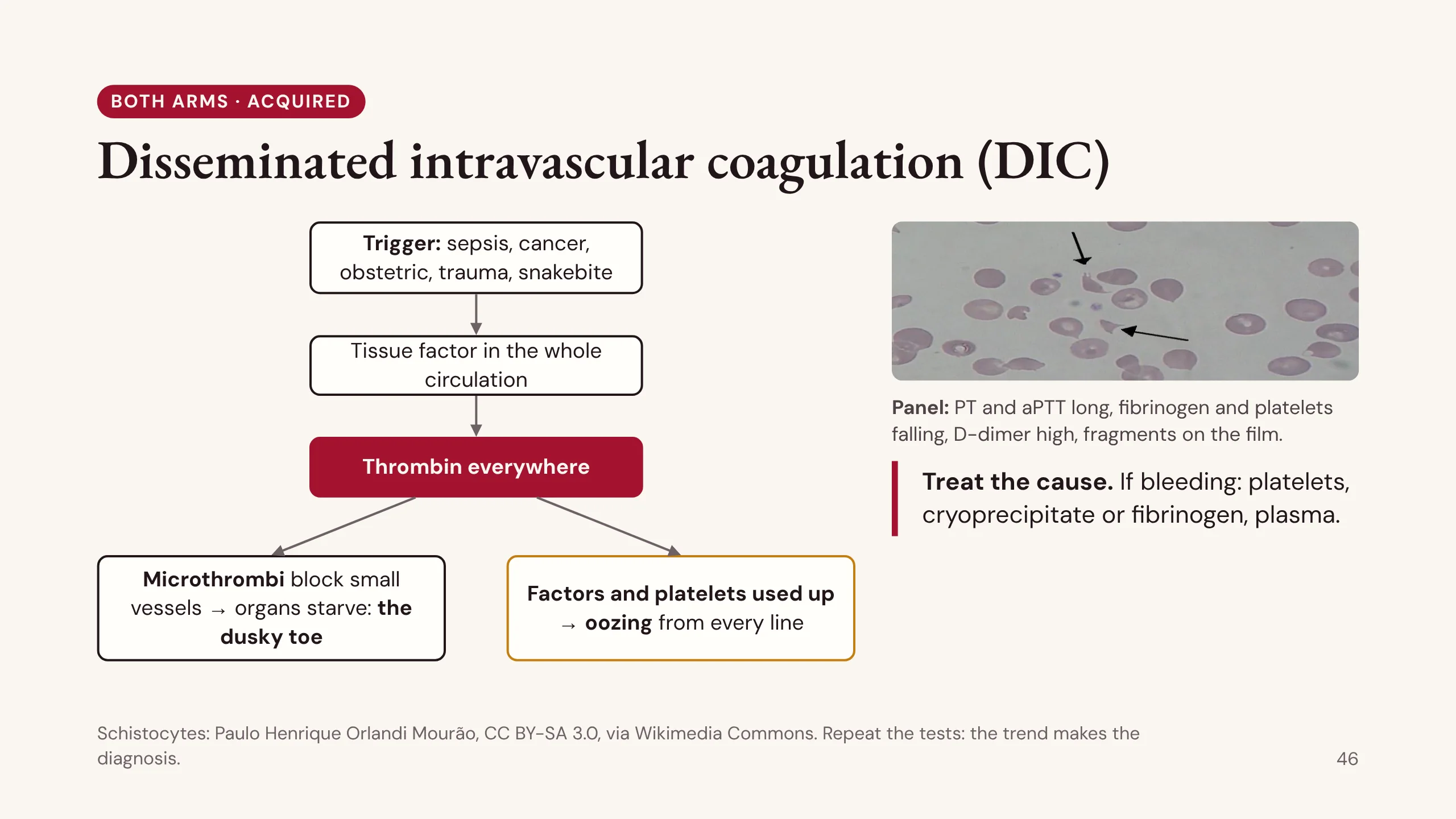
Disseminated intravascular coagulation (DIC)
Disseminated intravascular coagulation is never a diagnosis on its own. It is what a severe illness does to the clotting system, and it affects both arms of hemostasis.
Follow the diagram. A trigger exposes tissue factor to the whole circulation: sepsis above all, cancer (acute promyelocytic leukemia in particular), obstetric emergencies such as placental abruption or amniotic fluid embolism, major trauma, or snakebite. Thrombin is then made everywhere, not just at a wound.
Two things follow. Tiny clots block small vessels and organs starve: the dusky toe, and kidney and lung injury. And making all those clots uses up platelets, factors and fibrinogen faster than they can be replaced, so the same patient oozes from every line.
The panel follows the mechanism: PT and aPTT long, fibrinogen and platelets falling, D-dimer high, and fragmented red cells (schistocytes) on the film. Repeat the tests; the trend makes the diagnosis.
Treat the cause. If the patient is bleeding, support with platelets, cryoprecipitate or fibrinogen, and plasma. The schistocyte photograph is by Paulo Henrique Orlandi Mourão, CC BY-SA 3.0, via Wikimedia Commons.
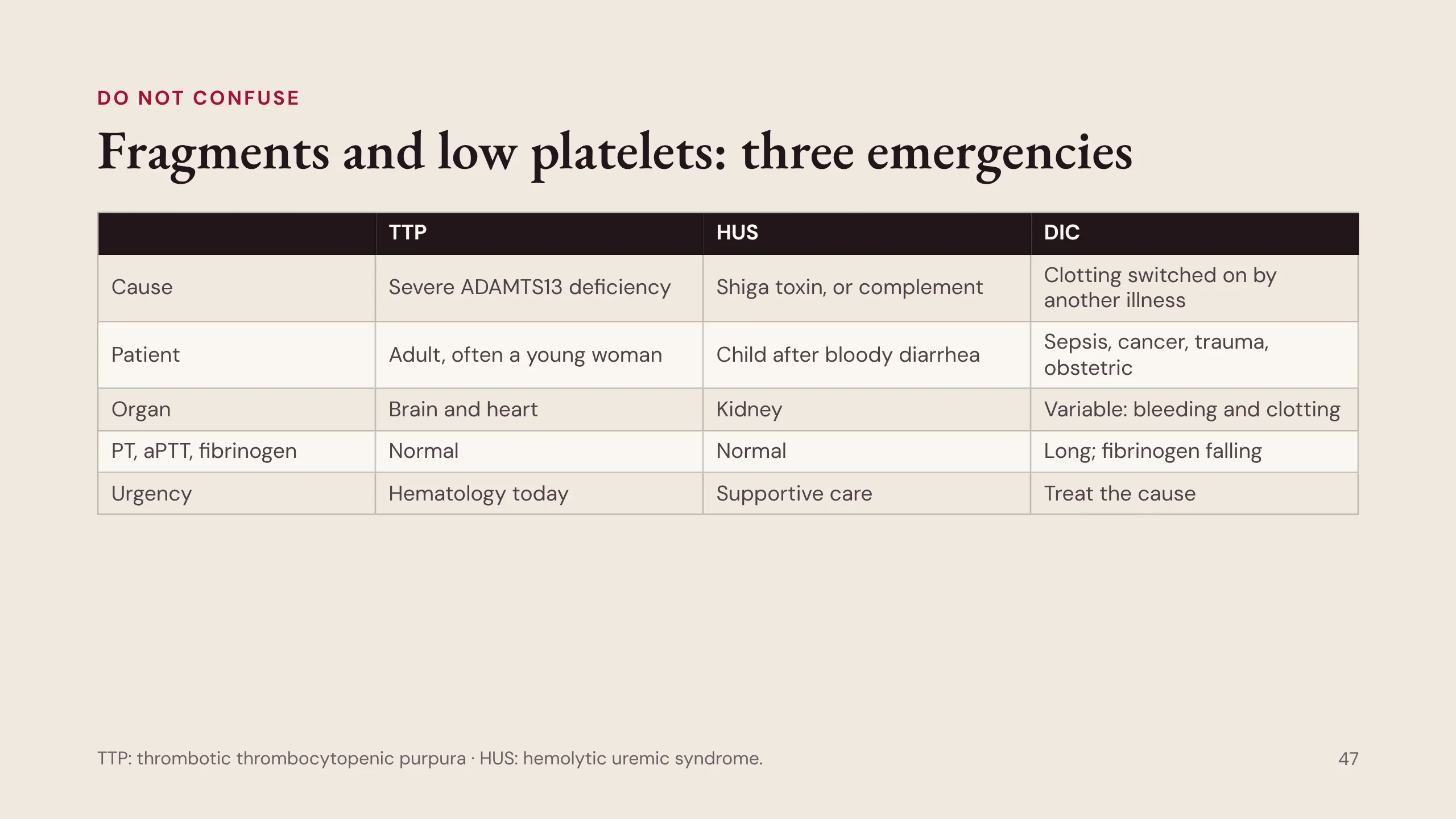
Fragments and low platelets: three emergencies
Fragmented red cells with a low platelet count can mean three different emergencies, and the clotting tests are the fork in the road.
In thrombotic thrombocytopenic purpura (TTP) and hemolytic uremic syndrome (HUS), small vessels fill with platelet clots, but the coagulation factors are not consumed, so the PT, aPTT and fibrinogen are normal. In DIC they are consumed: the times are long and the fibrinogen falls.
TTP is a severe lack of ADAMTS13, the enzyme that trims von Willebrand factor, usually from an autoantibody. Typically an adult, often a young woman; it affects the brain and heart. It is an emergency: call hematology today, before the ADAMTS13 result returns.
HUS is usually a child after bloody diarrhea, from Shiga toxin, and the kidney takes the damage. The atypical form is driven by complement.
DIC comes from another illness, and the answer is to treat that illness. These are covered fully in handbook chapter 29.
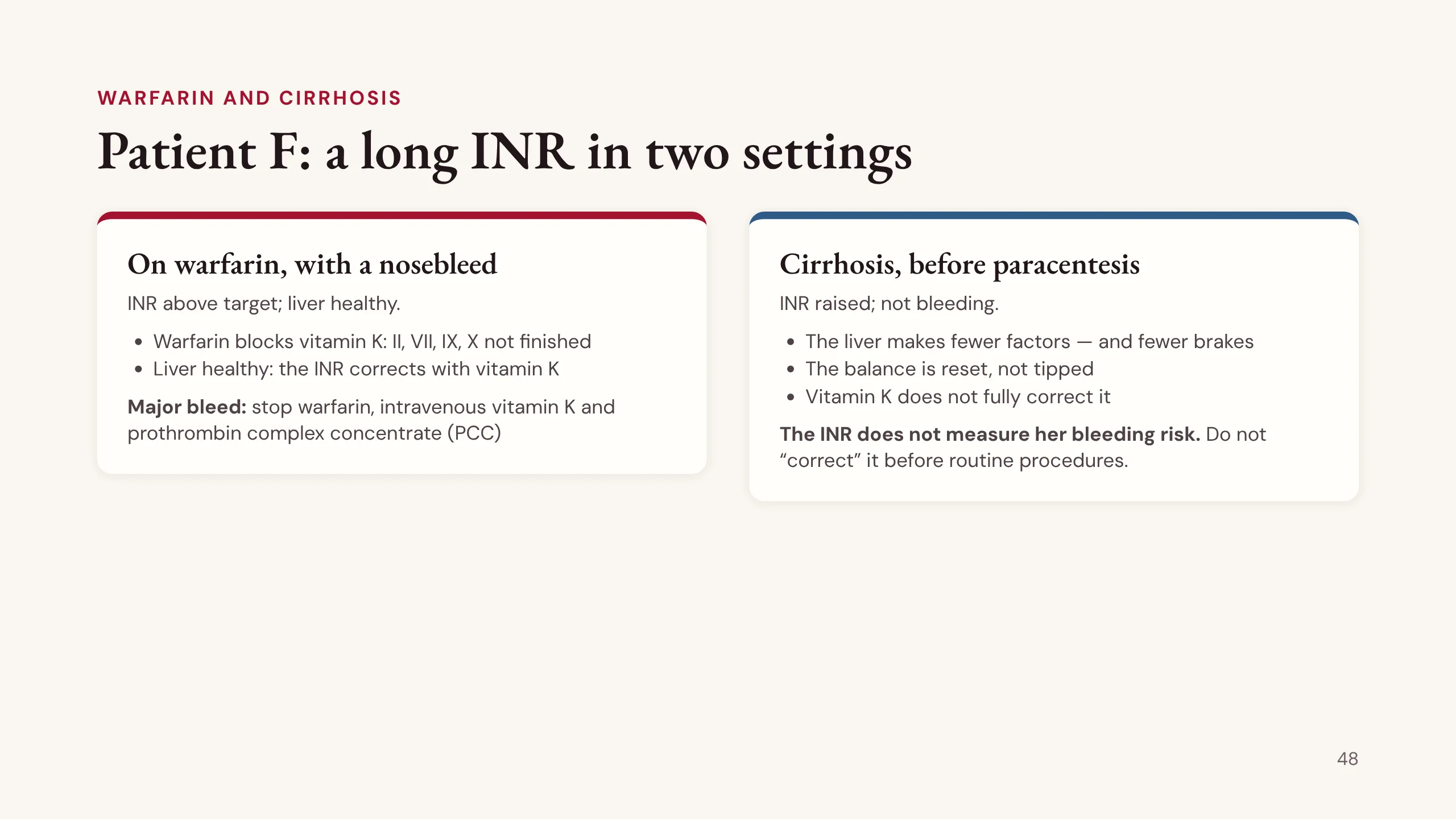
Patient F: a long INR in two settings
Patient F is two short cases with the same number and a different meaning.
On the left, a man on warfarin with a nosebleed and an INR above target. His liver is healthy. Warfarin blocks vitamin K, so the liver cannot finish factors II, VII, IX and X. Once vitamin K is available the factors are made again and the INR corrects. For a major bleed: stop warfarin, give intravenous vitamin K, which takes hours, and prothrombin complex concentrate (PCC), which works in minutes.
On the right, a woman with cirrhosis who needs a paracentesis. Her INR is raised but she is not bleeding. Her liver makes fewer factors, so vitamin K does not fully correct it. But it also makes fewer brakes: protein C, protein S and antithrombin. Her clotting is not simply tipped toward bleeding. It is reset to a new, fragile balance.
The INR measures only the factors, not the brakes. So in liver disease the INR does not measure the risk of bleeding, and it should not be “corrected” with plasma before routine procedures such as paracentesis.
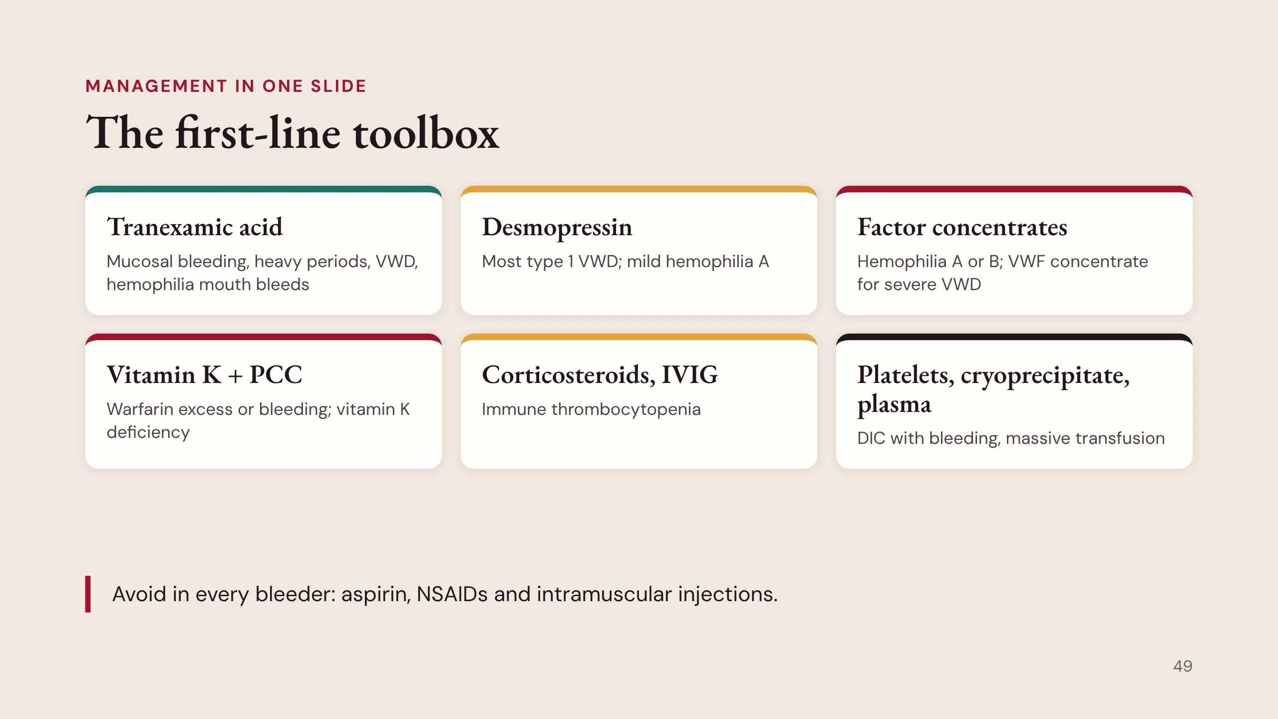
The first-line toolbox
First-line management in one slide. Match each tool to its disorder.
Tranexamic acid: mucosal bleeding, heavy periods, VWD, and mouth bleeds in hemophilia. Desmopressin: most type 1 VWD, and mild hemophilia A. Factor concentrates: hemophilia A or B, and VWF concentrate for severe VWD. Vitamin K with PCC: warfarin excess or bleeding, and vitamin K deficiency. Corticosteroids and IVIG: immune thrombocytopenia. Platelets, cryoprecipitate and plasma: DIC with bleeding, and massive transfusion.
And three things to avoid in every patient who bleeds: aspirin, NSAIDs and intramuscular injections.
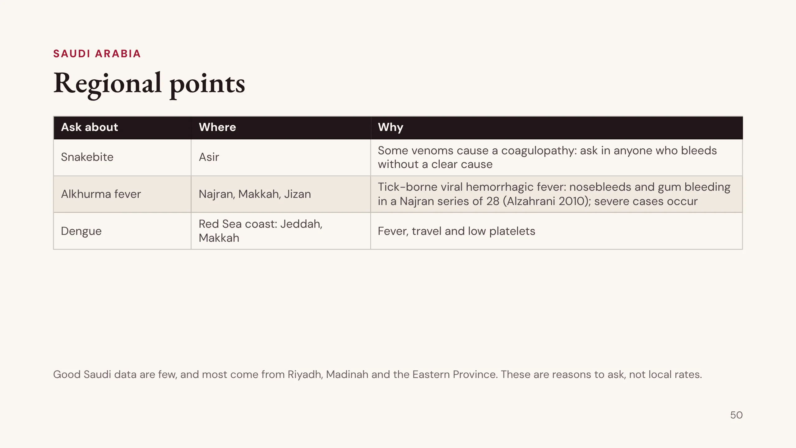
Regional points
Where you practise changes the history. In this region, ask about three more things.
Snakebite. Some venoms cause a coagulopathy. In anyone who bleeds without a clear cause, ask about a bite. A study from four tertiary hospitals in Asir reported coagulopathy after snakebite (Alfaifi and colleagues, 2024).
Alkhurma hemorrhagic fever, a tick-borne virus. In a Najran series of 28 patients from 2006 to 2009, 8 had nosebleeds and 5 gum bleeding (Alzahrani and colleagues, 2010). Severe and fatal cases are described elsewhere, so do not treat it as harmless. Ask about animal contact and tick bites.
Dengue, on the Red Sea coast, including Jeddah and Makkah. Ask about fever and travel in a patient with low platelets.
The honest position: good Saudi data on bleeding are few, and most come from Riyadh, Madinah and the Eastern Province. Use these as reasons to ask, not as local rates.
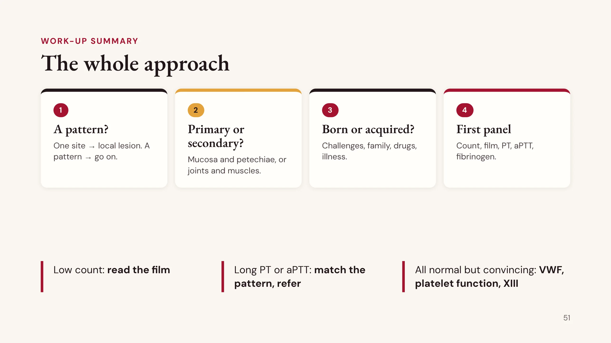
The whole approach
The whole lecture on one slide.
1. Is there a pattern? One site sends you to the lesion; a pattern means go on. 2. Primary or secondary? Mucosa and petechiae, or joints and muscles. 3. Born or acquired? Challenges, family, drugs and illness. 4. The first panel: count, film, PT, aPTT and fibrinogen.
Then three branches. A low count: read the film. A long PT or aPTT: match the pattern to the grid and refer if the history and drug chart do not explain it. Everything normal but the history convincing: von Willebrand studies, then platelet function, then factor XIII.
At every step read the drug chart, and watch for the patient who is also tired and feverish, whose problem is in the marrow.
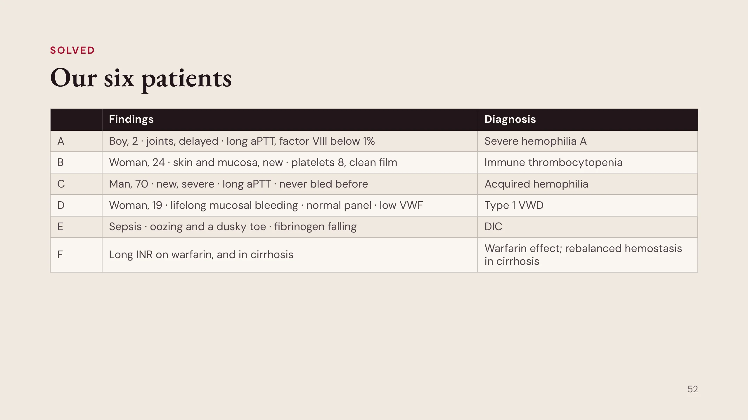
Our six patients
Back to the guesses you made at the start.
A, the boy: deep, delayed joint bleeding, a long aPTT alone, factor VIII below 1%. Severe hemophilia A: inherited, secondary hemostasis.
B, the young woman: new mucosal bleeding with petechiae, platelets 8, a clean film, everything else excluded. Immune thrombocytopenia: acquired, primary hemostasis.
C, the older man: new, severe bleeding in someone who never bled before, a long aPTT alone. Acquired hemophilia, after the drug chart is checked: call hematology today.
D, the 19-year-old: lifelong mucosal bleeding with a normal panel, and low VWF with activity in proportion. Type 1 von Willebrand disease: inherited, primary hemostasis.
E, the septic patient: oozing and a dusky toe, fibrinogen falling. DIC: both arms consumed.
F: the same long INR meaning warfarin effect in one patient and rebalanced hemostasis in cirrhosis in the other.
Five things to take home
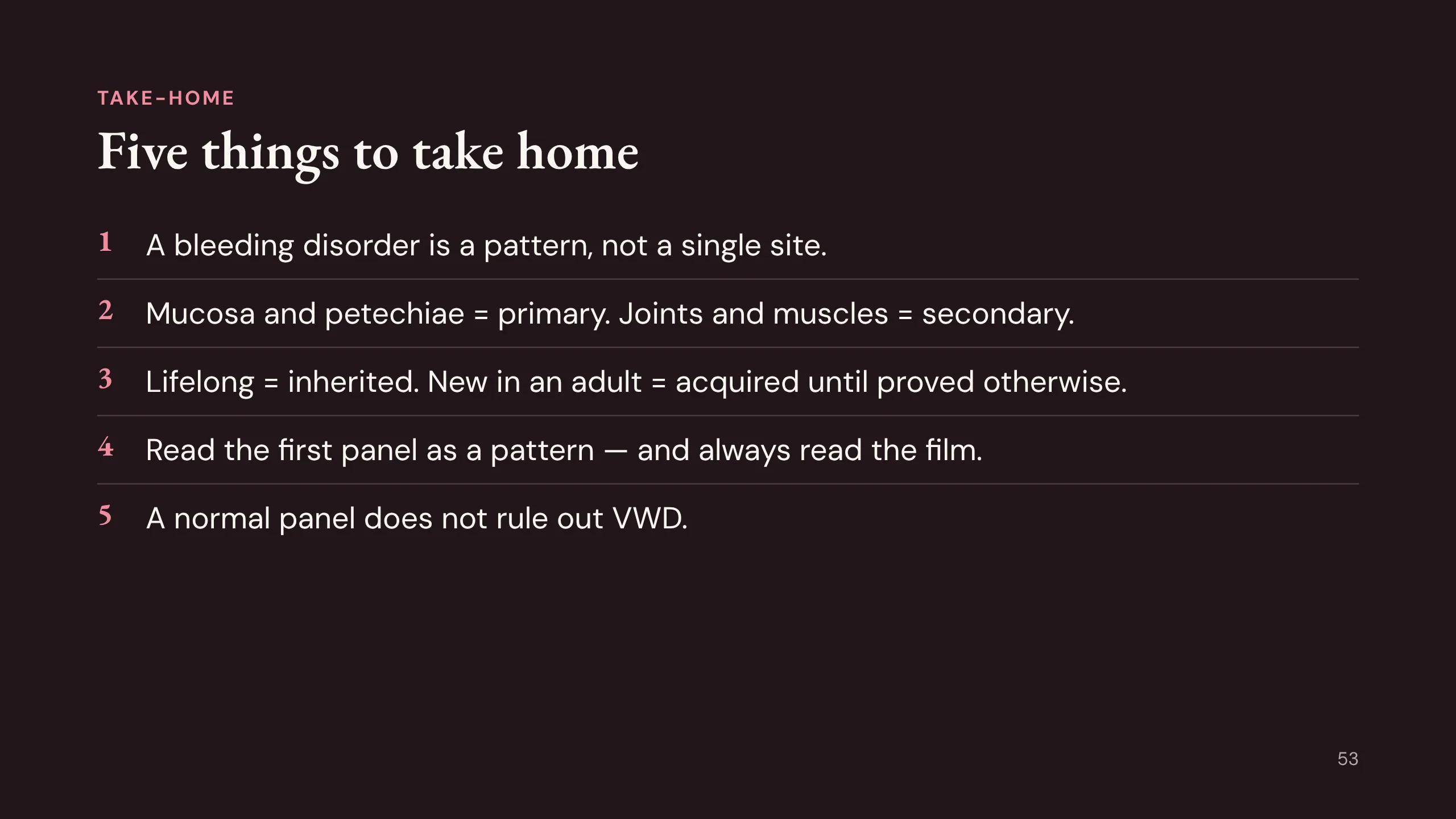
Five things to take home.
1. A bleeding disorder is a pattern, not a single site.
2. Mucosa and petechiae mean primary hemostasis. Joints and muscles mean secondary hemostasis.
3. Lifelong points to inherited. New in an adult is acquired until proved otherwise.
4. Read the first panel as a pattern, and always read the film.
5. A normal panel does not rule out von Willebrand disease. Ask every girl with heavy periods whether they have been heavy since her first period.
Read next
Read next: chapters 20 to 25 and 29 of the Hematology Handbook; Case 53, The Bleeding Patient, and section 6 of Case 51 (DIC) in the Internal Medicine Casebook; and items 46 to 59 of the Hematology Question Bank.
A short, clinical and free book: Bloody Easy Coagulation Simplified, 3rd edition (2026), from the Ontario Regional Blood Coordinating Network.
Coming later: how the laboratory works up a long PT or aPTT, including sample problems, mixing studies and factor assays. Thrombosis, the other side of the balance, is in handbook chapter 26, and venous thromboembolism is taught again in Med-2.
Image credits. All other figures were drawn for this lecture.
- Slides 6 and 7: 1909 Blood Clotting (OpenStax, Anatomy & Physiology) by OpenStax, via Wikimedia Commons, CC BY 3.0 (cropped).
- Slides 11, 13 and 14 are from Husain Alkhaldy's earlier version of this lecture.
- Slide 30: Platelet clumping (Platelet Clumping 8.jpg) by Erhabor Osaro, via Wikimedia Commons, CC BY-SA 4.0.
- Slides 30 and 46: Schistocytes (Schizocyte smear 2009-12-22.JPG) by Paulo Henrique Orlandi Mourão, via Wikimedia Commons, CC BY-SA 3.0.
- Slide 30: Blasts in acute myeloid leukemia by Armed Forces Institute of Pathology, via Wikimedia Commons, public domain.