Disorders of Hemostasis
Lecture 2 of 2 — the bleeding patient from history to diagnosis: history, screen, mix and confirm.
These slides are an outline only. Read the notes under each slide below with them, and chapters 20 to 29 of the Hematology Handbook. Then work through the Internal Medicine Casebook and the Hematology Question Bank. Start with the first lecture, Introduction to Bleeding Disorders.
Disorders of Hemostasis
These slides are an outline only. Read the notes under each slide on this page with them, and chapters 20 to 29 of the Hematology Handbook. Then work through the Internal Medicine Casebook and the Hematology Question Bank.
This is the second of the two bleeding lectures. The first lecture taught the physiology, hemophilia and ITP. This one picks up the patient who was left at the end of it, and follows her all the way to a diagnosis.
The route for today is history, screen, mix and confirm. That is the order a clinician meets these patients in, and slide 3 sets it out. Treatment is not a core topic for Med-1, so it appears only as a single line where it is needed.
These slides are an outline. The full reasoning is in these notes, and in chapters 20 to 29 of the Hematology Handbook.
Case 1
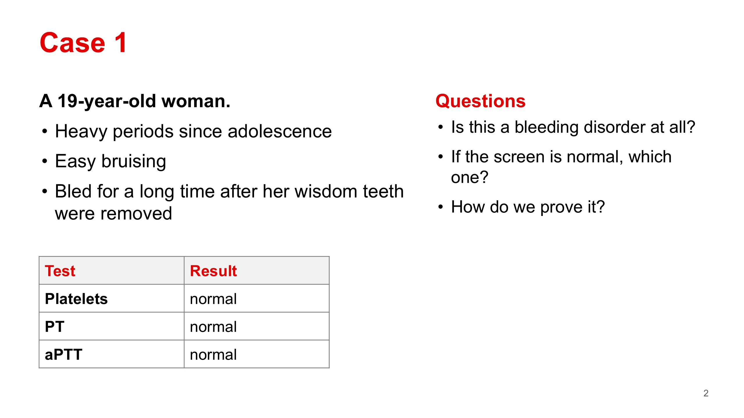
At the end of the first lecture we met this young woman. She is 19. Her periods have been heavy since she was a teenager, she bruises easily, and when her wisdom teeth came out she kept bleeding. Her platelet count, PT and aPTT are all normal.
This patient comes from chapter 21 of the handbook, and she is the reason this lecture exists. Many students, and many doctors, would look at three normal results and tell her she has no bleeding disorder. That would be wrong.
So we will carry three questions through the hour. Is this a real bleeding disorder, or is she one of the many women who bleed heavily for other reasons? If it is a disorder and the screen is normal, which disorders does the screen miss? And how do we prove the diagnosis and treat it?
Ask the room before moving on: what would you send next? Keep their answers. We will come back to them at the end.
Outline
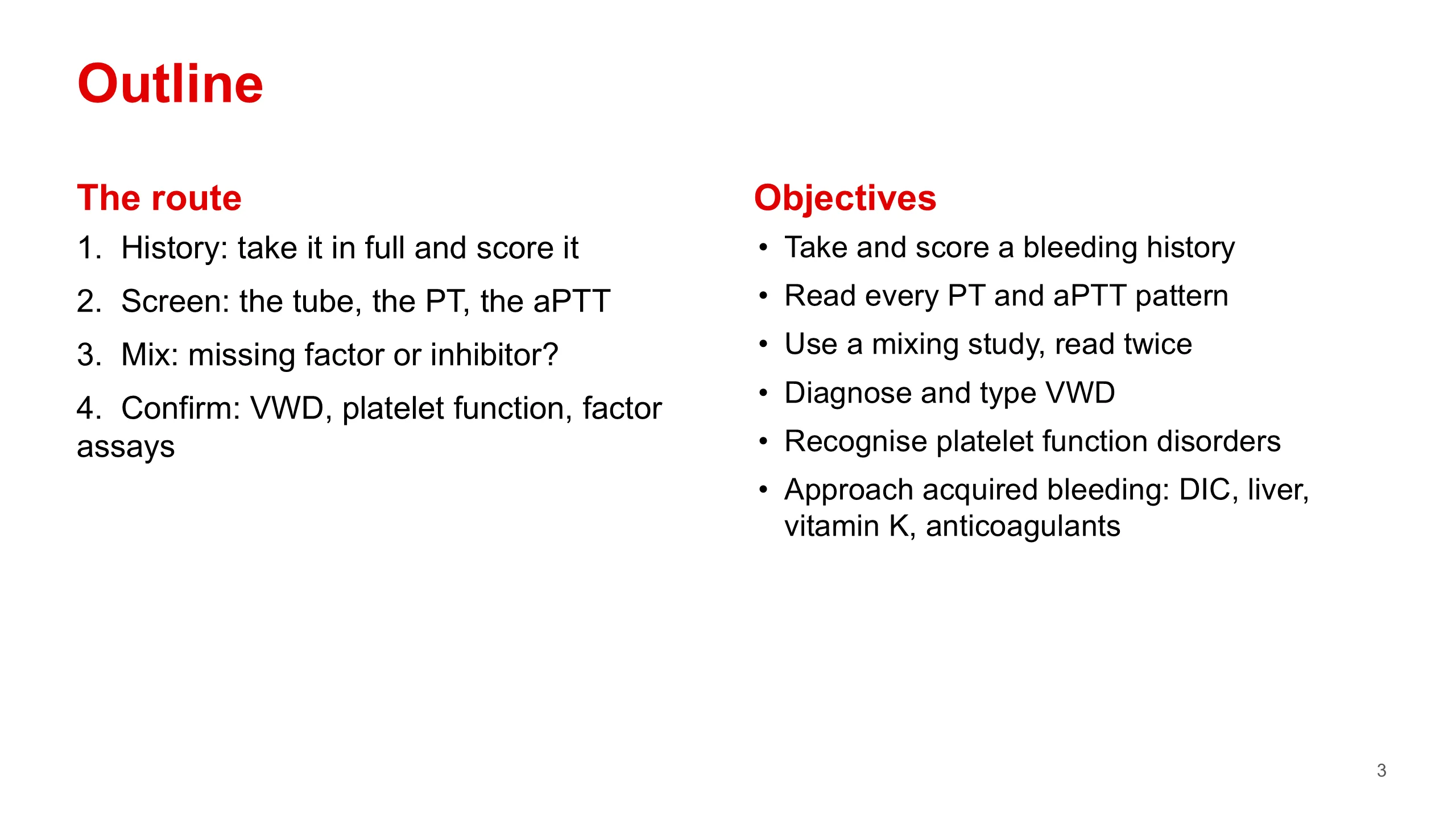
Here is the route. History first, because the history decides which tests you send and how you read them. Then the screen: the PT and aPTT, and the tube they come in. Then the mixing study, which tells a missing factor from an inhibitor. Then the confirmatory tests.
Treatment is not core at this stage of your course. Where it matters, it appears as one short line, and the details belong to later years.
The objectives are on the slide. Two things are deliberately not here. Hemophilia and ITP were taught in the first lecture, so today I only point back to them. And the physiology was also taught there, so I use it without teaching it again.
Local bleeding or bleeding disorder?
The first lecture gave you the rule in one line. Here it is in full, because this lecture owns the history.
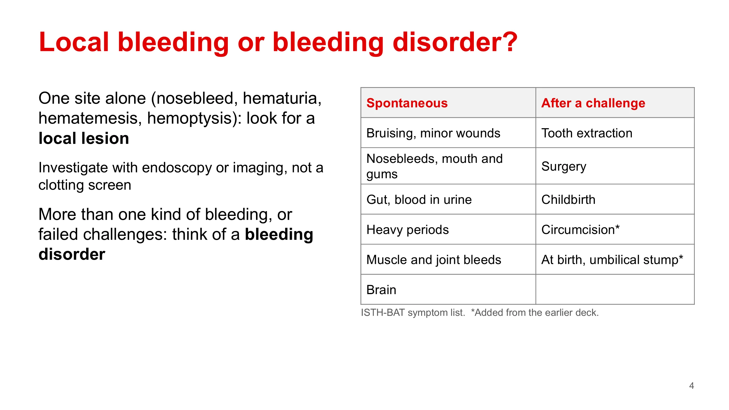
Most people who bleed do not have a bleeding disorder. They have a lesion: an ulcer, a polyp, a tumour, a vessel in the nose. A single kind of bleeding on its own, such as blood in the urine or a nosebleed, should send you looking for that lesion, with endoscopy or imaging. A clotting screen will not find it.
A bleeding disorder shows itself as a pattern: more than one kind of bleeding, or bleeding after the natural tests of life, such as a tooth extraction, an operation or childbirth.
The two groups of boxes are the symptoms that the ISTH bleeding assessment tool asks about. The left group is spontaneous bleeding. The right group is bleeding after a challenge. On the right I have added two questions from my own history slide that matter early in life: bleeding at birth or from the umbilical stump, and bleeding after circumcision. In this country almost every boy has had circumcision, so it is a test that most male patients have already taken.
Bleeding history
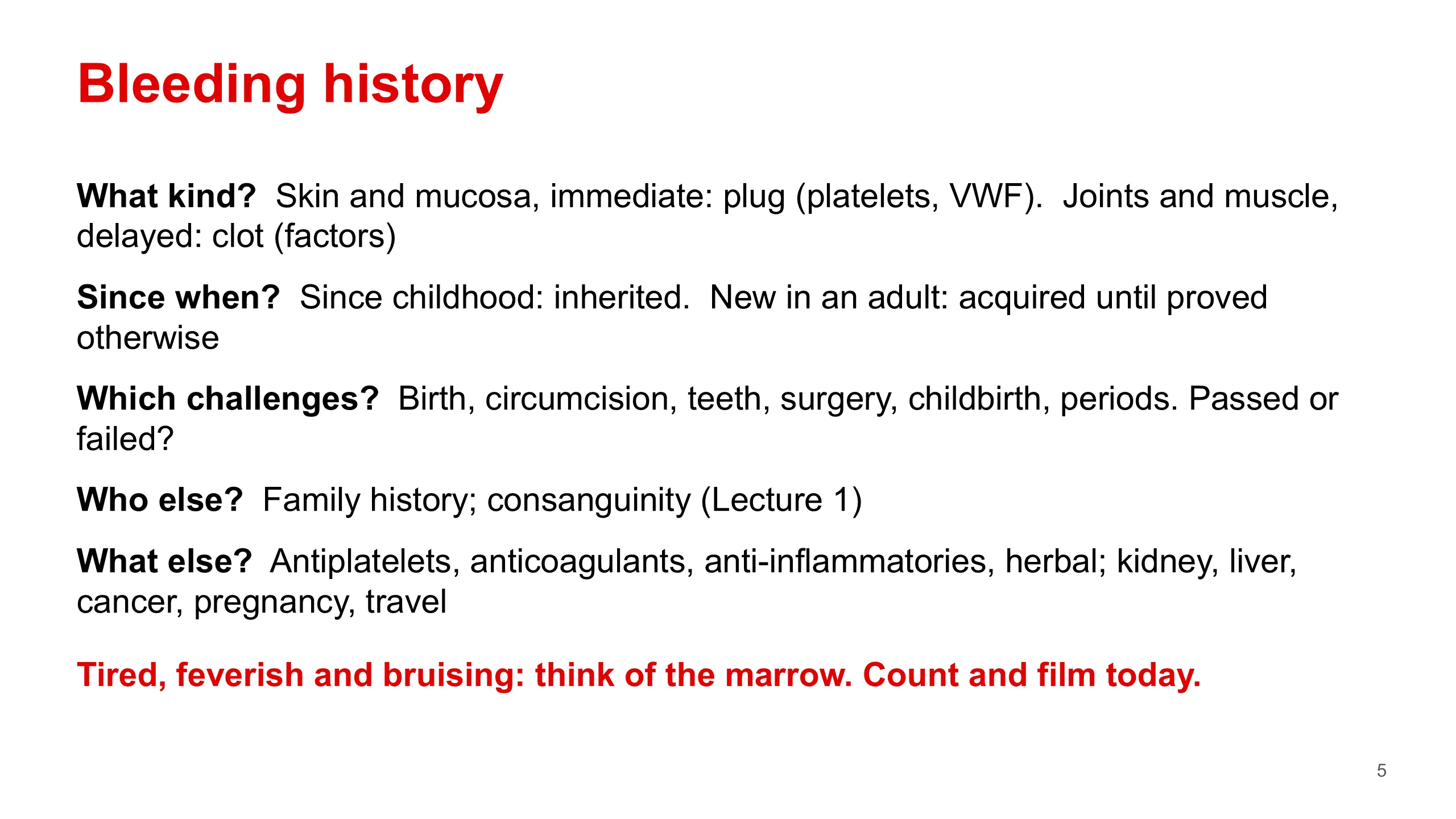
These five questions are the whole bleeding history.
First, what kind of bleeding? Bleeding into skin and mucosa that starts straight away means the platelet plug failed: platelets or von Willebrand factor. Bleeding into joints and muscle that starts late, after the plug has formed, means the fibrin clot failed: the coagulation factors.
Second, since when? A story that goes back to childhood points to an inherited disorder. New bleeding in an adult is acquired until you prove otherwise.
Third, which challenges has the patient already faced, and did they pass? Each one is a natural test. A patient who has had teeth out and an operation without trouble is unlikely to have a significant inherited disorder.
Fourth, the family. Lecture 1 showed you why consanguinity matters here: it makes the recessive disorders more common.
Fifth, the drug chart and the illnesses. Antiplatelets and anticoagulants are the commonest cause of bleeding of all. Ask about over-the-counter aspirin and herbal products by name, because patients do not count them as medicines.
The amber box is the one question that changes the urgency. If the patient also has symptoms of anemia and fever or infections, all three cell lines may be low. That is a marrow problem, perhaps leukemia, and the count and film are needed the same day. This links to the acute leukemia lecture.
Menstrual history
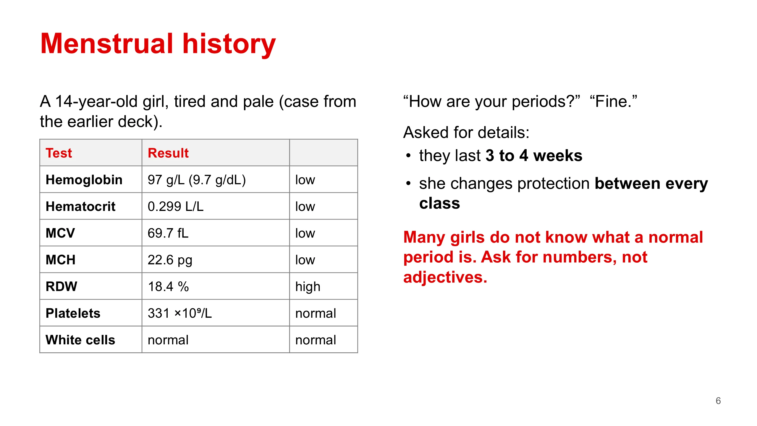
This short case comes from the earlier version of this lecture, with thanks to the colleague credited there (@acweyand). It is the best single lesson about heavy periods I know.
A 14-year-old girl sees her doctor because she is tired. She is pale. Read the count. Her hemoglobin is 97 grams per litre, which is 9.7 in grams per decilitre. Her red cells are small and pale: an MCV of 69.7 and an MCH of 22.6. The RDW is high at 18.4, so the cells vary in size. The platelets and white cells are normal. This is the picture of iron deficiency.
Where did the iron go? In a girl of this age who eats normally, the usual answer is menstrual loss. So the doctor asks about her periods, and she says they are fine.
"Fine" should open the question, not close it. Asked for details, she says they last three to four weeks, and she changes her pad or tampon between every class at school. That is heavy menstrual bleeding by any measure, and it explains her anemia.
The lesson: many girls and women have nothing to compare their periods with. Their mothers and sisters may bleed the same way, because the same disorder runs in the family. Always ask for numbers: how many days, how many products, how often they are changed, whether there are clots, whether she has to change at night, and whether blood has ever soaked through her clothes.
Heavy menstrual bleeding
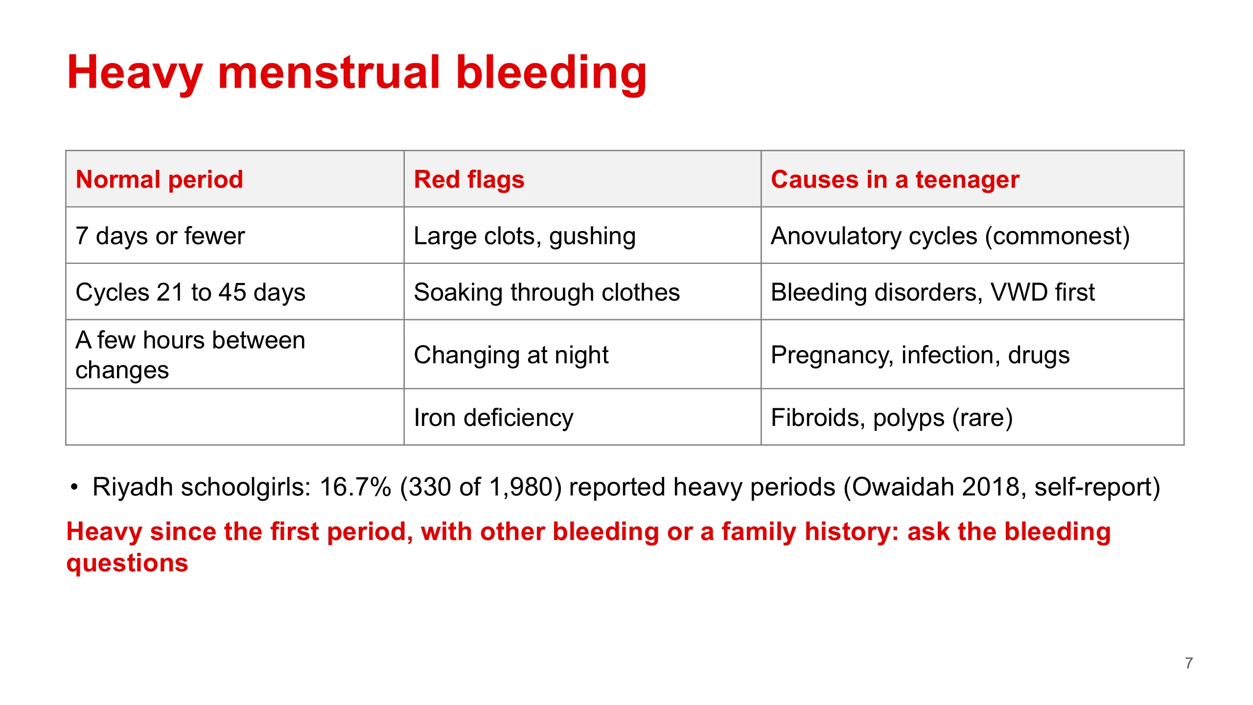
To call a period heavy you need a standard. A normal period lasts seven days or fewer, cycles come 21 to 45 days apart, and a woman can go at least a few hours without changing protection. These figures are from the earlier deck.
The red flags mark the heavy end: large clots, a feeling of gushing, blood soaking through clothes, needing to change at night, and iron deficiency, which is the flag our 14-year-old raised.
Heavy periods are common. The earlier deck quoted a figure of up to 37 percent of adolescents from its international source. A Riyadh school survey of 1,980 girls found 16.7 percent reported heavy periods (Owaidah 2018). The two numbers measure different things in different groups, and the Saudi one is self-report, so do not compare them directly. The point is the same: a bleeding disorder that shows itself this way is easy to lose among many girls who bleed heavily for other reasons.
In a teenager the commonest cause by far is anovulatory cycles, because the hormonal axis is still immature. Bleeding disorders come next, von Willebrand disease above all. They are in red because they are the subject of this lecture, not because they are the commonest. Fibroids and polyps, which dominate in older women, are rare at this age.
The rule at the bottom is the practical point. Heavy bleeding since the first period, in a girl with other bleeding or a family story, must not be put down to anovulation until you have asked the bleeding questions.
ISTH bleeding assessment tool
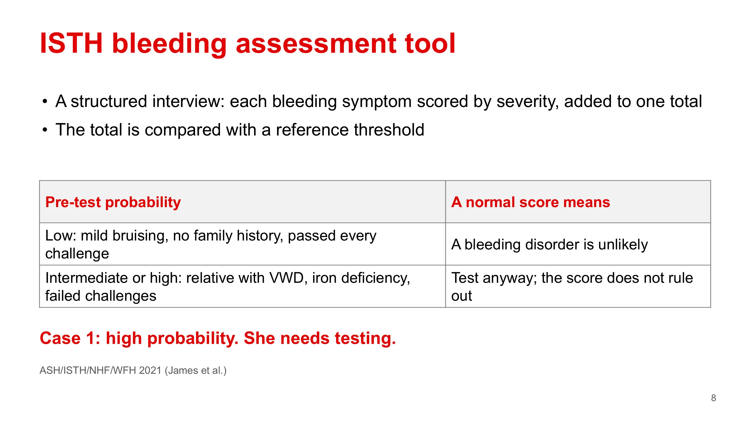
A bleeding history from memory is vague. The ISTH bleeding assessment tool makes it structured. It walks through every symptom on slide 4, gives each one a score for how severe it was, for example whether it needed a doctor, a transfusion or an operation, and adds them up. The total is compared with a reference threshold.
Now the limit, and it matters. The 2021 guideline on von Willebrand disease from ASH, ISTH, NHF and WFH (James and colleagues) says the score is useful to rule out a bleeding disorder when the chance of one is low to begin with, for example in a general clinic, a patient with mild bruising, no family history, and who has passed every challenge. There a normal score is reassuring.
When the chance is intermediate or high, for example a patient with a relative who has von Willebrand disease, or with heavy periods that have caused iron deficiency, the guideline advises going straight to blood tests and not relying on the score. A normal score in that patient does not rule a disorder out.
Our 19-year-old has lifelong heavy periods, bruising, and bleeding after her wisdom teeth. Her probability is high. She needs testing, whatever the score says.
I have not put the threshold numbers on the slide. They differ by age and sex. Your handbook, chapter 21, uses the same principle: a convincing history justifies specialised testing even when the screen is normal.
Sample collection
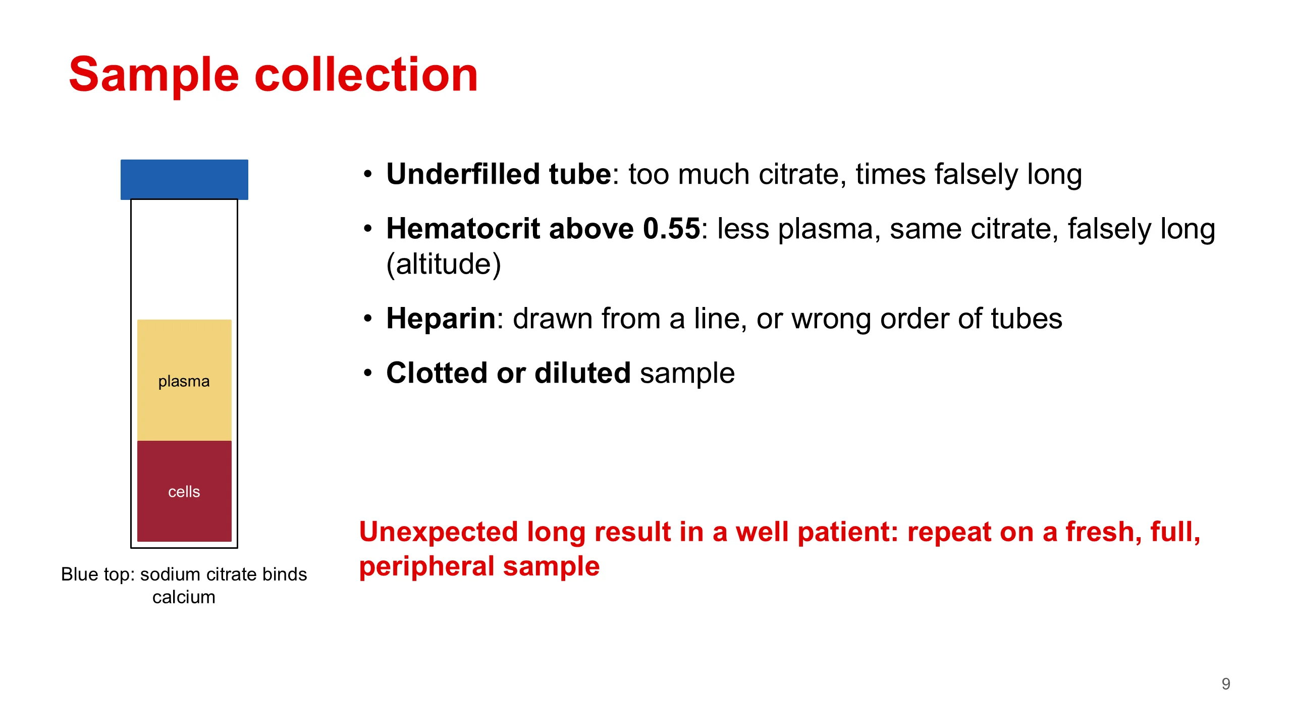
Before you interpret a PT or an aPTT, think about the tube. Coagulation tests are done on plasma collected into a blue-top tube that contains sodium citrate. Citrate binds calcium. Without calcium the cascade cannot run, so the blood stays liquid on the way to the laboratory. In the lab the tube is spun, the plasma is taken off, and calcium is added back to start the clock.
The amount of citrate is fixed for a full tube of blood. So anything that changes the balance between citrate and plasma changes the result.
An underfilled tube has too much citrate for the blood in it, and the times come out falsely long. A patient with a hematocrit above 0.55 has less plasma in the same volume of blood, so the citrate is again too much, and again the times are falsely long. This matters in Abha, where high hematocrits are more common at altitude. The investigations lecture covers the correction.
Heparin contamination, from a sample drawn through a line or from filling the tubes in the wrong order, prolongs the aPTT. A sample that partly clotted during a difficult draw, or one taken near a drip, gives wrong results too.
So the rule: if a well patient has an unexpected long result, repeat it on a fresh, correctly filled sample from a peripheral vein before you do anything else.
PT and aPTT
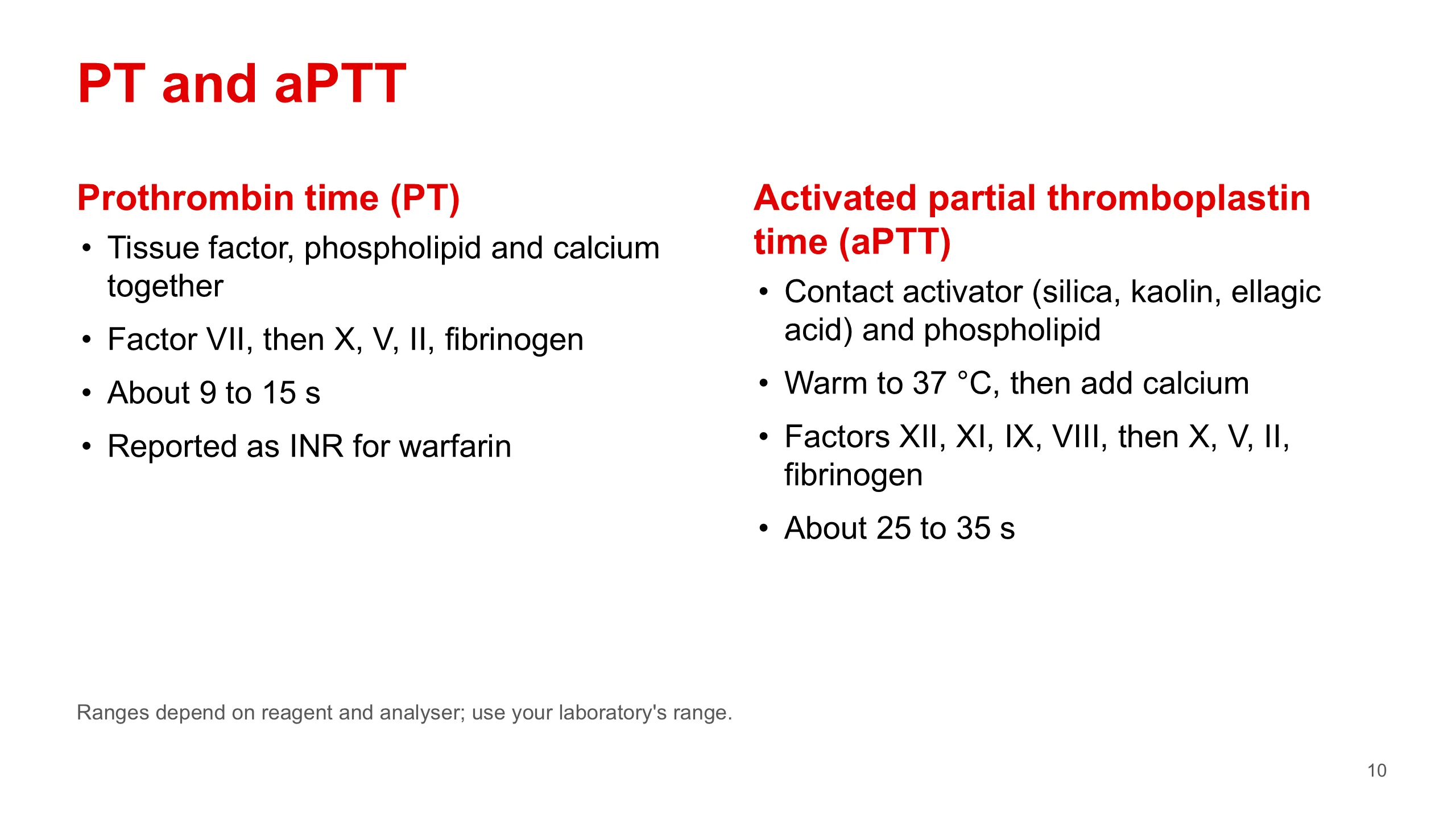
A clotting test measures, in seconds, how long plasma takes to clot after the reaction is started in a tube. The PT and the aPTT differ only in what starts it.
The prothrombin time uses tissue factor, also called thromboplastin, together with phospholipid and calcium, all in one reagent. It is added to warmed plasma and the clot is timed. It tests factor VII and then the common pathway: factors X, V, II and fibrinogen. For patients on warfarin it is reported as the INR, which makes results comparable between laboratories.
The activated partial thromboplastin time uses a contact activator, such as silica, kaolin or ellagic acid, with phospholipid but no tissue factor. That is why it is called a partial thromboplastin. Plasma and reagent are warmed at 37 degrees so the contact factors can be activated, then calcium is added and the clock starts. It tests factors XII, XI, IX and VIII, and the same common pathway.
The normal ranges on the slide, about 9 to 15 seconds and about 25 to 35 seconds, are the ones in my earlier deck. They vary with the reagent and the machine, so always use the range printed on your own laboratory's report.
Notice that the PT is shorter. Tissue factor drives clotting faster than contact activation. That is the same lesson the cell-based model taught you in Lecture 1.
Reading the screen
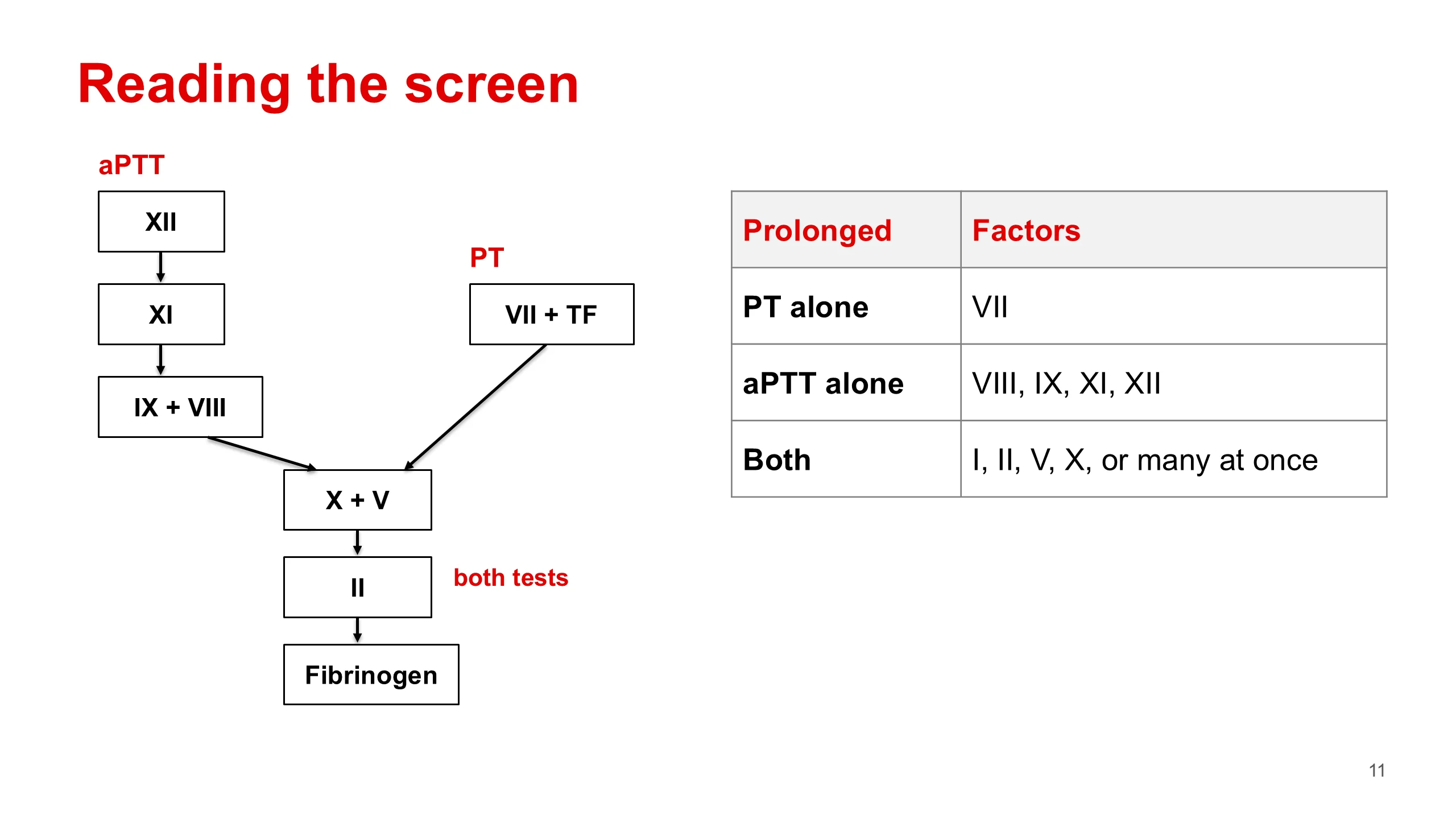
This is the one map to carry in your head. The old Y-shaped cascade is not how clotting works in the body. Lecture 1 showed you that. But it is exactly how the tests work in the tube, so it is the right map for reading results.
Read it from the bottom up. The common pathway, factors X, V, II and fibrinogen, is shared by both tests. A defect there prolongs both. Above the junction the arms split. The PT arm has only factor VII with tissue factor. The aPTT arm has XII, XI, IX and VIII.
So there are three patterns. PT long alone: think factor VII. aPTT long alone: think VIII, IX, XI or XII. Both long: a common pathway factor, I, II, V or X, or, much more often, many factors at once.
A note for those who have the old slides: the old slide 28 printed "XI" twice and left out IX. The correct list for the aPTT alone is VIII, IX, XI and XII.
The next three slides give the causes behind each pattern.
Prolonged PT, normal aPTT
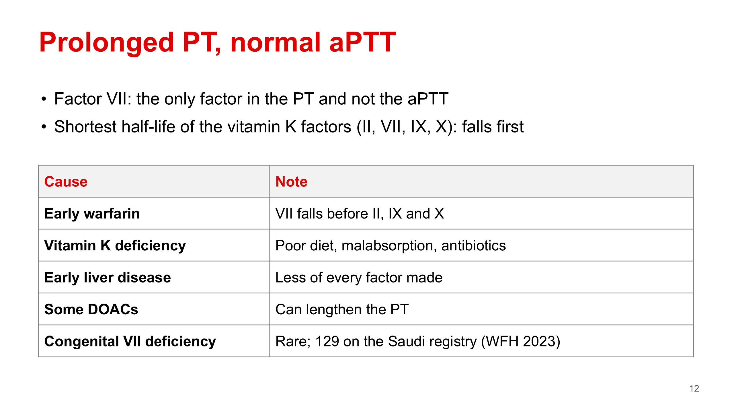
A long PT with a normal aPTT points to the one factor the PT tests and the aPTT does not: factor VII.
Inherited factor VII deficiency exists but is rare. It is on the Saudi registry: the World Federation of Hemophilia survey for 2023 lists 129 people. That is a count of registered patients, not a prevalence.
Much more often, the cause is acquired, and the reason is the half-life. Factors II, VII, IX and X all need vitamin K to be finished in the liver. Of the four, VII has the shortest half-life. So when the supply of new factors falls, VII runs out first and the PT lengthens before the aPTT does. That happens in the first days of warfarin, in early vitamin K deficiency, from poor diet, malabsorption, or antibiotics that clear the gut bacteria, and in early liver disease.
Some direct oral anticoagulants can also lengthen the PT. An antibody against factor VII is exceptionally rare.
In practice: check the drug chart, ask about diet and the gut, and look for liver disease. If none explains it, a mixing study and a factor VII level come next.
Prolonged aPTT, normal PT
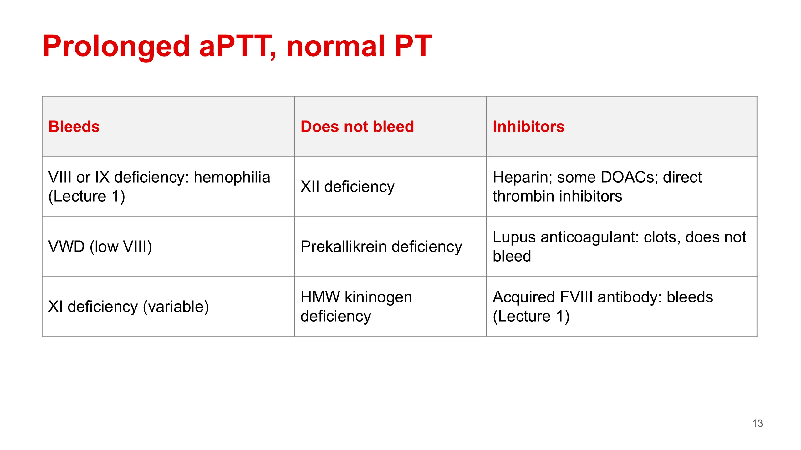
A long aPTT with a normal PT points to the intrinsic arm. Here the list is longer, and the most useful way to sort it is by asking: does this patient bleed?
Left column, those who bleed. Deficiency of factor VIII or IX: hemophilia A or B, which you met in Lecture 1. Von Willebrand disease, because von Willebrand factor carries factor VIII, and when the carrier is low the passenger is cleared faster. And factor XI deficiency, where bleeding varies from person to person and is often linked to injury or surgery.
Middle column, those who do not bleed. Deficiency of factor XII, prekallikrein or high-molecular-weight kininogen can make the aPTT very long, yet the patient does not bleed. The tube needs the contact factors to start clotting. The body does not, because in the body clotting starts with tissue factor. Lecture 1 explained why.
Right column, inhibitors. Heparin is the commonest cause of a long aPTT in hospital. Some DOACs and the direct thrombin inhibitors do it too. The lupus anticoagulant is the trap: it prolongs the aPTT in the tube, but in the patient it causes thrombosis, not bleeding. And the acquired antibody against factor VIII, acquired hemophilia, causes severe bleeding in someone who never bled before. Lecture 1 covered it.
So a long aPTT is not the same as a bleeding tendency. The history tells you which column the patient is in.
Prolonged PT and aPTT
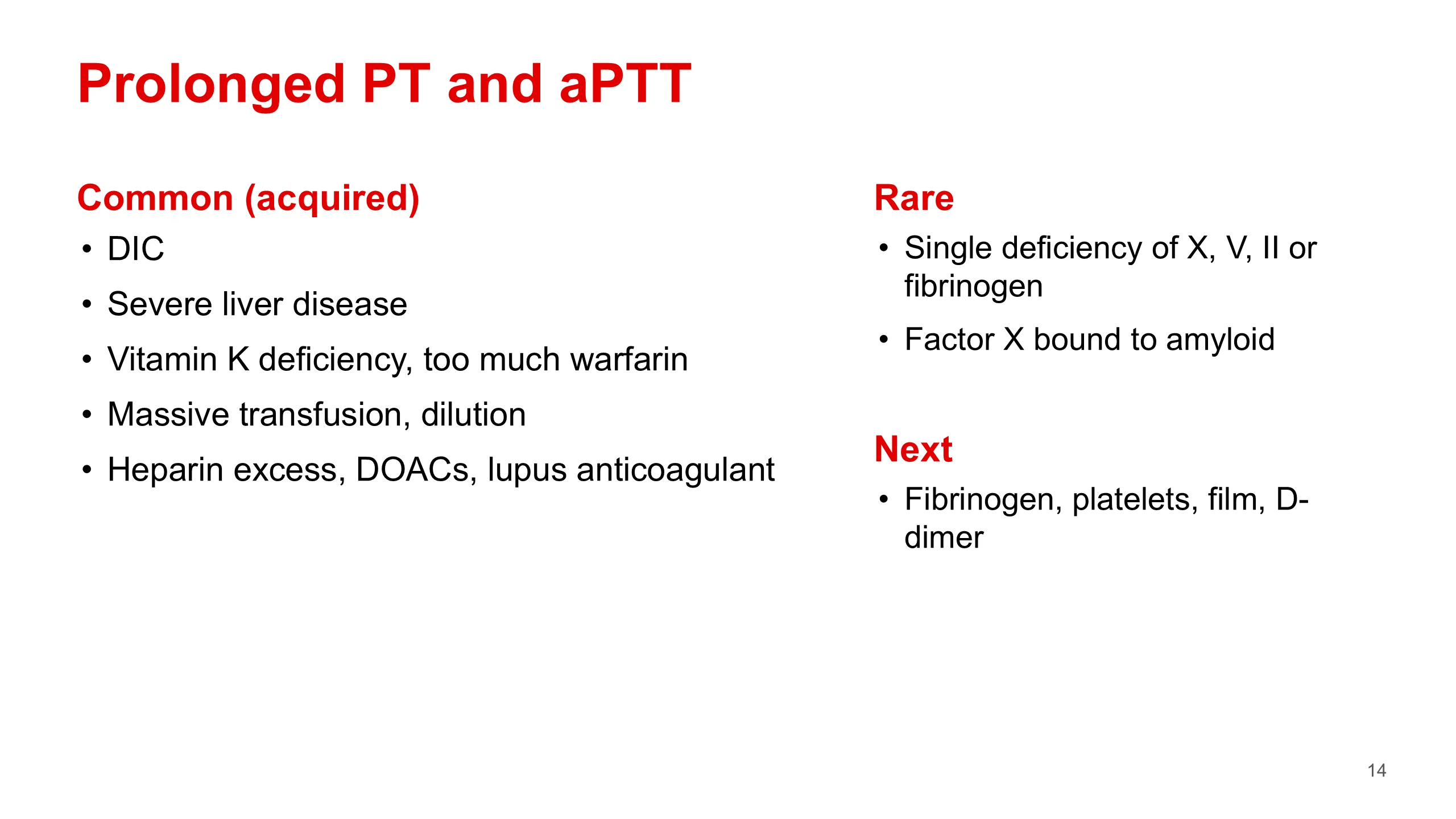
When both the PT and the aPTT are long, there are two possibilities. Either one factor in the common pathway is missing, X, V, II or fibrinogen, which is rare. Or many factors across both arms are low at once, which is common, and is everyday hospital medicine.
The common causes are all acquired. DIC uses factors up faster than the liver can replace them. Severe liver disease means the factors are not made. Vitamin K deficiency or too much warfarin lowers all four vitamin K factors, II, VII, IX and X, so both arms are hit. Massive transfusion and dilution lower everything. And too much heparin, the DOACs and the lupus anticoagulant can lengthen both tests.
The rare causes are single inherited deficiencies of a common pathway factor, and one association worth remembering: in systemic amyloidosis factor X can stick onto the amyloid and be lost.
What next? Send a fibrinogen, look at the platelet count and the film, and check a D-dimer. A falling fibrinogen and platelet count with a high D-dimer and fragments on the film point to DIC, which is our second case. A mixing study helps less here, because when many factors are low it only gives a clue. The history, and the factor assays, settle it.
Normal screen, still bleeding
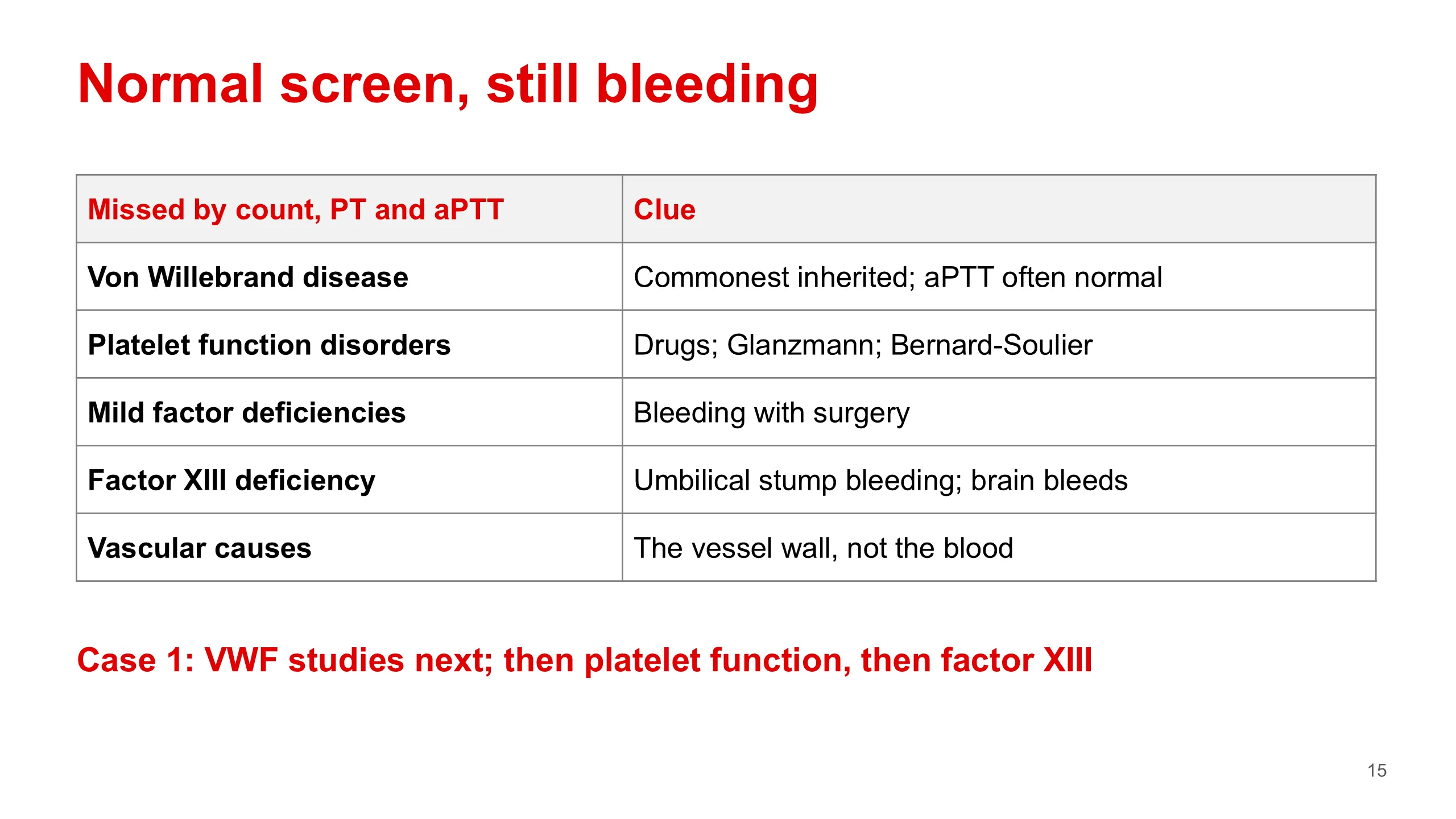
This is the most important slide for our patient. A normal platelet count, PT and aPTT do not exclude a bleeding disorder. Your handbook says it in capitals, in chapter 21.
What do the three tests miss? First, von Willebrand disease. It is the commonest inherited bleeding disorder, and in mild disease the factor VIII is high enough to keep the aPTT normal. Second, disorders of platelet function, where the count is normal but the platelets do not work: aspirin and other drugs most often, and rarely the inherited disorders, Glanzmann thrombasthenia and Bernard-Soulier syndrome. Third, mild factor deficiencies, where there is enough factor to clot in the tube but not enough to stop bleeding after surgery. Fourth, factor XIII deficiency. Factor XIII cross-links fibrin after the clot forms, so the clotting tests, which stop the clock when the clot first appears, are normal. It classically bleeds from the umbilical stump and carries a risk of bleeding into the brain. And last, causes in the vessel wall rather than in the blood.
Our patient has mucocutaneous bleeding, a normal count and a normal screen. So the next step is von Willebrand studies. If those are normal and the history is still convincing, platelet function testing and a factor XIII level follow.
Mixing study
A long PT or aPTT tells you the cascade is not working. It does not tell you why. There are two reasons, and they have different causes and opposite treatments: a factor that is missing, or a factor that is present but blocked by an inhibitor.
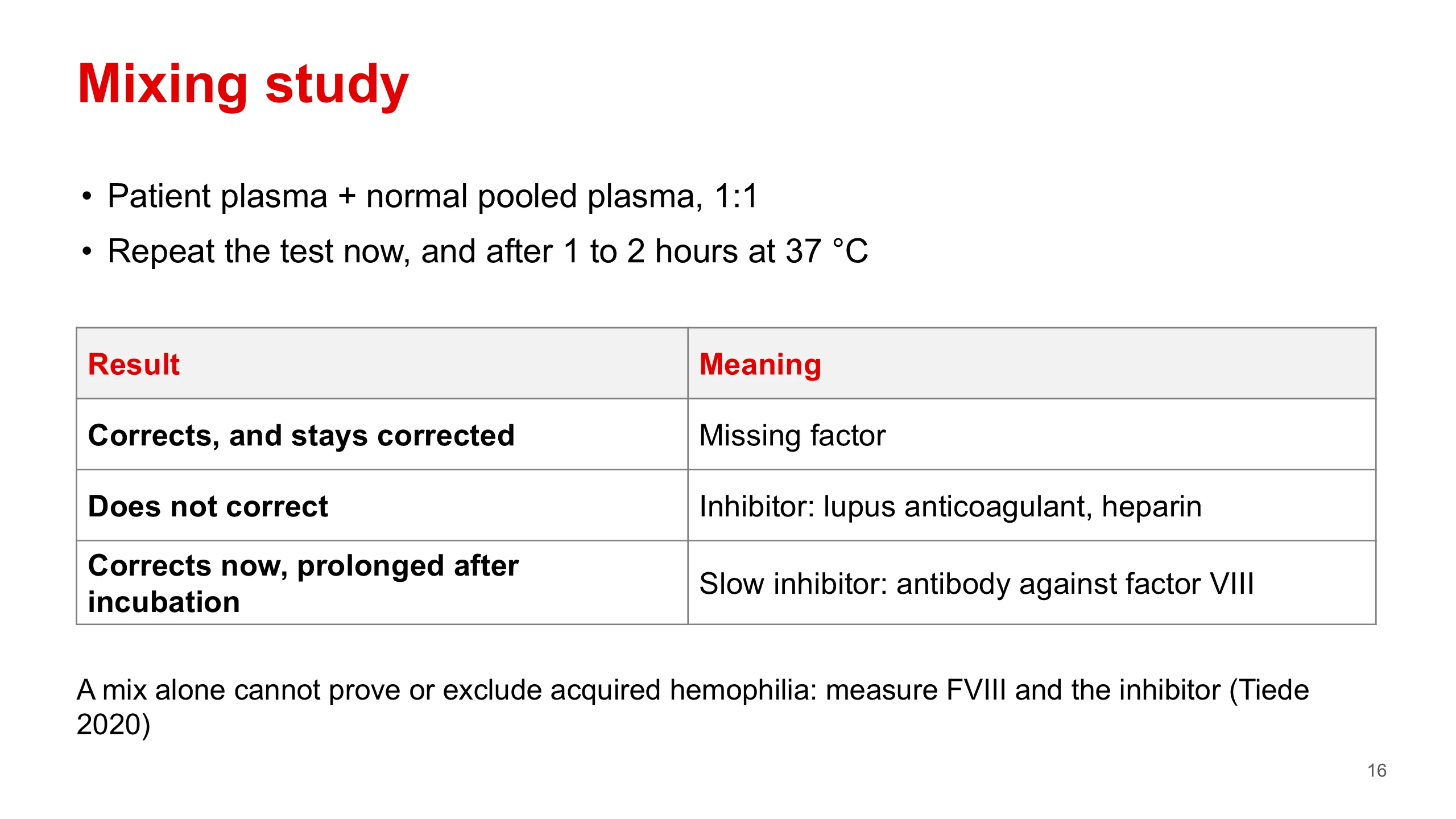
The mixing study separates them, and you can reason it out rather than memorise it. Mix the patient's plasma one to one with normal pooled plasma, and repeat the long test on the mixture.
If a factor was missing, the normal plasma supplies at least half of it, which is enough to clot in normal time. The test corrects.
If an inhibitor is present, it is still in the mixture and it blocks the factors in the normal plasma too. The test does not correct. The lupus anticoagulant and heparin behave like this.
Now the refinement. Some inhibitors act slowly, and the classic one is the antibody against factor VIII in acquired hemophilia. Straight after mixing, the test may correct. After the mixture has been incubated at 37 degrees for one to two hours, the antibody has had time to work, and the aPTT is long again. So the mix is read twice: straight away and after incubation.
The international recommendations on acquired hemophilia (Tiede and colleagues, 2020) go one step further. They say a mixing test cannot establish or exclude the diagnosis on its own. If you suspect it, measure factor VIII and the inhibitor. Some older texts, including an older version of our handbook chapter, say flatly that the aPTT does not correct on mixing. An immediate mix can correct, and that is why this matters.
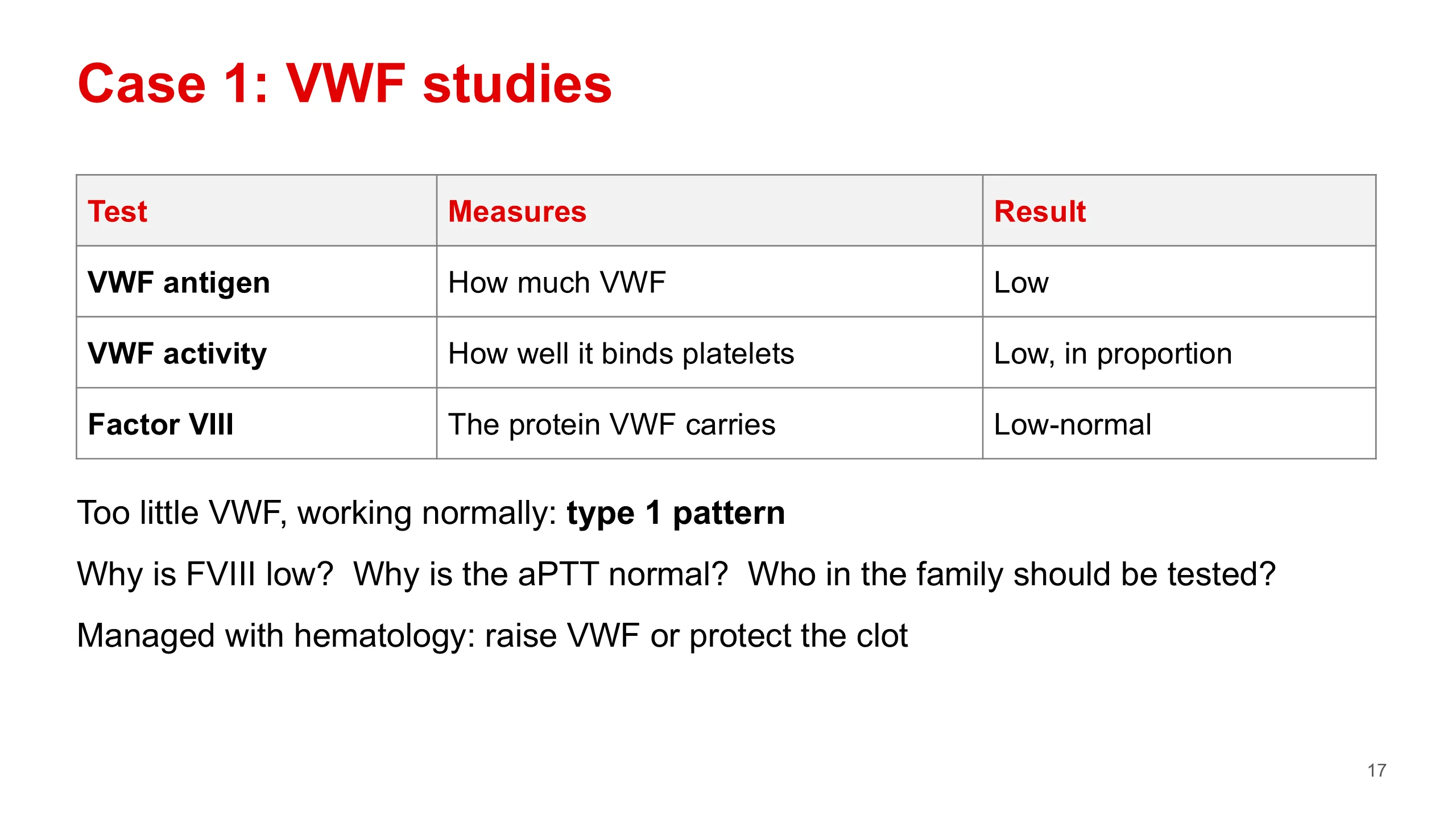
Case 1: VWF studies
Our patient's history fits a disorder of primary hemostasis: lifelong mucocutaneous bleeding. Her count is normal, so this is not a shortage of platelets. The commonest diagnosis that hides behind a normal screen is von Willebrand disease, so the next tests are the three von Willebrand studies. Platelet function tests come later if these are normal, because the inherited platelet disorders are rarer.
The three tests are the VWF antigen, which measures how much von Willebrand factor there is; the VWF activity, which measures how well it binds platelets; and factor VIII.
Her antigen is low. Her activity is low in proportion to the antigen, so what she has works normally. Her factor VIII is low-normal. This is the pattern your handbook gives in its chapter 24 case. Too little of a protein that works normally is a quantitative defect: the pattern of type 1 von Willebrand disease.
Three questions for the room. Why is factor VIII low? Because von Willebrand factor carries and protects it, and that is the next slide. Why is the aPTT still normal? Because her factor VIII is only slightly low, still enough for the tube. And who else should be tested? Her family. Von Willebrand disease is usually autosomal, so it affects her brothers as well as her sisters, and her mother or father.
Treatment is not core for you. In one line: it raises her von Willebrand factor or protects the clot, it is planned with the hematology team, and her heavy periods and iron deficiency are managed with gynecology.
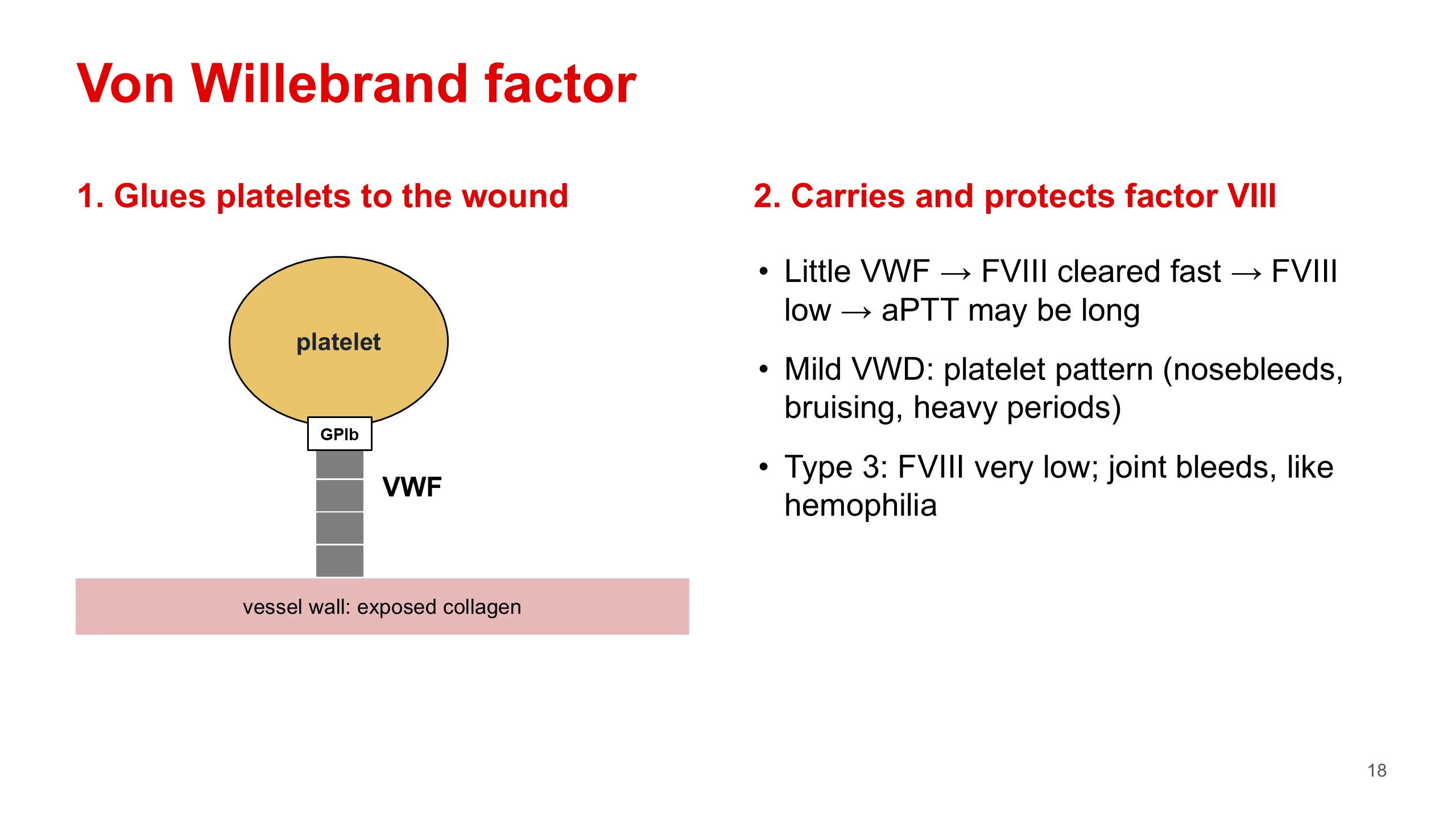
Von Willebrand factor
To understand von Willebrand disease you need two facts about the protein.
Job one. Von Willebrand factor is the glue between the injured vessel wall and the platelet. It binds exposed collagen at one end and the platelet receptor glycoprotein Ib at the other. That is primary hemostasis, the START step from Lecture 1. When the glue is short, platelets do not stick, and the bleeding is the plug type: nosebleeds, bruising, gum bleeding and heavy periods.
Job two. Von Willebrand factor carries factor VIII in the blood and protects it from being cleared. When there is little von Willebrand factor, factor VIII is cleared faster and its level falls. If it falls far enough, the aPTT becomes long.
That explains the whole clinical range. In mild disease, which is most patients, the bleeding is the platelet type and the screen is normal, as in our patient. In severe disease, type 3, there is almost no von Willebrand factor, factor VIII is very low as well, and the patient bleeds into joints and muscle like a person with hemophilia.
It also explains a trap. Type 2N, which we meet on slide 20, cannot carry factor VIII. It looks exactly like mild hemophilia A, but it is inherited autosomally and affects women. Test von Willebrand factor before you label a patient as having hemophilia.
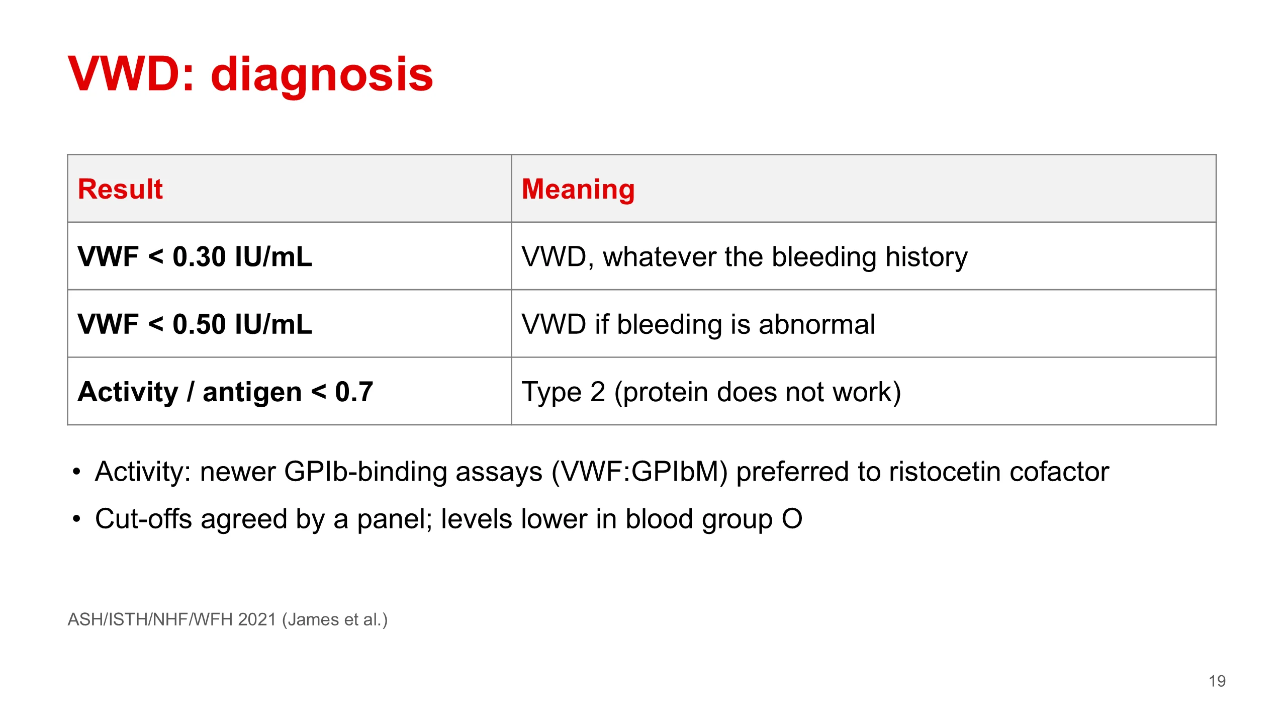
VWD: diagnosis
How low is low? Since 2021 there has been an international guideline, from ASH, ISTH, NHF and WFH, written by James and colleagues. It gives three numbers.
A von Willebrand factor level below 0.30 international units per millilitre confirms von Willebrand disease, whatever the bleeding history. A level below 0.50 confirms it in a patient who has abnormal bleeding. The panel decided against a separate "low VWF" label for these patients. Our patient has abnormal bleeding, so a level anywhere below 0.50 would confirm her diagnosis.
The third number is a ratio. Divide the activity by the antigen. If the ratio is below 0.7, the protein is present but does not work well: think of type 2. Older teaching used a ratio of 0.5; the guideline moved it to 0.7.
On the activity test itself, the guideline prefers the newer assays that measure binding to glycoprotein Ib, such as the one called VWF:GPIbM, over the older ristocetin cofactor assay. You may see either on a report.
Now the point about evidence. 0.30 and 0.50 are agreements made by a panel. They are useful and they are what you should use, but they are not a line in nature. Von Willebrand factor levels are also lower in people with blood group O, so a borderline result needs care.
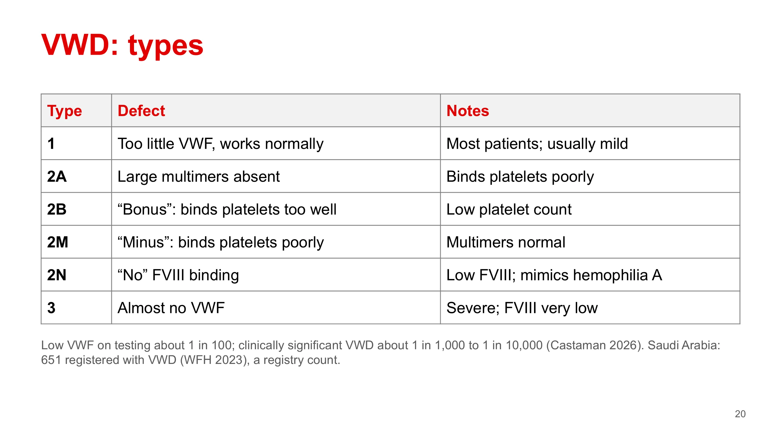
VWD: types
There are three types. Type 1 is too little of a normal protein: a partial quantitative defect. It is the commonest type and is usually mild. Type 2 is a protein that is present but does not work: a qualitative defect. Type 3 is an almost complete absence. It is severe, factor VIII is very low too, and these patients bleed like hemophiliacs.
How common is type 1? Sources differ. Your handbook gives about 75 percent of cases. A 2026 review (Castaman) gives 60 to 70 percent, and about 5 percent for type 3. The old deck said 80 percent. I have written "most patients" on the slide because the sources do not agree.
The letters of type 2 are hard to keep apart, so attach a word to each. 2A: the large multimers, which do most of the platelet binding, are absent. 2B, think "bonus": a gain of function. The factor binds platelets too well, even in the circulation, and the platelet-factor complexes are cleared, so the platelet count falls. 2M, think "minus": binding to platelets is reduced, but the multimers are normal. 2N, think "no factor VIII binding": factor VIII is low and the patient looks like mild hemophilia A, but the inheritance is autosomal.
How common is the disease? About 1 in 100 people have a low level on testing, but clinically significant disease is about 1 in 1,000 to 1 in 10,000 (Castaman 2026). The old slide said that only 1 percent of those with the disease have symptoms. That was wrong, and it has been removed.
For Saudi Arabia, the 2023 World Federation of Hemophilia survey lists 651 people registered with von Willebrand disease. That is the number on a registry, not how common the disease is. A single referral centre in Riyadh reported a much higher share of type 3 than international series; referral bias is a likely reason.
Platelet function disorders
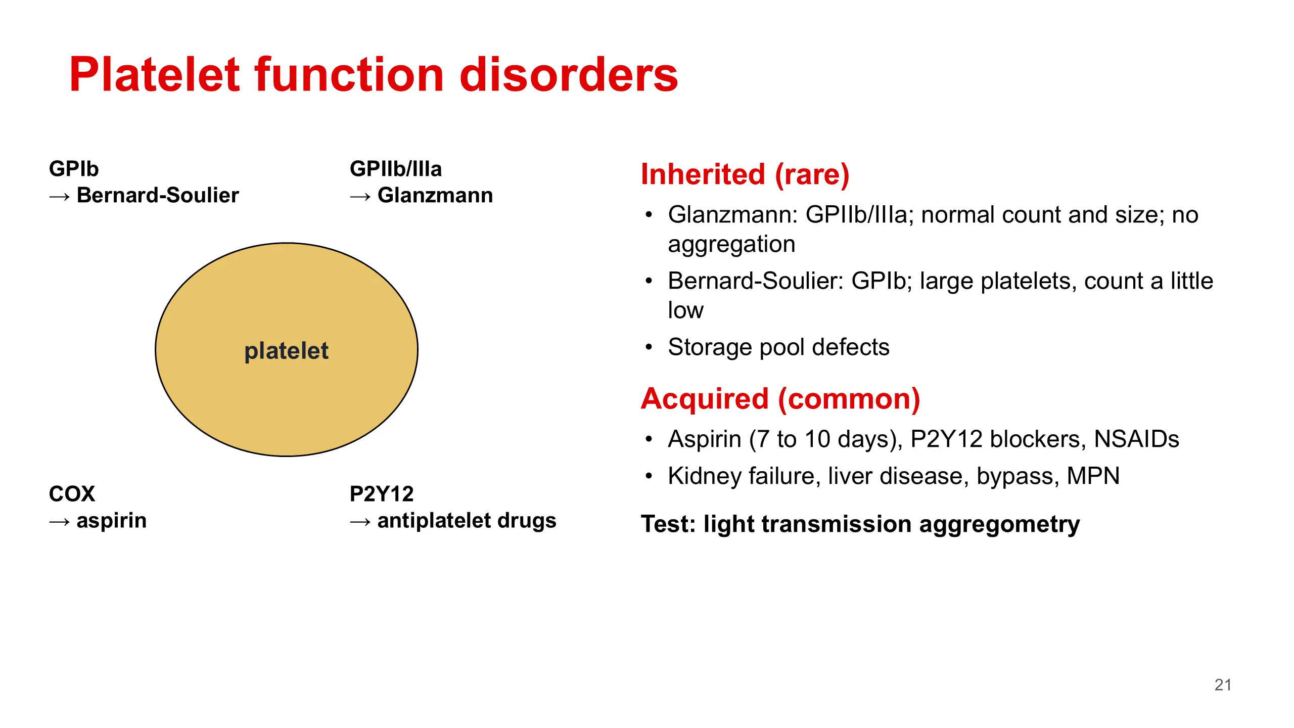
Sometimes the platelets are there in normal numbers but do not work. The bleeding is the plug type, and the count and the clotting screen are both normal, so it is easy to miss if you only look at numbers.
Each receptor on the drawing has a disease or a drug. Glycoprotein Ib binds von Willebrand factor: its absence is Bernard-Soulier syndrome, with large platelets and a count that is a little low. Glycoprotein IIb/IIIa binds fibrinogen and links platelets to each other: its absence is Glanzmann thrombasthenia, with a normal count and normal-sized platelets that do not aggregate at all, and severe bleeding. Cyclooxygenase makes thromboxane: aspirin blocks it for the whole life of the platelet, 7 to 10 days. The P2Y12 receptor responds to ADP, and a group of antiplatelet drugs blocks it.
The acquired causes are far more common than the inherited ones. Drugs are the leading cause: aspirin, the P2Y12 blockers and anti-inflammatory drugs. Kidney failure impairs platelets. Liver disease, cardiopulmonary bypass and the myeloproliferative neoplasms do it too.
The diagnosis is made with platelet function testing. The reference method is light transmission aggregometry, a specialist test. In Glanzmann the platelets fail to aggregate with all the usual agonists.
Glanzmann thrombasthenia
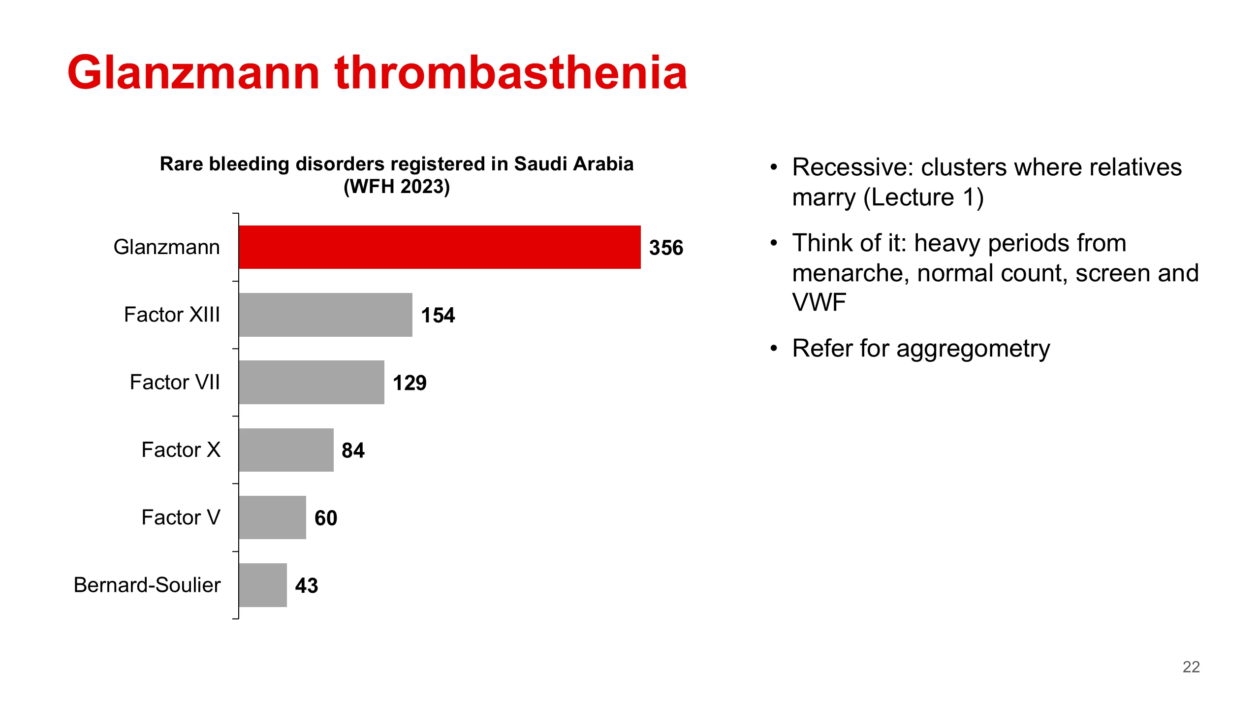
This chart is the Saudi data that makes this disorder matter for you. The 2023 Global Survey of the World Federation of Hemophilia lists the rare bleeding disorders registered in Saudi Arabia. Glanzmann thrombasthenia is the largest group, with 356 patients. Then factor XIII deficiency with 154, factor VII with 129, factor X with 84, factor V with 60 and Bernard-Soulier syndrome with 43.
Two cautions. These are counts of registered patients, not the number of people who have the disorder. And the survey's own columns do not add up to its total for other bleeding disorders, so treat the exact numbers with care. The order is what matters: Glanzmann is not a curiosity here.
Why so many? Glanzmann is inherited recessively, so a child needs a faulty gene from both parents, and that happens more often when parents are related. Lecture 1 covered consanguinity in this population. A study from Madinah found that almost all the families it recorded had related parents, and that most affected girls had heavy periods. Estimates of how common Glanzmann is in Saudi Arabia differ about four-fold between sources, so I have not put a prevalence on the slide.
The practical point: think of Glanzmann in a girl with heavy periods from her first period, a normal platelet count, a normal screen, and normal von Willebrand studies. Refer her for platelet aggregometry.
Case 2
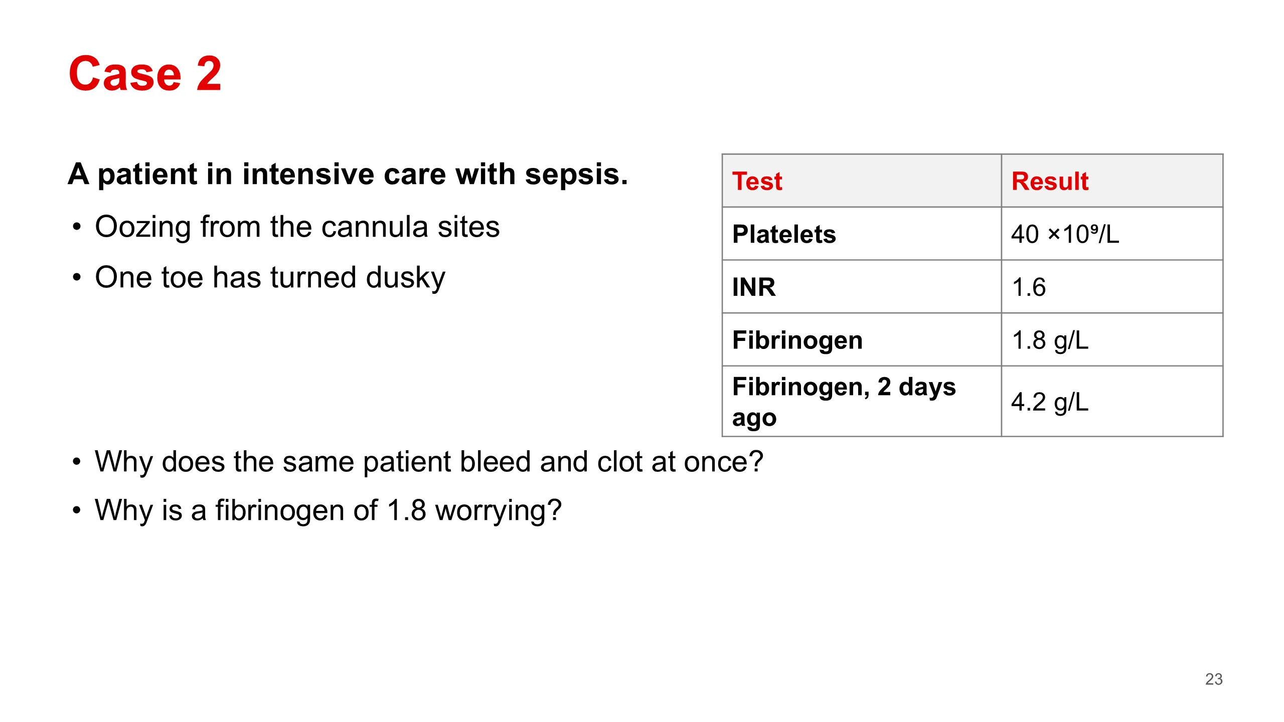
Our second case is acquired, and it is the kind of bleeding you will meet on every night on call. It comes from the casebook, Case 51.
A patient in intensive care with sepsis starts to ooze from the cannula sites. One toe has turned dusky. The platelet count is 40 times ten to the nine per litre. The INR is 1.6. The fibrinogen is 1.8 grams per litre, and two days ago it was 4.2.
Ask the room two questions before moving on.
First: how can the same patient bleed from the lines and clot in a toe at the same time? It sounds like two diseases. It is one.
Second: why is a fibrinogen of 1.8 more worrying than it looks? The answer is the earlier value. Fibrinogen rises in inflammation, as an acute phase protein, so a septic patient should have a high fibrinogen. A fall from 4.2 to 1.8 in two days means it is being used up. The trend tells you more than any single value.
DIC: mechanism
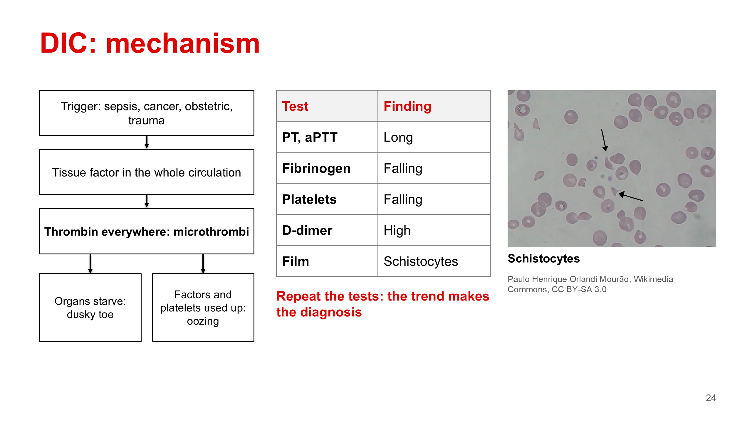
Disseminated intravascular coagulation is never a diagnosis on its own. It is what a severe illness does to the clotting system.
Follow the chain on the left. Something exposes tissue factor to the whole circulation at once: sepsis above all, cancer, an obstetric emergency or major trauma. Thrombin is then made everywhere, not just at a wound, and tiny clots form in small vessels throughout the body.
Two things follow. The tiny clots block small vessels, so organs are starved of blood: that is the dusky toe, and the kidney and lung injury. And making all those clots uses up platelets, clotting factors and fibrinogen faster than the liver and marrow can replace them. So the same patient bleeds from every line. One process explains both.
The tests follow the mechanism. The PT and aPTT are long because the factors are used up. Fibrinogen and platelets fall. The body's clot-dissolving system breaks down the tiny clots and releases D-dimer, so the D-dimer is high. On the film you may see schistocytes, red cells cut into fragments as they pass through vessels lined with fibrin strands. The photograph shows them.
No single result makes the diagnosis. It is a pattern moving in one direction over time, so serial tests are better than any single set. That is also why the ISTH scoring system is built on repeated measurement. The ISTH revised its definition and score in 2025.
Image credit.
- Schistocytes on a blood film (Schizocyte smear 2009-12-22.JPG) Wikimedia Commons · CC BY-SA 3.0 · Paulo Henrique Orlandi Mourão
DIC: causes
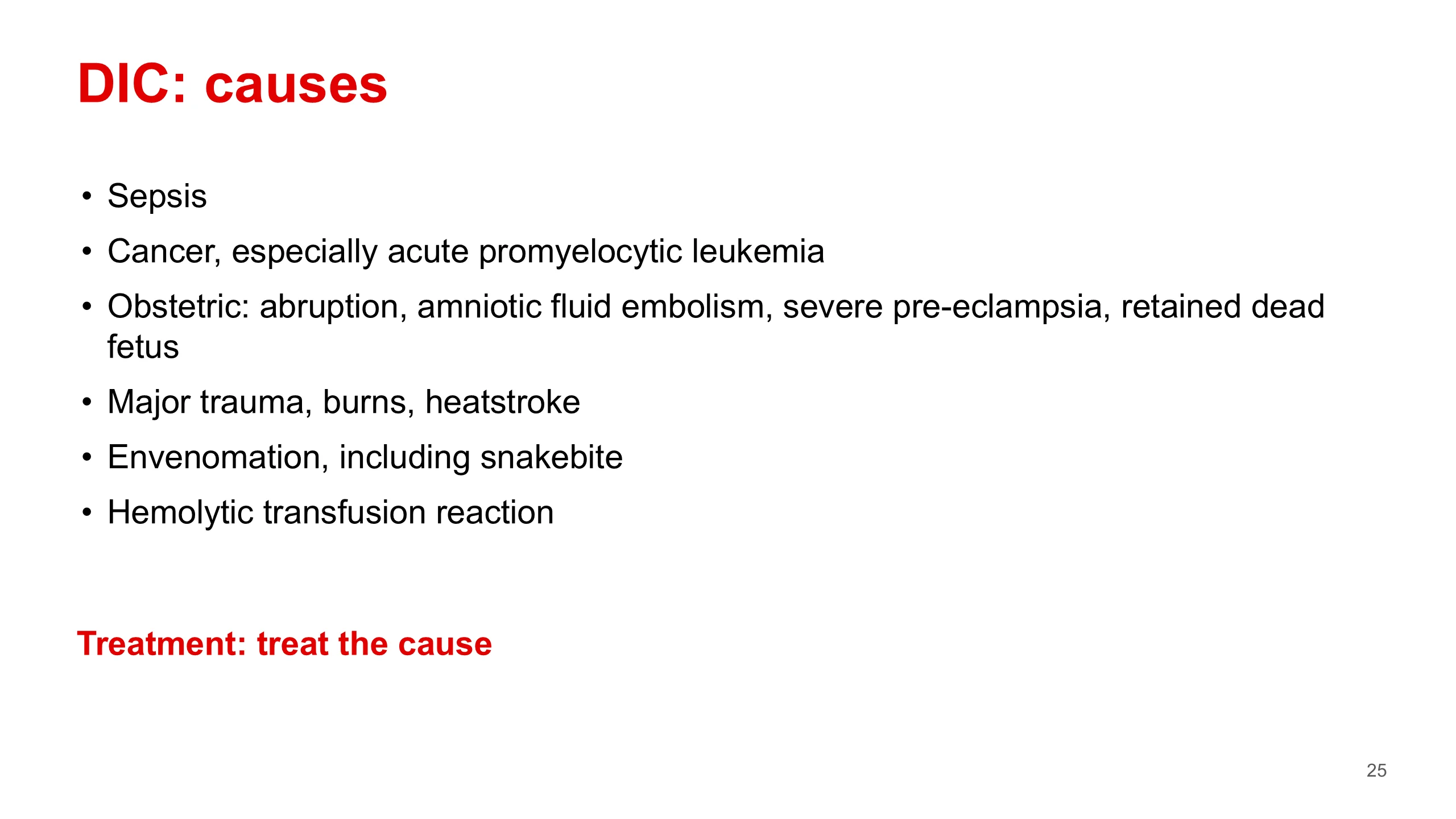
DIC is treated by treating its cause. That is the one line you need about treatment.
The causes on the slide are the things that expose tissue factor to the whole circulation: sepsis above all; cancer, and acute promyelocytic leukemia in particular, which you met in the leukemia lecture; obstetric emergencies such as placental abruption, amniotic fluid embolism, severe pre-eclampsia or a retained dead fetus; major trauma, burns and heatstroke; severe envenomation; and a severe hemolytic transfusion reaction.
Envenomation matters in Asir. Snakebite coagulopathy is seen in our hospitals. A study from four tertiary hospitals in the region reported coagulopathy in a large share of snakebite admissions.
Schistocytes and low platelets
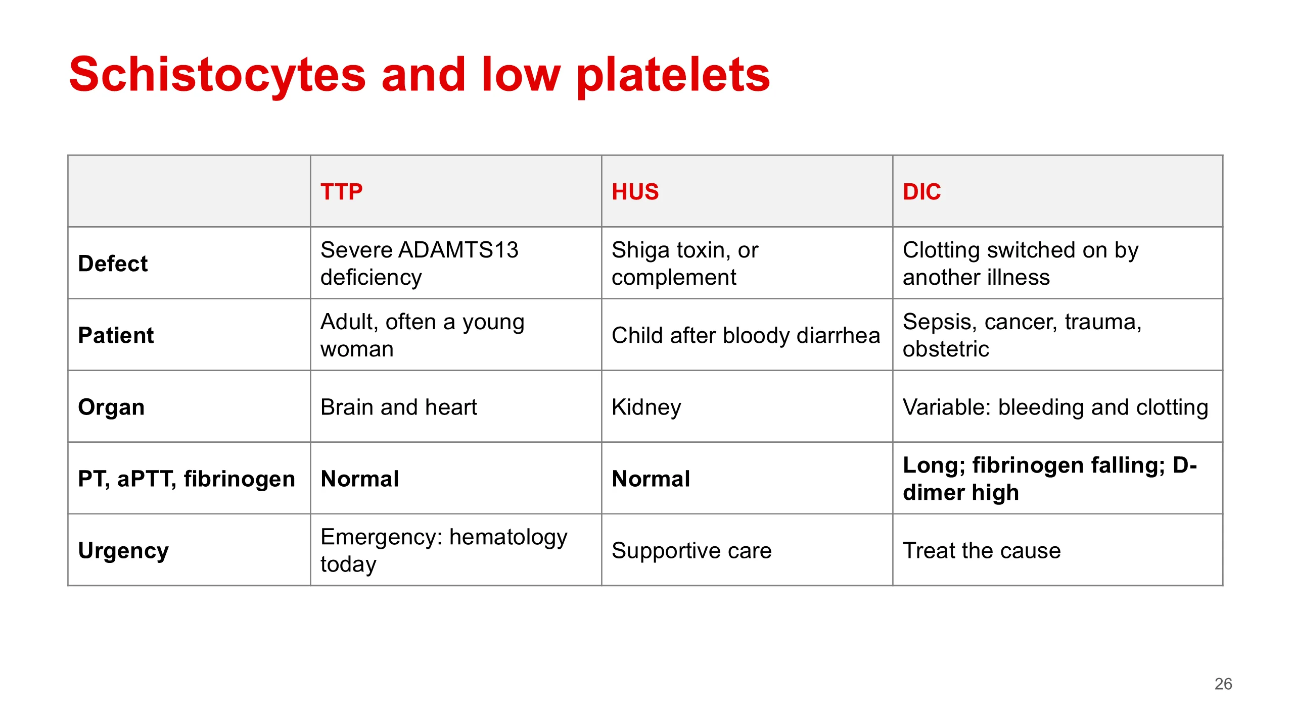
Schistocytes with a low platelet count can mean three different diseases. The clotting screen is the fork in the road. This table is from Case 51 in the casebook.
In TTP and HUS the small vessels fill with platelet clots, but the clotting cascade itself is not consumed, so the PT, aPTT and fibrinogen are normal. In DIC the cascade is consumed, so the times are long, the fibrinogen falls and the D-dimer is high. That one row, in bold, tells you which way to go.
TTP is caused by a severe lack of ADAMTS13, the enzyme that cuts von Willebrand factor down to size. It is usually an acquired antibody, typically in an adult, often a young woman, and it affects the brain and heart. It is an emergency: call hematology the same day, before the ADAMTS13 result comes back.
HUS is usually a child after bloody diarrhea, from Shiga toxin, and the kidney takes the damage. The atypical form is driven by complement.
DIC, as we have just seen, comes from another illness. TTP and HUS are taught fully in the handbook, chapter 29. Here you need only the fork.
Case 3
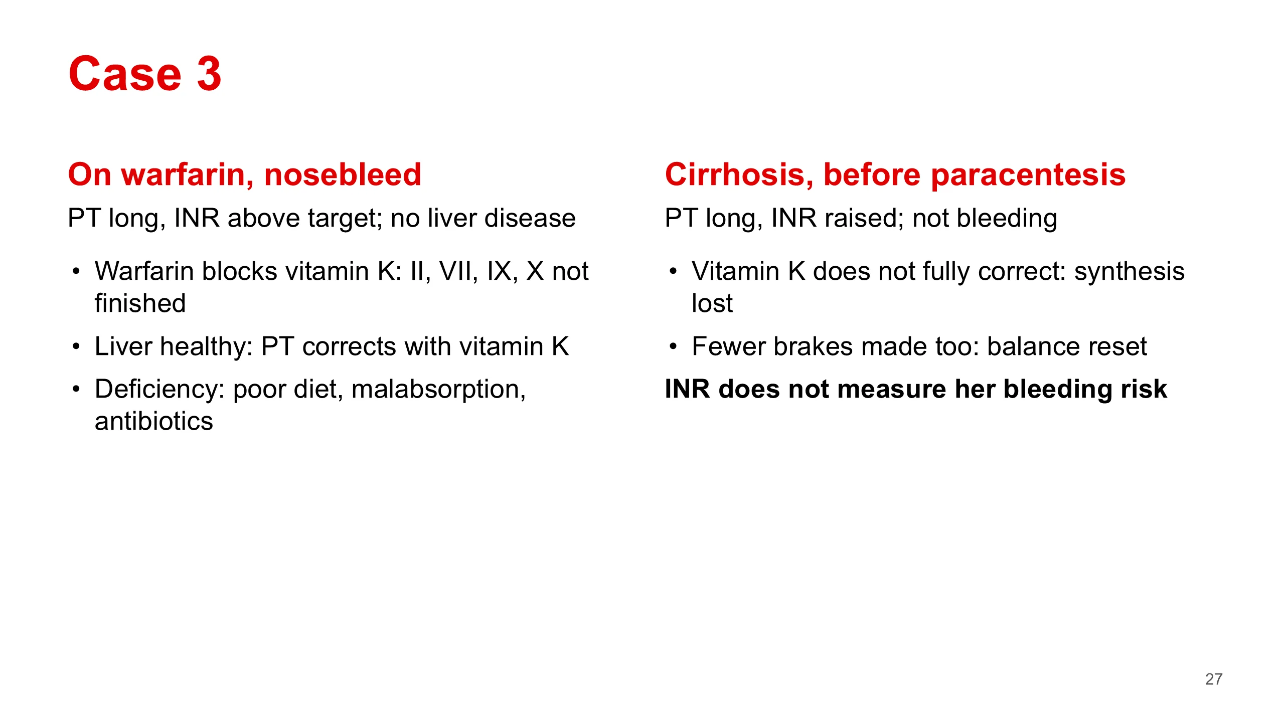
Our third case is two short patients side by side. Both have a long PT. The question is why.
The man on the left is on warfarin, has a nosebleed, and his INR is above his target. He has no liver disease. Warfarin blocks vitamin K, and without vitamin K the liver cannot finish making factors II, VII, IX and X. Vitamin K deficiency from poor diet, malabsorption or antibiotics does the same. The liver itself is healthy, so once vitamin K is available the factors are made again and the PT corrects.
The woman on the right has cirrhosis and needs a paracentesis. Her INR is raised. She is not bleeding. Her liver cannot make enough of the factors, so vitamin K will not fully correct her PT. Synthesis is lost.
But here is the important point. Her liver also makes less of the natural brakes, such as protein C, protein S and antithrombin. So her clotting system is not simply tipped towards bleeding. It is reset to a new, fragile balance. The PT and INR measure only the factors, not the brakes, and so the casebook says plainly: in liver disease the INR does not measure the risk of bleeding.
Work-up summary
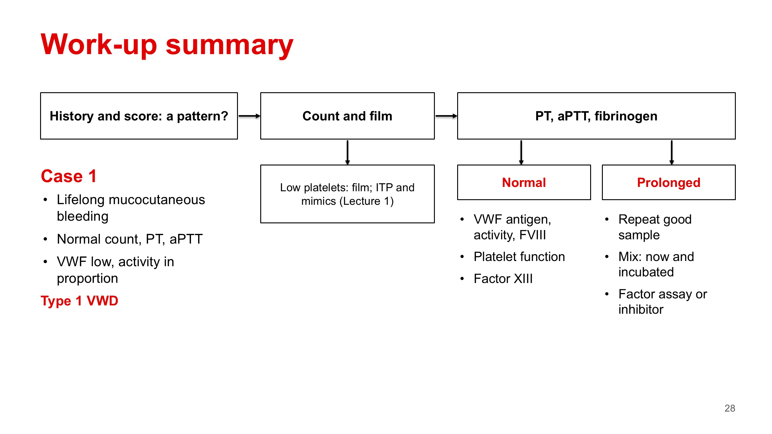
Here is the whole lecture as one flow chart, and with it the answer for our patient.
Start with the history and the score. Is there a pattern of bleeding? Then the count and the film. If the platelets are low, look at the film first and think of ITP and its mimics, from Lecture 1. If the count is normal, the PT, aPTT and fibrinogen come next, on a good sample.
If the screen is long, repeat it on a good sample, mix it straight away and after incubation, and then measure the factors or the inhibitor.
If the screen is normal and the history is convincing, do not stop. Send von Willebrand antigen, activity and factor VIII. If those are normal, go on to platelet function testing, and then factor XIII.
At every step, read the drug chart, ask whether the problem is inherited or acquired, and watch for the patient who is also tired and feverish, whose problem is in the marrow.
And our 19-year-old? Lifelong mucocutaneous bleeding. A normal count, PT and aPTT. A low von Willebrand factor with activity in proportion. She has type 1 von Willebrand disease. Go back to the answers the room gave on slide 2 and see how many were right.
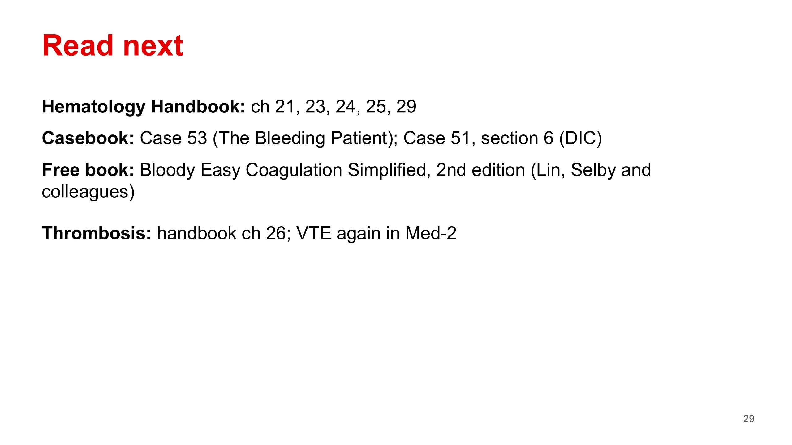
Read next
Tonight, read chapters 21, 23, 24, 25 and 29 of the Hematology Handbook. Then work through Case 53, The Bleeding Patient, and section 6 of Case 51 in the casebook, which covers the microangiopathies and DIC. Then try the bank items on chapters 20 to 25.
The book on the slide, Bloody Easy Coagulation Simplified, second edition, edited by Yulia Lin, Rita Selby and colleagues from the Toronto teaching hospitals, is short, clinical and free to download. It was recommended in my earlier deck.
The other side of the balance is thrombosis: what happens when the brakes fail and blood clots where it should not. It is in the handbook, chapter 26. Venous thromboembolism is taught again in Med-2. The old slides pointed to a "thrombosis seminar"; there is no such session in the Med-1 timetable, so the handbook chapter is the right place.
Image credits. All other figures were drawn for this lecture.
- Slide 24: Schistocytes on a blood film (Schizocyte smear 2009-12-22.JPG) by Paulo Henrique Orlandi Mourão, via Wikimedia Commons, CC BY-SA 3.0.