Myeloid Neoplasms
Myeloproliferative neoplasms and myelodysplastic syndromes — classification and management.
These slides are an outline only. Read the accompanying material with them: the notes under each slide below, and the chapters of the Hematology Handbook on chronic myeloid leukemia, the myeloproliferative neoplasms and the myelodysplastic syndromes. Then work through the Internal Medicine Casebook and the Hematology Question Bank.

What this lecture is about
These slides are an outline only. Read the accompanying material with them: the notes under each slide on this page, and the chapters of the Hematology Handbook on chronic myeloid leukemia, the myeloproliferative neoplasms and the myelodysplastic syndromes. Then work through the Internal Medicine Casebook and the Hematology Question Bank.
Most students meet the myeloproliferative neoplasms and the myelodysplastic syndromes as a list of unfamiliar names. This lecture asks you instead to do one thing, printed on the title slide: classify. Both groups of disease are clonal disorders of the myeloid stem cell, and everything about them falls into place once you can answer two questions about a patient's blood count. Is the abnormality reactive or clonal? And if it is clonal, is the marrow producing effectively, giving high counts, or ineffectively, giving low counts?
The lecture builds that framework in order. It first takes the word myeloproliferative neoplasm apart, then reviews the normal physiology of proliferation so that the mutation which breaks it makes sense. It sets out the WHO classification, and then puts it to work in three short cases: a high hemoglobin, a high white count with a big spleen, and a high platelet count, with a closer look at CML after the second. It then covers the complications and the management of the MPNs, and closes with the counterpoint of myelodysplasia.
The deck is based on student notes from 2022 and is intended as a clinical guide rather than a reference; the entities are introduced through the patients who have them.
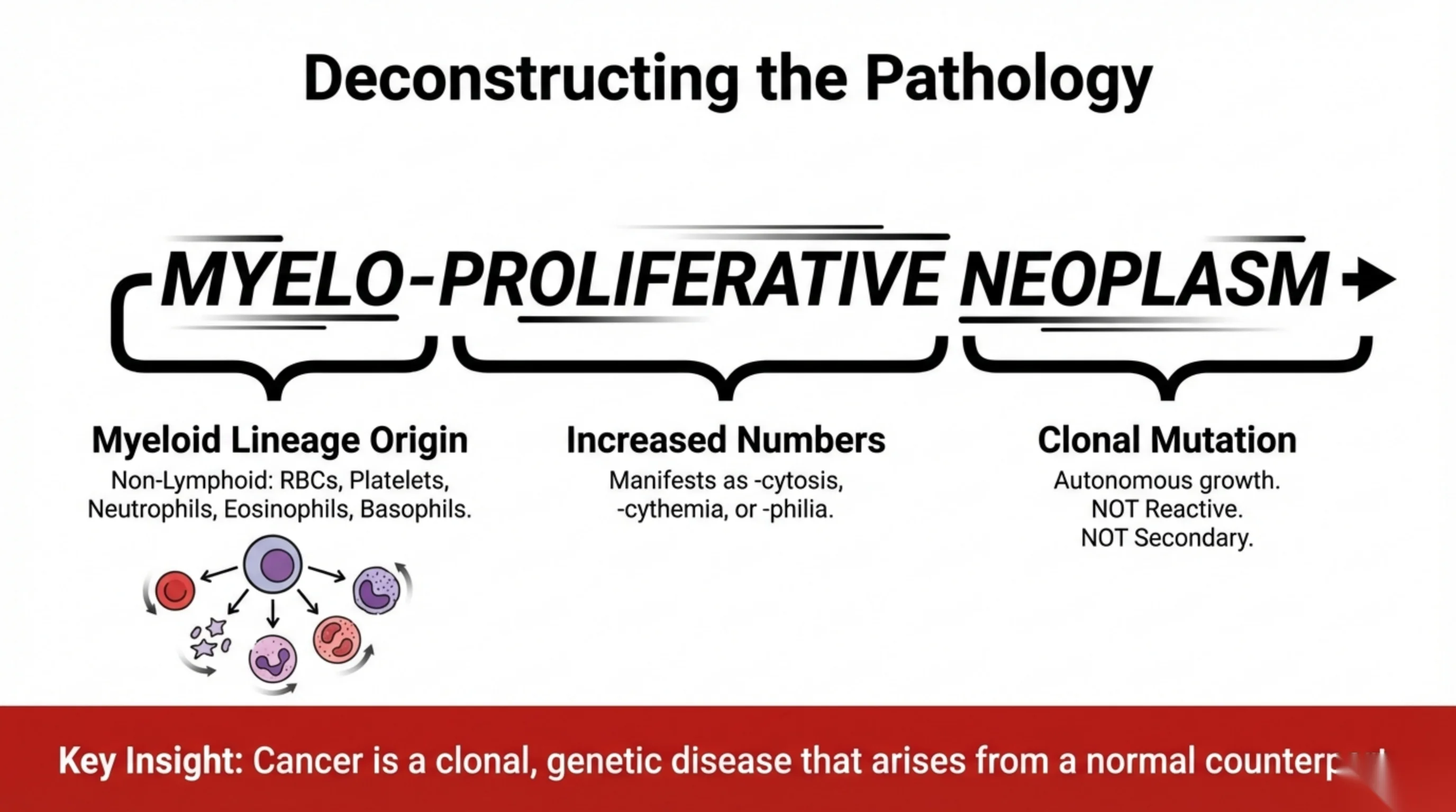
Deconstructing the name: three words, three ideas
The term myeloproliferative neoplasm carries its whole definition inside it, and the slide breaks it into its three parts.
Myelo names the lineage. Myeloid means every blood cell that is not a lymphocyte: red cells, platelets, neutrophils, eosinophils and basophils, all descended from the common myeloid progenitor shown in the lineage illustration. Proliferative names the direction of the abnormality: too many cells. In the language of the blood count this appears as a -cytosis (leukocytosis, thrombocytosis), a -cythemia (erythrocythemia, polycythemia) or a -philia (neutrophilia, eosinophilia). Neoplasm is the word that matters most. It means the excess is caused by a clonal mutation in a stem cell, so that the cells grow autonomously. They are not responding to anything. The proliferation is not reactive and not secondary to some other disease.
The key insight at the foot of the slide generalises this: cancer is a clonal, genetic disease that arises from a normal counterpart. A myeloproliferative neoplasm is the normal process of blood production, running without its controls. The next slide shows what those controls are.
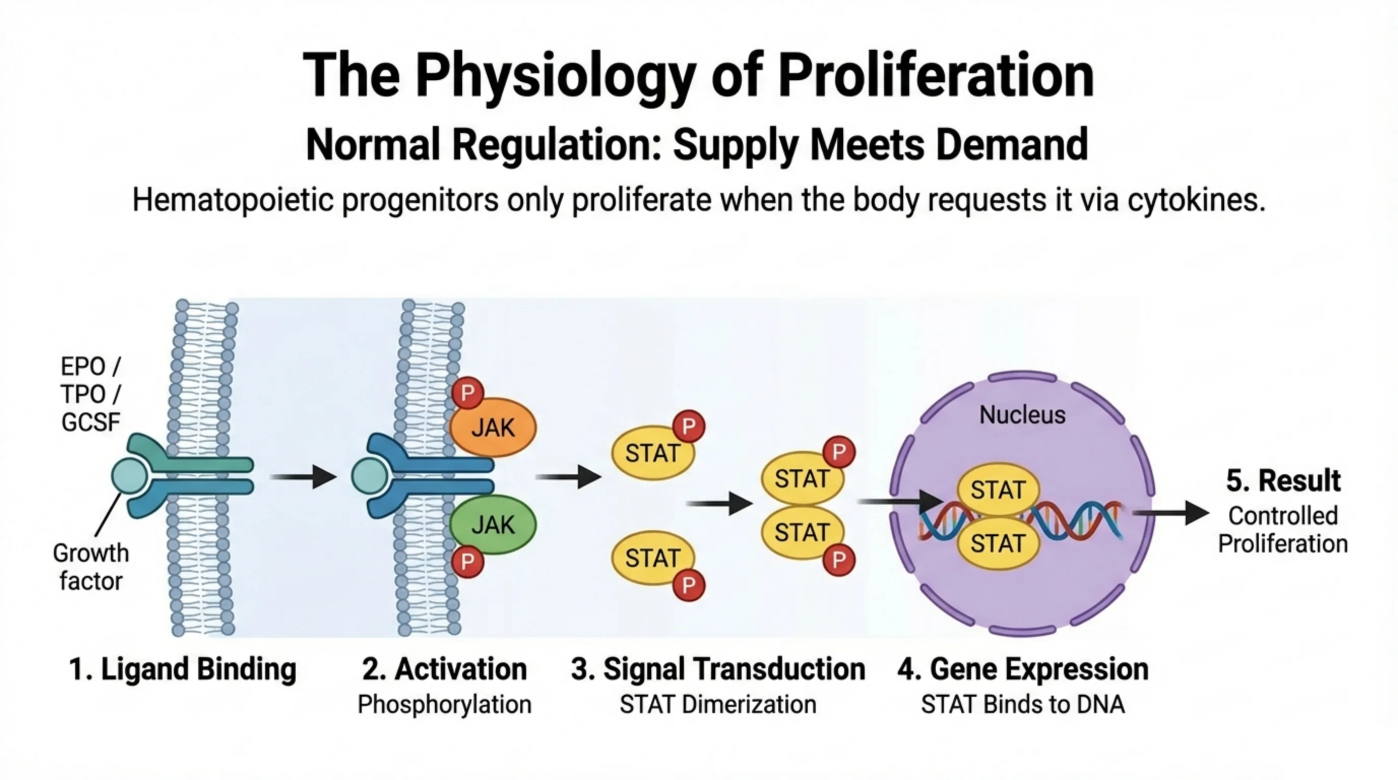
Normal proliferation: supply meets demand
Healthy hematopoietic progenitors do not divide on their own initiative. They divide when the body asks them to, and the request arrives as a cytokine: erythropoietin for red cells, thrombopoietin for platelets, granulocyte colony-stimulating factor for neutrophils. Remove the signal and production falls back to baseline. This is the principle of supply meeting demand, and the five-step diagram shows how the signal is delivered inside the cell.
The growth factor, the ligand, binds its receptor on the cell surface. Binding brings the receptor's two halves together and activates the enzyme attached to them, JAK, a Janus kinase, which phosphorylates itself and the receptor. The phosphorylated receptor recruits STAT proteins, which are themselves phosphorylated, pair up as dimers, and travel to the nucleus. There the STAT dimer binds DNA and switches on the genes for growth and division. The result is proliferation, but controlled proliferation, lasting only as long as the ligand is present.
Hold onto the JAK-STAT pathway. The next slide shows that the classical myeloproliferative neoplasms are, almost all of them, diseases of one mutation in one component of it.
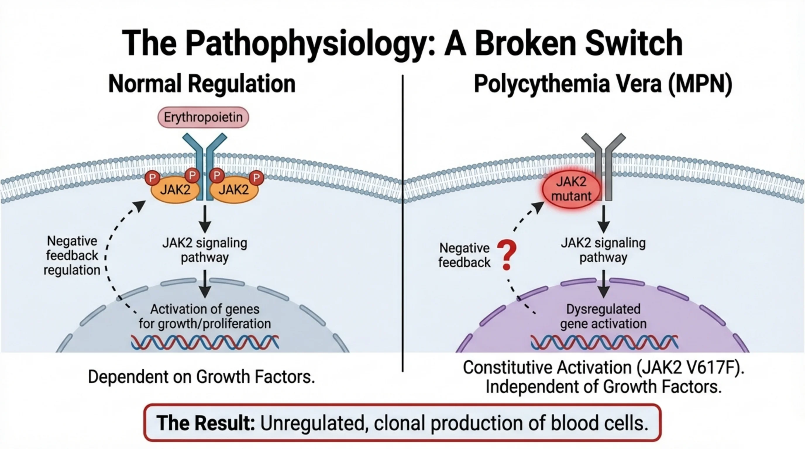
The broken switch: JAK2 that never turns off
The two panels contrast the normal red cell precursor with the precursor in polycythemia vera, and the difference is a single amino acid.
On the left, erythropoietin binds its receptor, the paired JAK2 molecules phosphorylate each other, the signalling pathway runs, and the genes for growth are activated. Two features keep this under control: the pathway is dependent on the growth factor, so it stops when erythropoietin falls, and there is intact negative feedback that damps the signal even while ligand is present.
On the right, the JAK2 protein carries the V617F mutation, a substitution of phenylalanine for valine at position 617. The mutant kinase is locked in the active state. It drives the pathway without any ligand at all, the gene activation is dysregulated, and the negative feedback is lost, which the diagram marks with a question mark. This is constitutive activation: the switch is stuck on, and proliferation has become independent of growth factors.
The result is the unregulated, clonal production of blood cells that defines the disease. One consequence should be anticipated now, because it becomes the diagnostic test on slide 7: if the red cell precursors are producing on their own, the body will detect the excess red cells and switch off erythropoietin. In polycythemia vera the erythropoietin level is low.
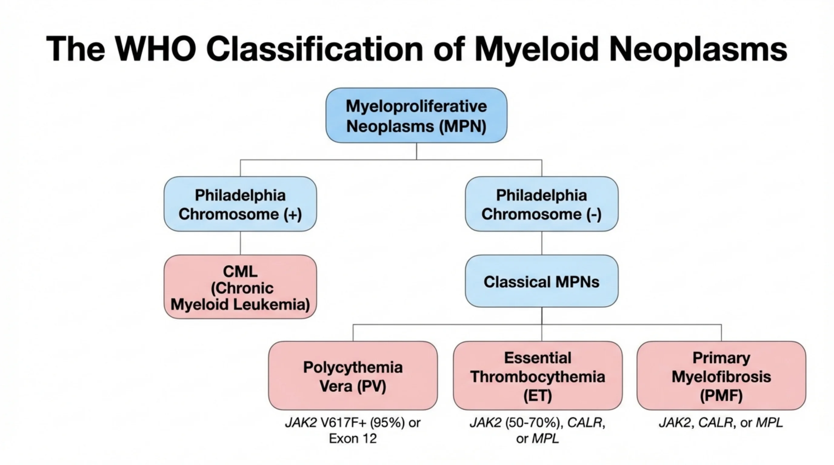
The WHO classification: one chromosome divides the family
The myeloproliferative neoplasms are classified first by a single test: the presence or absence of the Philadelphia chromosome, the translocation that produces the BCR-ABL fusion gene.
Philadelphia-positive disease is chronic myeloid leukemia, CML, and it stands alone because its driver is a fusion kinase with its own targeted treatment, the tyrosine kinase inhibitors on slide 11. Philadelphia-negative disease comprises the three classical MPNs, distinguished by which lineage predominates. Polycythemia vera is the disease of red cells and is driven by JAK2 V617F in about 95 percent of cases, with mutations in JAK2 exon 12 accounting for most of the rest. Essential thrombocythemia is the disease of platelets; JAK2 is mutated in 50 to 70 percent, and the remainder carry mutations in CALR or MPL. Primary myelofibrosis is the disease in which the marrow becomes scarred, and it carries the same three drivers, JAK2, CALR or MPL.
The practical consequence is a short diagnostic panel. A patient with a persistently high count and no reactive cause is tested for BCR-ABL and for JAK2; if both are negative and the count is platelets or fibrosis is suspected, for CALR and MPL. Finding one of these mutations proves that the proliferation is clonal, which is the question the next slide poses.
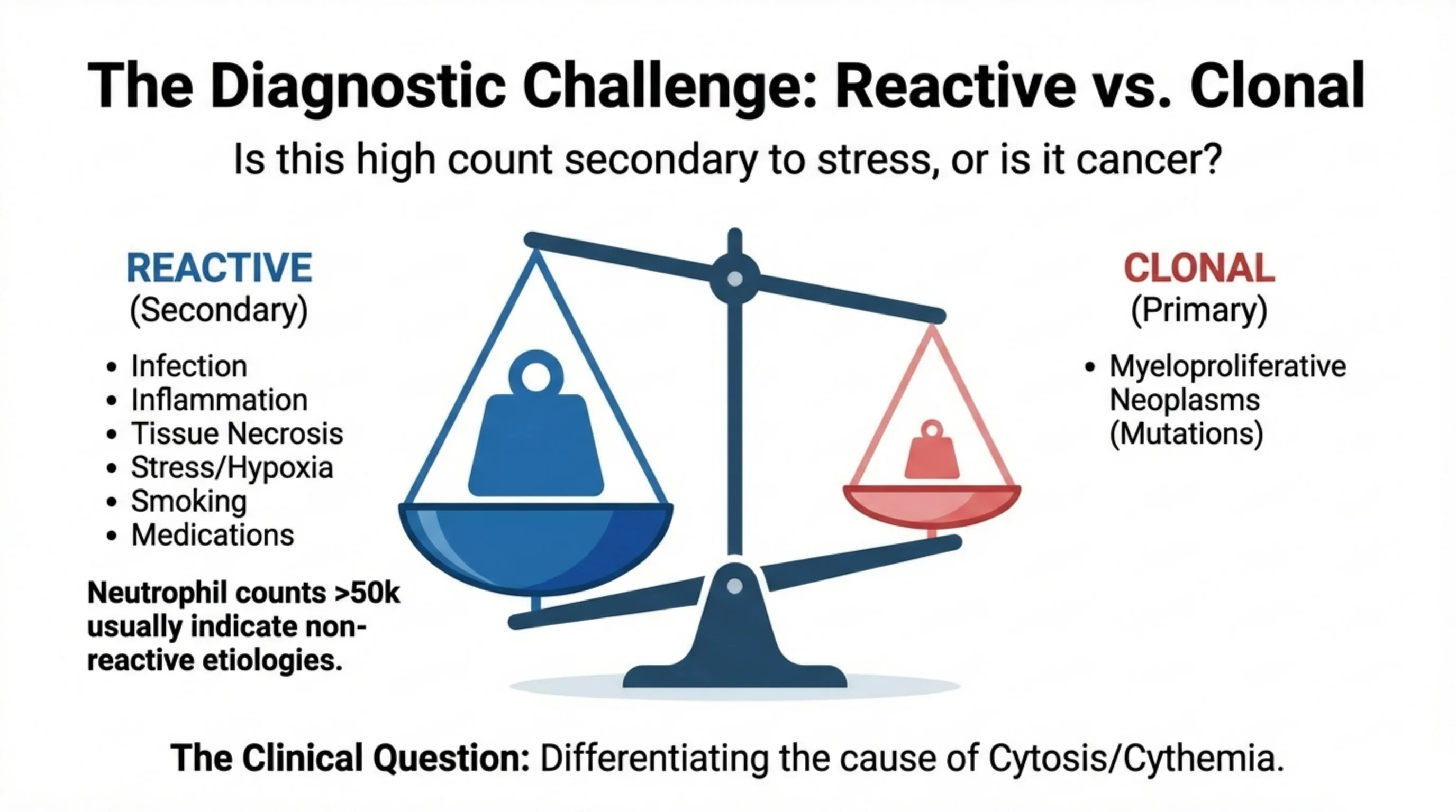
The diagnostic challenge: is this high count a reaction or a cancer?
Every myeloproliferative neoplasm presents as a high blood count, but the great majority of high blood counts are not neoplasms. The balance on the slide weighs the two possibilities, and the clinical question printed at the bottom is the one you must ask before anything else.
On the reactive side sit the common causes of a raised count: infection, inflammation, tissue necrosis, physiological stress and hypoxia, smoking and medications. In each the marrow is doing what it was asked to do, responding to a cytokine signal from a body under stress. The count is secondary to something else, and it settles when that something is treated.
On the clonal side sits the myeloproliferative neoplasm, where the count is primary, driven by a mutation and independent of any signal. The two sides are not always easy to tell apart at first sight, but the slide gives one useful threshold: a neutrophil count above 50,000 per microlitre usually indicates a non-reactive cause. Below that, the history, the examination and the pattern of the other lineages guide you, and the three cases that follow show how.
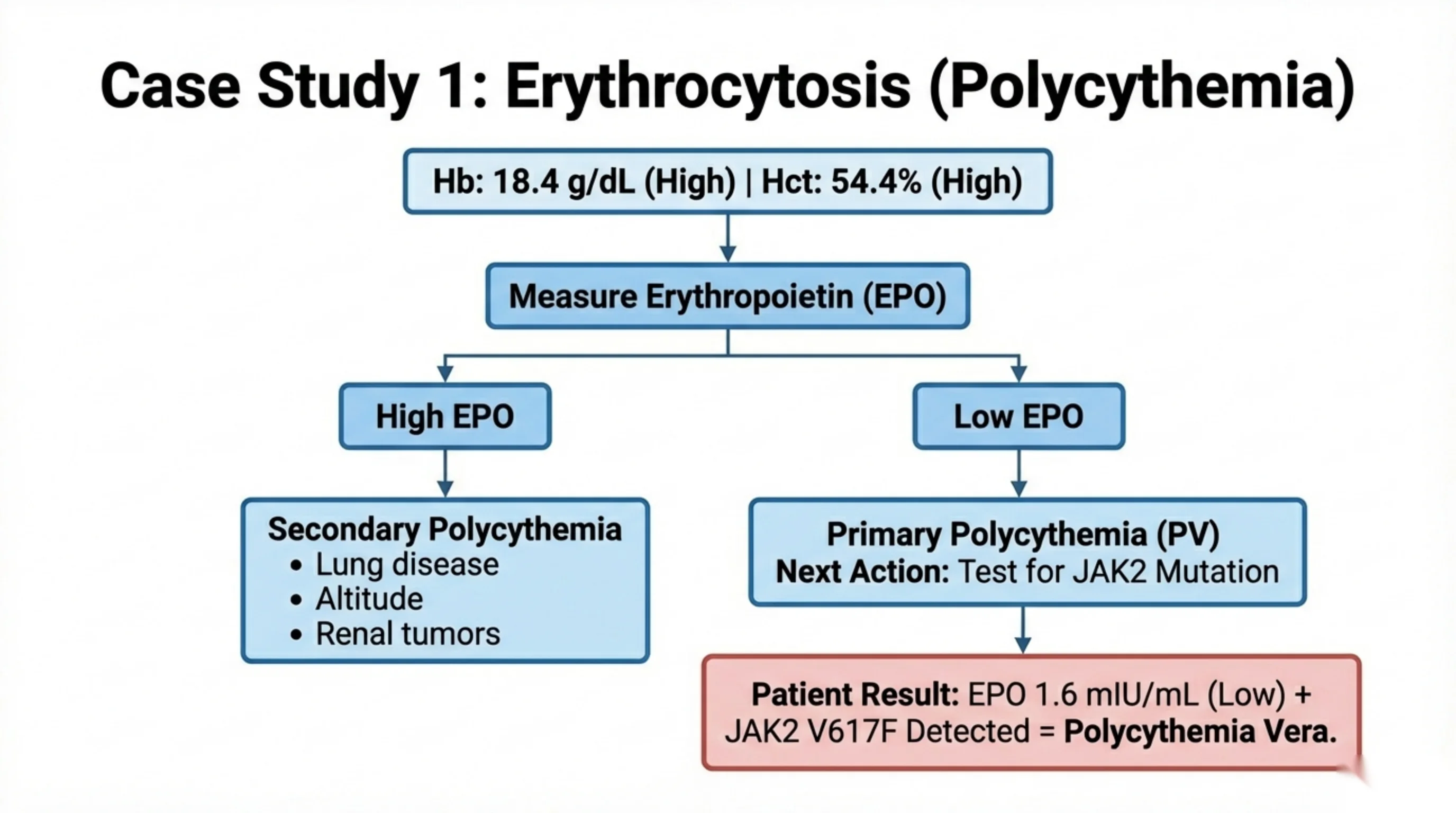
Case 1: a high hemoglobin, and the one test that sorts it
The patient has a hemoglobin of 18.4 g/dL and a hematocrit of 54.4 percent, both high. The flowchart on the slide is the approach to erythrocytosis, and it turns on the physiology of slide 4.
The first step is to measure erythropoietin. Red cell production is normally driven by erythropoietin released from the kidney in response to hypoxia. If erythropoietin is high, the marrow is responding appropriately to a signal, and the erythrocytosis is secondary: the causes listed are chronic lung disease, life at altitude, and tumours of the kidney that secrete the hormone. Living in Abha, you should expect to see the altitude version. If erythropoietin is low, the body is trying to switch production off and the marrow is ignoring it, which means the production is autonomous. That is primary erythrocytosis, polycythemia vera, and the next action is to test for the JAK2 mutation.
This patient's erythropoietin was 1.6 mIU/mL, low, and JAK2 V617F was detected. The combination of a suppressed physiological signal and a clonal driver mutation is the diagnosis of polycythemia vera.
Read next. Case 58 · The Myeloproliferative Neoplasms in the casebook is this slide in one patient: a high hemoglobin, a low erythropoietin and a JAK2 mutation. Its first table sorts the other causes of a high hemoglobin, and it lists residence at altitude as a routine local cause to establish before a marrow is considered.
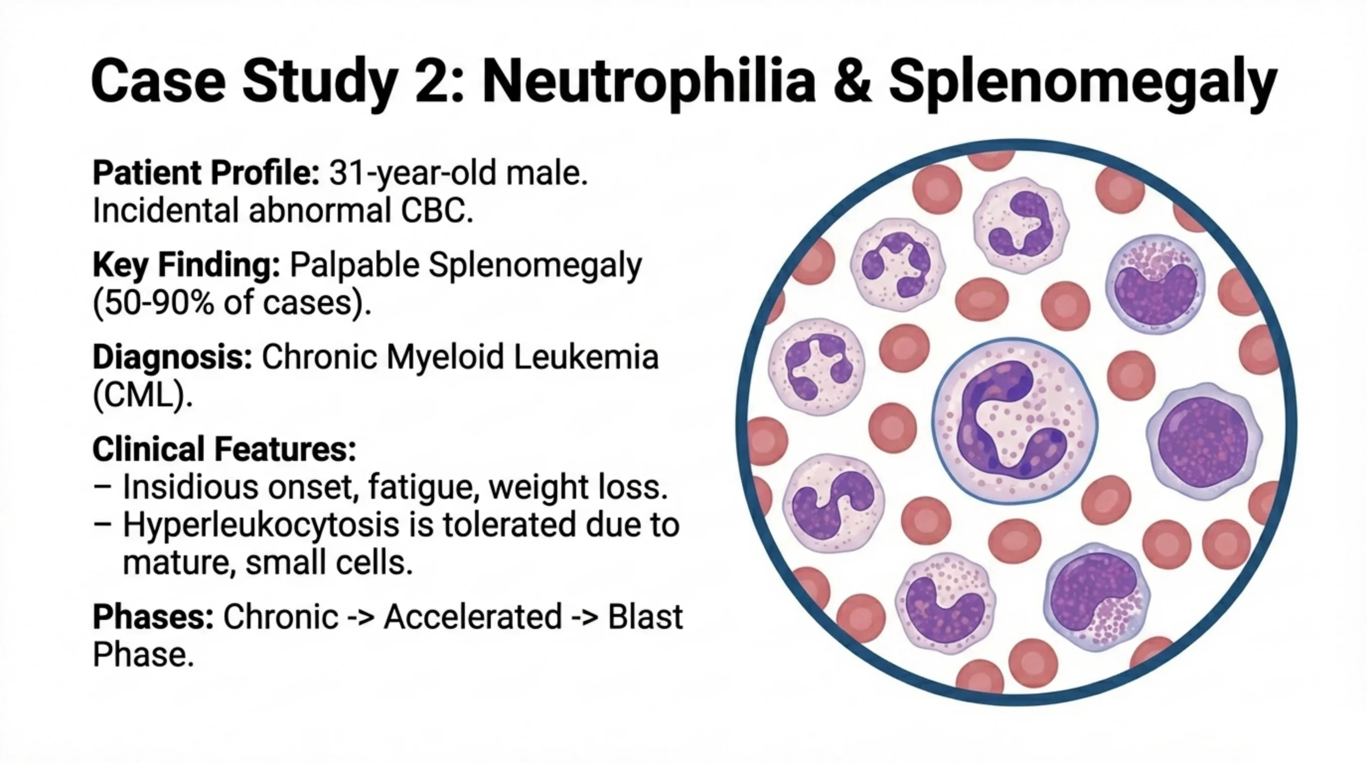
Case 2: a high white count, a large spleen, and a disease found by accident
A 31-year-old man has an abnormal blood count found incidentally. The key finding on examination is a palpable spleen, present in 50 to 90 percent of cases of this disease. The diagnosis is chronic myeloid leukemia, the Philadelphia-positive branch of the classification.
The clinical features explain why it is so often found by accident. Onset is insidious, with fatigue and weight loss developing over months if there are any symptoms at all. The white cell count may be very high, yet the patient tolerates it, and the smear on the slide shows why: the excess cells are mature, small granulocytes at every stage of development, not blasts. Contrast this with acute leukemia, where a count of the same magnitude made up of large, rigid blasts causes leukostasis. Mature cells flow; blasts sludge. This is the same acute-versus-chronic distinction drawn in the leukemia lecture: proliferation with maturation, rather than proliferation with maturation arrest.
The disease is nevertheless not benign. Untreated, it passes through three phases: a chronic phase that can last years, an accelerated phase, and finally a blast phase in which it transforms into an acute leukemia. The tyrosine kinase inhibitors on slide 11 are what prevent that progression.
Read next. Case 55 · The Chronic Leukaemias in the casebook opens with this patient: a man found on an insurance medical to have a very high white count and a large spleen. It takes him from the film to the BCR-ABL1 test and on to treatment.
CML on the blood film
This is a real blood film from a patient with chronic myeloid leukemia, the disease of Case 2. The drawing on the previous slide is a simpler version of it.
Look first at how crowded it is. In a normal film you move across many red cells to find one white cell. Here the white cells are everywhere. Then look at the range of white cells. Some are large, with a round nucleus and a rim of cytoplasm; these are the younger granulocytes. Beside them are band forms and segmented neutrophils, which are mature. Every stage from myelocyte to neutrophil is present. That is the left shift named on the next slide, and it is why the count is so high and yet so well tolerated: the cells still mature. Mature cells flow; blasts sludge.
The next slide adds the second clue on the film, basophilia, which points away from a reactive neutrophilia.
Image: “Chronic Myeloid Leukemia smear 2009-04-09” by Paulo Henrique Orlandi Mourão, via Wikimedia Commons, CC BY-SA 3.0.
CML up close: presentation and diagnosis
This slide widens Case 2. CML is worth the extra time because one abnormal gene explains the whole disease. In the translocation t(9;22), the ABL1 gene moves from chromosome 9 to sit next to the BCR gene on chromosome 22. The shortened chromosome 22 that carries the join is the Philadelphia chromosome. The fused gene, BCR::ABL1 (the older name is BCR-ABL), makes a tyrosine kinase that is always switched on. That one fact explains the disease, is the test that proves it, and is the target of the drug.
Presentation. Many patients have no symptoms, and the disease is found on a routine blood count. When there are symptoms, they are fatigue, weight loss, sweats, anemia, and a feeling of fullness after small meals because the enlarged spleen presses on the stomach. The spleen can be massive. The film shows why the count is so high and yet so well tolerated: the marrow makes too many myeloid cells, but they still mature, so every stage from myelocyte to neutrophil is present. Basophilia is the clue that points away from a reactive neutrophilia; eosinophils are often raised too. Most patients are diagnosed in the chronic phase. Untreated, the disease moves on to a blast phase that behaves like acute leukemia, either through an accelerated phase or directly.
Diagnosis. The job is to prove the fusion. The European LeukemiaNet (ELN) 2020 recommendations set out the work-up. A marrow aspirate is needed at diagnosis, because the proportion of blasts and basophils decides whether the disease is in chronic, accelerated or blast phase. The same sample gives the karyotype, which looks at whole chromosomes in dividing marrow cells. It shows the Philadelphia chromosome, and it also shows any additional chromosome abnormalities, which change the risk. A qualitative RT-PCR on blood detects the messenger RNA made by the fused gene and names the transcript type. This matters later: a few patients have unusual transcripts that the routine monitoring test cannot see, and without this step such a patient could falsely look as if the disease had gone. FISH uses fluorescent probes for BCR and ABL1 and shows them joined. ELN keeps it for the patient whose PCR finds BCR::ABL1 but whose karyotype shows no Philadelphia chromosome. Many laboratories also use FISH on blood as a quick first test, so practice here differs between centres.
See it online.
CML up close: treatment and monitoring
The previous slide proved the fusion. This slide shows how to switch it off, and how to tell whether that is working.
Treatment. If the patient has symptoms from a very high count, a short course of hydroxyurea can bring it down while the diagnosis is being confirmed. The real treatment is a tyrosine kinase inhibitor (TKI), which blocks the fusion kinase. Four are approved for first-line use: imatinib, dasatinib, nilotinib and bosutinib. ELN names generic imatinib as the cost-effective start. The second-generation drugs give faster and deeper responses than imatinib, but in the trials survival was similar. The choice also depends on the patient's other illnesses and on each drug's side effects: dasatinib, for example, can cause pleural effusion, and an ECG to check the QTc interval is done before starting any TKI. TKIs are withheld during pregnancy. Response is followed by qPCR on blood at least every 3 months, and this continues after major molecular response is reached. Results are reported as BCR::ABL1 percent on the International Scale (IS) so that results from different laboratories can be compared. As a rough guide, a level of 1 percent IS matches a complete cytogenetic response, meaning no Philadelphia-positive cells on the karyotype.
The table gives the ELN milestones at 3, 6 and 12 months. An optimal response is 10 percent or less at 3 months, 1 percent or less at 6 months, and 0.1 percent or less at 12 months; 0.1 percent or less is major molecular response (MMR). Failure is more than 10 percent at 3 months once confirmed within 1 to 3 months, more than 10 percent at 6 months, or more than 1 percent at 12 months. Between the two is a warning zone, where the patient is watched closely and the decision to switch depends on the patient. Failure at any time also includes resistance mutations and high-risk additional chromosome abnormalities. ELN also counts two findings at diagnosis as warnings from the start: high-risk additional chromosome abnormalities and a high-risk ELTS score, a risk score worked out from age, spleen size, blasts and platelet count. These cut-offs are agreed conventions, chosen because they predict outcome; they are not natural boundaries.
On failure, the next step is to test for mutations in the kinase domain and change the TKI; the mutation found guides the choice. ELN recommends prompt consideration of allogeneic stem cell transplant when the response to two or more TKIs is not good enough, and transplant remains an option above all in advanced phase. HLA typing of the patient and a donor search should start early, so that no time is lost if a transplant is needed. Finally, in some patients whose response is deep and has lasted for years, the TKI can be stopped under close qPCR monitoring. Some then stay in remission without treatment; this is called treatment-free remission.
Read next. The CML section of Case 55 · The Chronic Leukaemias makes one point about monitoring that this slide does not. When a patient misses a milestone, the commonest reason is not resistance but missed tablets. Ask about missed doses at every visit, without accusation, before sending a mutation screen.
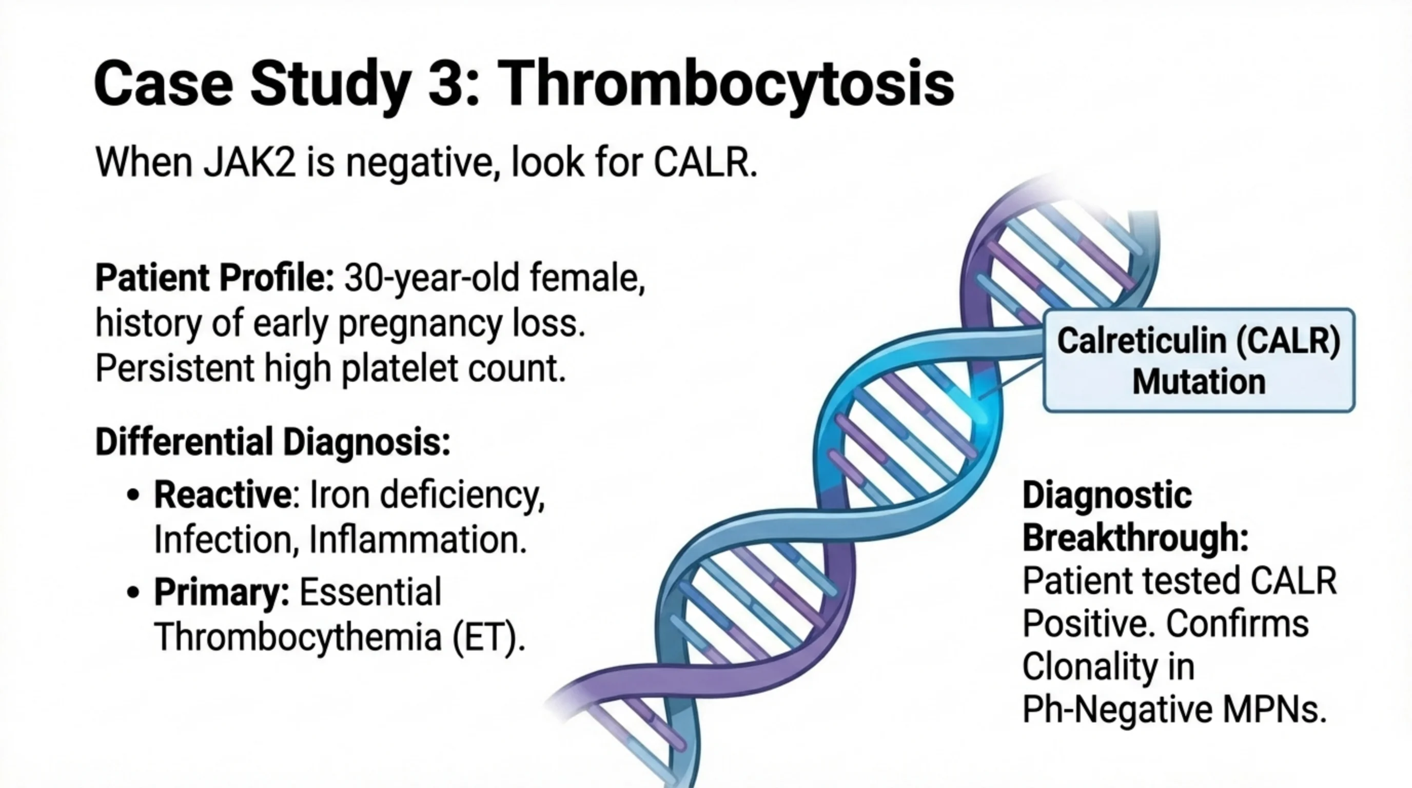
Case 3: a high platelet count in a young woman, and what to test when JAK2 is negative
A 30-year-old woman has a persistently high platelet count and a history of early pregnancy loss. The history is the clue: recurrent pregnancy loss can be a manifestation of thrombosis in the placental circulation, and thrombosis is the principal complication of a clonal platelet excess.
The differential is the reactive-versus-clonal question again. Reactive thrombocytosis is common and is caused by iron deficiency, infection and inflammation; in a young woman iron deficiency in particular must be excluded, because it is frequent and it raises the platelet count. If those are absent and the count persists, the primary diagnosis is essential thrombocythemia.
Proving it means finding the clone. JAK2 is tested first, but it is present in only 50 to 70 percent of essential thrombocythemia, so a negative result does not exclude the disease. The next test is for a mutation in calreticulin, CALR, the gene illustrated on the slide. This patient was CALR positive. The diagnostic breakthrough that the slide records is that the CALR mutation, like JAK2 and MPL, confirms clonality in the Philadelphia-negative neoplasms, turning what used to be a diagnosis of exclusion into a positive finding.
Clinical features and complications: the disease beyond the blood count
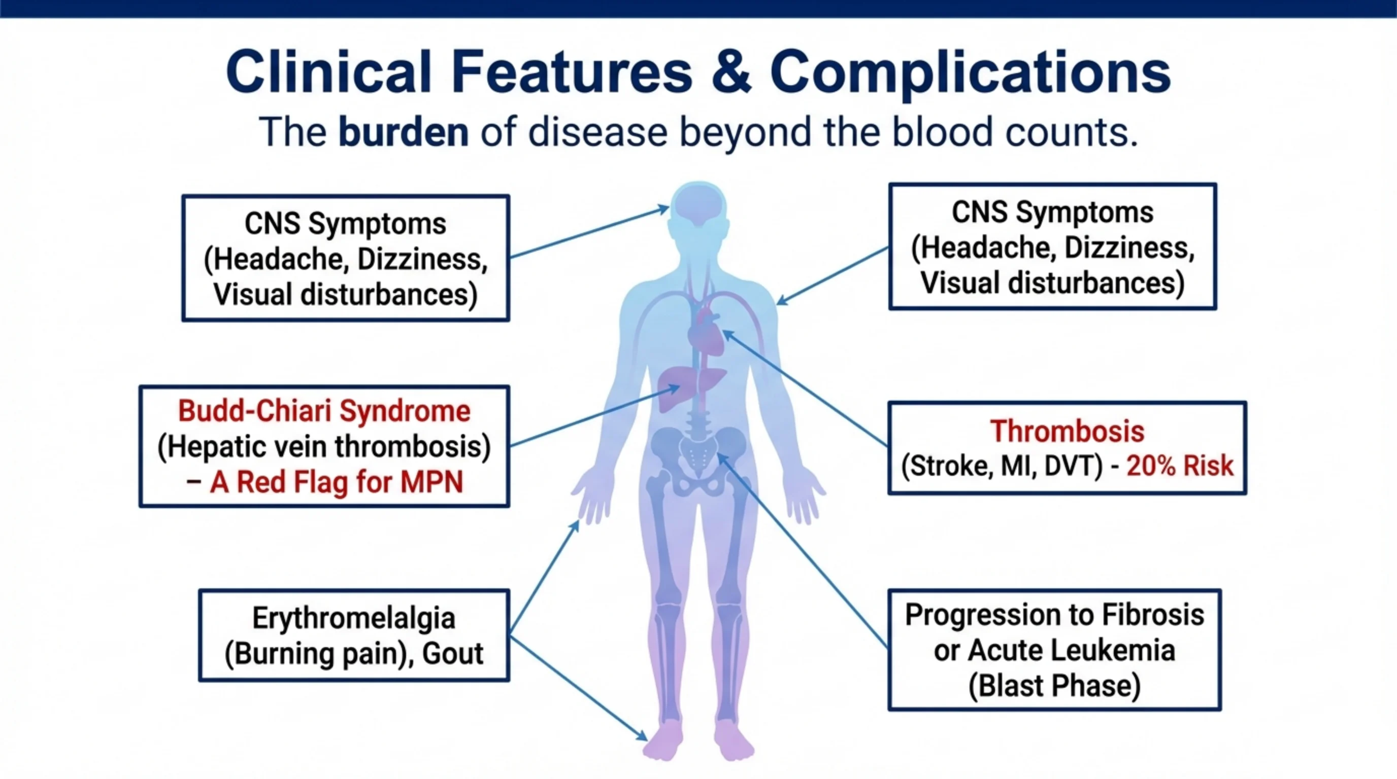
The body map collects the ways a myeloproliferative neoplasm harms a patient, and almost all of them come down to one mechanism: blood that is too thick or too rich in platelets clots where it should not.
The central nervous system symptoms, headache, dizziness and visual disturbance, come from hyperviscosity and from small-vessel occlusion. Erythromelalgia is a burning pain and redness in the hands and feet caused by platelet aggregates in the small vessels of the skin; it is characteristic enough that its presence should prompt a blood count. Gout follows from the high turnover of cells and the uric acid they release. The major thromboses, stroke, myocardial infarction and deep vein thrombosis, carry a risk of about 20 percent and are the main cause of morbidity.
One complication deserves special emphasis. Budd-Chiari syndrome, thrombosis of the hepatic veins, is a red flag for a myeloproliferative neoplasm. Thrombosis in an unusual site, the hepatic, portal or splanchnic veins, in a patient without an obvious cause should prompt testing for JAK2 even when the blood count is normal. Finally, the diseases evolve: over years they can progress to marrow fibrosis or transform into acute leukemia, the blast phase.
Management: preventing the clot, reducing the count, targeting the mutation
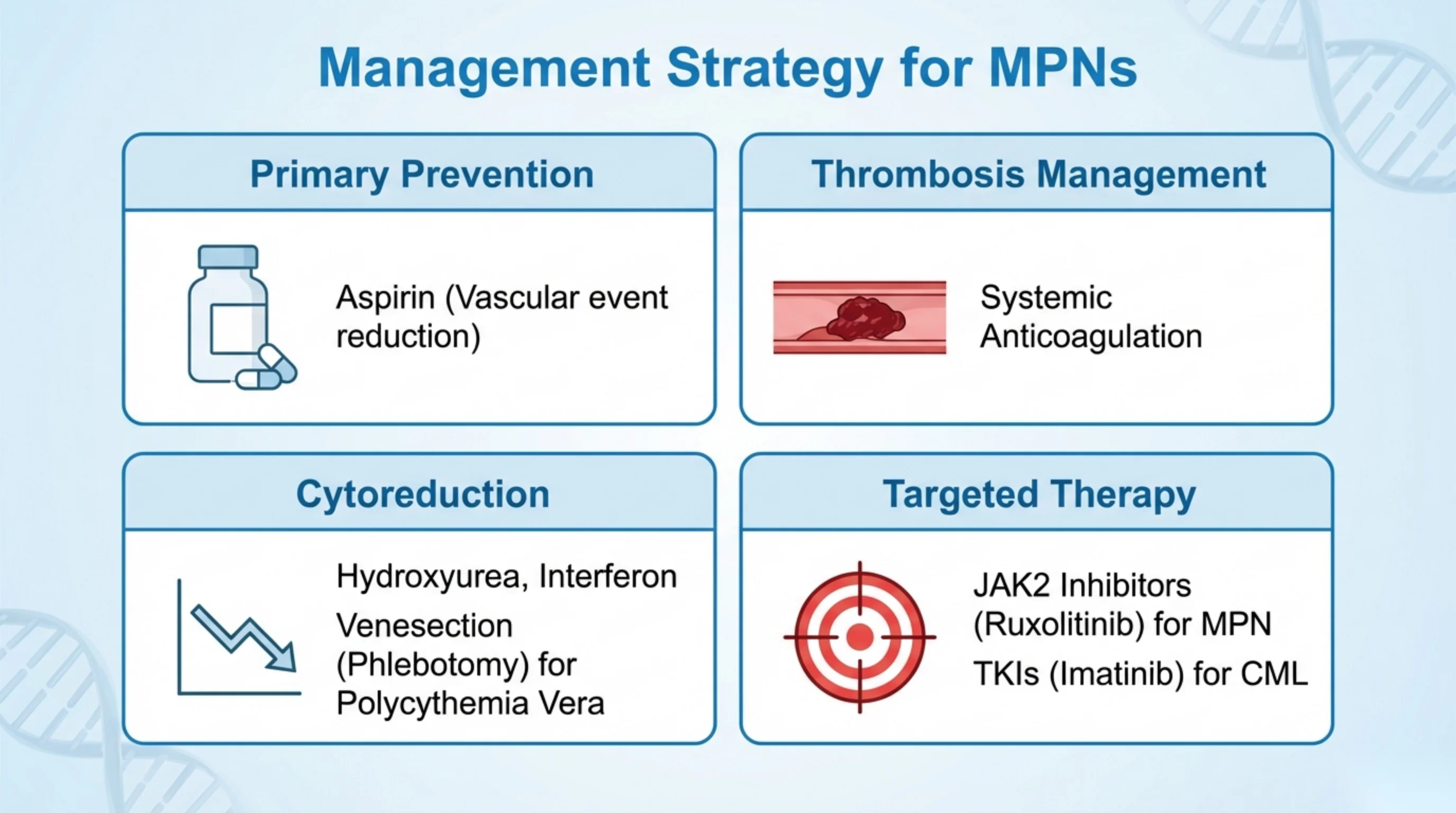
Treatment of the myeloproliferative neoplasms is organised by the four quadrants on the slide, and it follows directly from the complications on slide 13. The goal for most patients is not cure but the prevention of thrombosis and the control of symptoms.
Primary prevention is low-dose aspirin, which reduces vascular events by damping platelet activation. Thrombosis management, once a clot has occurred, is systemic anticoagulation. Cytoreduction lowers the counts themselves: hydroxyurea and interferon suppress the overactive marrow, and for polycythemia vera the simplest and oldest treatment is venesection, removing blood at intervals to keep the hematocrit down and the blood less viscous.
Targeted therapy is where the molecular classification pays off. For Philadelphia-positive disease, CML, the tyrosine kinase inhibitors such as imatinib block the BCR-ABL kinase and have turned a fatal disease into a chronic one. For the Philadelphia-negative neoplasms, the JAK2 inhibitor ruxolitinib blocks the pathway that slide 4 showed stuck in the on position; it is used above all to control the spleen and symptoms of myelofibrosis. In each case the drug was designed against the mutation that defines the disease.
Read next. Case 58 · The Myeloproliferative Neoplasms in the casebook gives the target this slide leaves out: in polycythemia vera, venesect to a hematocrit below 0.45. It also sets out management separately for polycythemia vera, essential thrombocythemia and primary myelofibrosis.

The counterpoint: myelodysplastic syndromes, clonal but ineffective
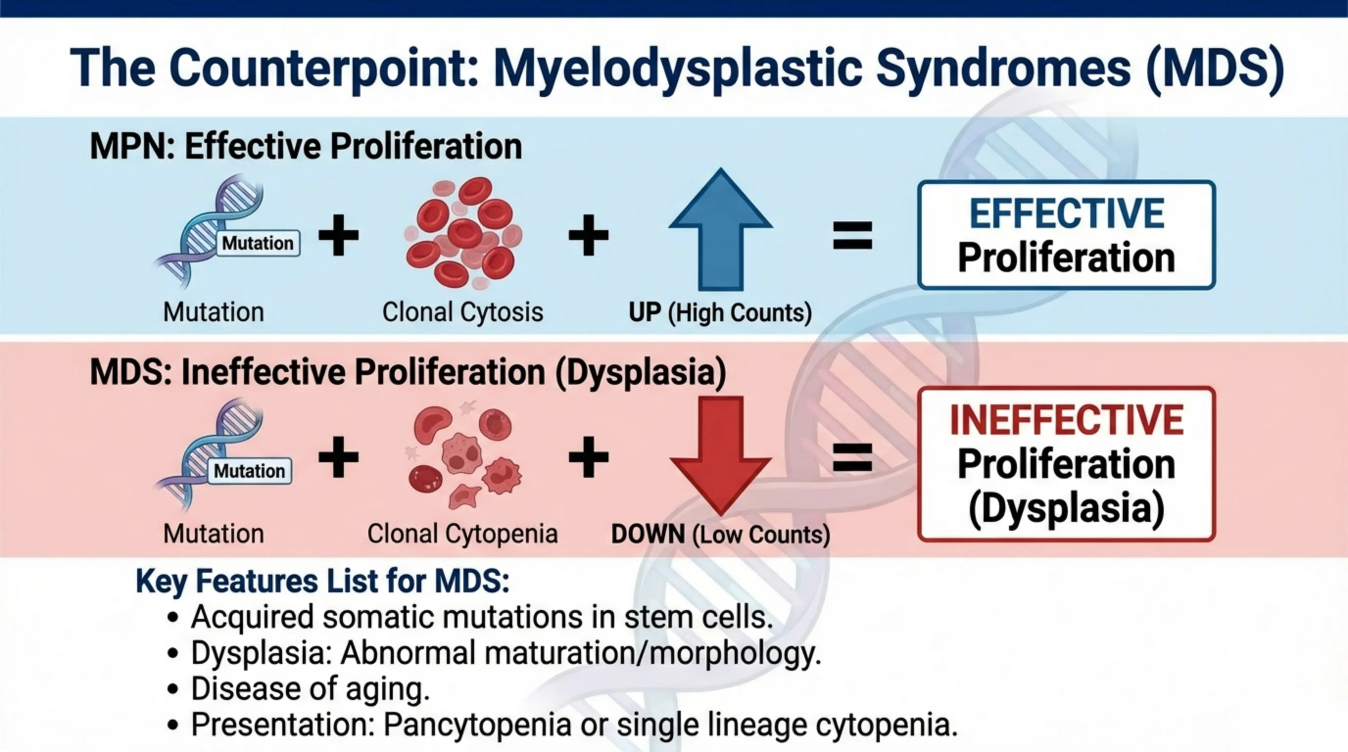
The two equations on the slide are the heart of the lecture's framework, and they should be learned as a pair.
In a myeloproliferative neoplasm: a mutation produces a clone, the clone produces a cytosis, and the counts go up. The proliferation is effective; the cells that are made are released and work. In a myelodysplastic syndrome: a mutation produces a clone, but the clone produces cytopenias, and the counts go down. The proliferation is ineffective. The marrow is busy, often hypercellular, but the cells it makes are abnormal, dysplastic, and most of them die before they leave. This is the paradox of MDS: a full marrow and an empty blood.
The key features listed on the slide follow. MDS arises from acquired somatic mutations in stem cells. It is defined by dysplasia, abnormal maturation and morphology visible on the smear and in the marrow. It is a disease of ageing, seen mainly in the elderly. And it presents as cytopenia, either of a single lineage, most commonly an anemia that does not respond to the usual treatments, or as pancytopenia. The next slide shows a typical patient.
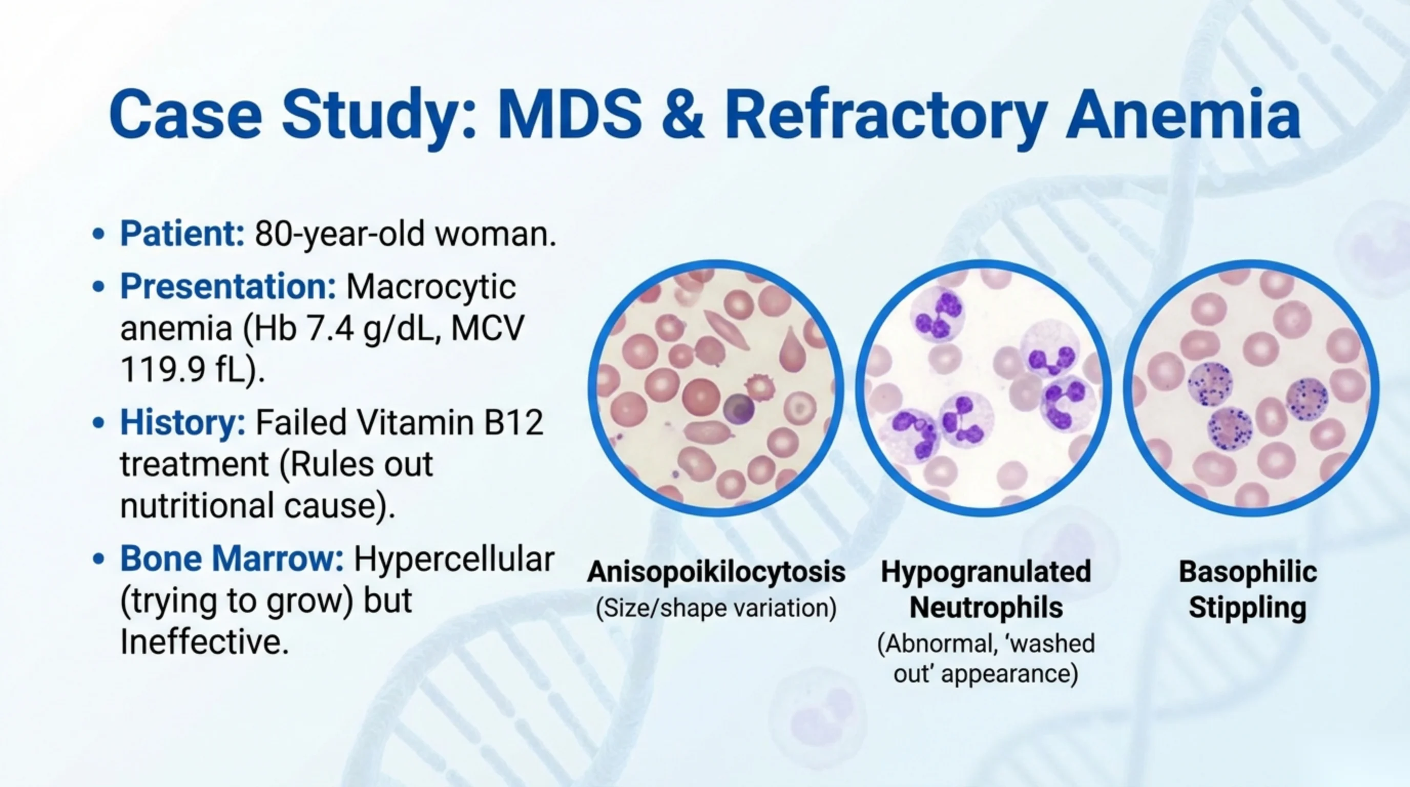
Case: an 80-year-old woman with an anemia that vitamin B12 did not fix
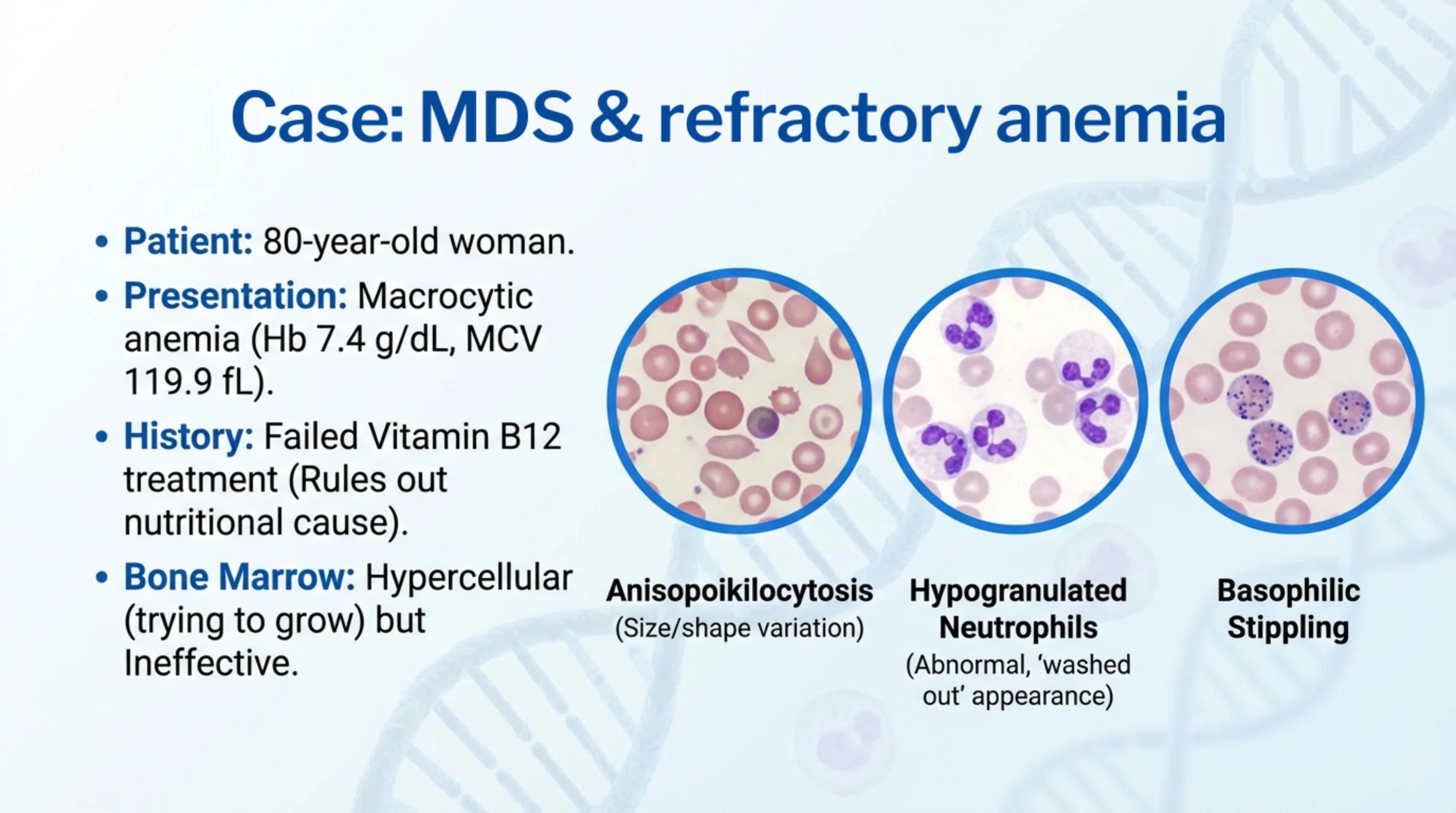
The patient is 80 and has a macrocytic anemia: hemoglobin 7.4 g/dL, mean cell volume 119.9 fL. A macrocytic anemia in an elderly patient is first assumed to be nutritional, and she was treated with vitamin B12. It failed. That failure is the most important fact in the case, because it rules out the nutritional cause and leaves a marrow that cannot make normal red cells even when it is given what it needs.
The bone marrow was hypercellular: it is trying to grow. Yet the blood is empty. This is the ineffective hematopoiesis of the previous slide, and the diagnosis is a myelodysplastic syndrome.
The three micrographs show the dysplasia that gives the disease its name, and they are worth learning because the smear is often the first clue. Anisopoikilocytosis means red cells of widely varying size and shape. Hypogranulated neutrophils have pale, washed-out cytoplasm because the granules that should fill it were never made properly; a neutrophil that looks as though it has been bleached is a strong pointer to MDS. Basophilic stippling, fine blue dots in the red cell, is residual ribosomal material and marks disordered red cell maturation. When you see a macrocytic anemia in an older patient with normal B12 and folate, or one that fails to respond to them, ask the laboratory to look for these changes.
See it online.
- A hypogranular, dysplastic neutrophil ASH Image Bank
- Basophilic stippling, with anisopoikilocytosis ASH Image Bank
MDS on the blood film
These two photographs show two of the three findings named on the previous slide. They are real films, not drawings.
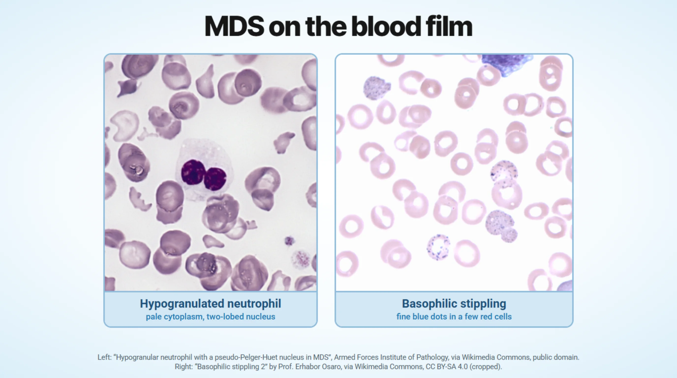
Left: a hypogranulated neutrophil. Look at the cytoplasm. It is pale and almost empty, because the granules that should fill it were never made properly. This is the neutrophil that looks as though it has been bleached. Its nucleus is abnormal too: it has only two round lobes, and the atlas that published the picture calls it a pseudo-Pelger-Huët nucleus. The film comes from a woman whose myelodysplastic syndrome followed radiotherapy and chemotherapy for Hodgkin disease. Her red cells are very irregular in shape, but the source says this is partly because her spleen had been removed. So do not read the red cells in this picture as a pure MDS finding.
Right: basophilic stippling. A few red cells carry fine blue dots; the cells around them have none. The dots are the residual ribosomal material described on the previous slide, and they mark disordered red cell maturation. The source does not say which disease this film came from. It is shown for what the finding looks like.
The third finding, anisopoikilocytosis, is not shown here.
Left: “Hypogranular neutrophil with a pseudo-Pelger-Huet nucleus in MDS”, Armed Forces Institute of Pathology, via Wikimedia Commons, public domain. Right: “Basophilic stippling 2” by Prof. Erhabor Osaro, via Wikimedia Commons, CC BY-SA 4.0, cropped.
Summary and recap
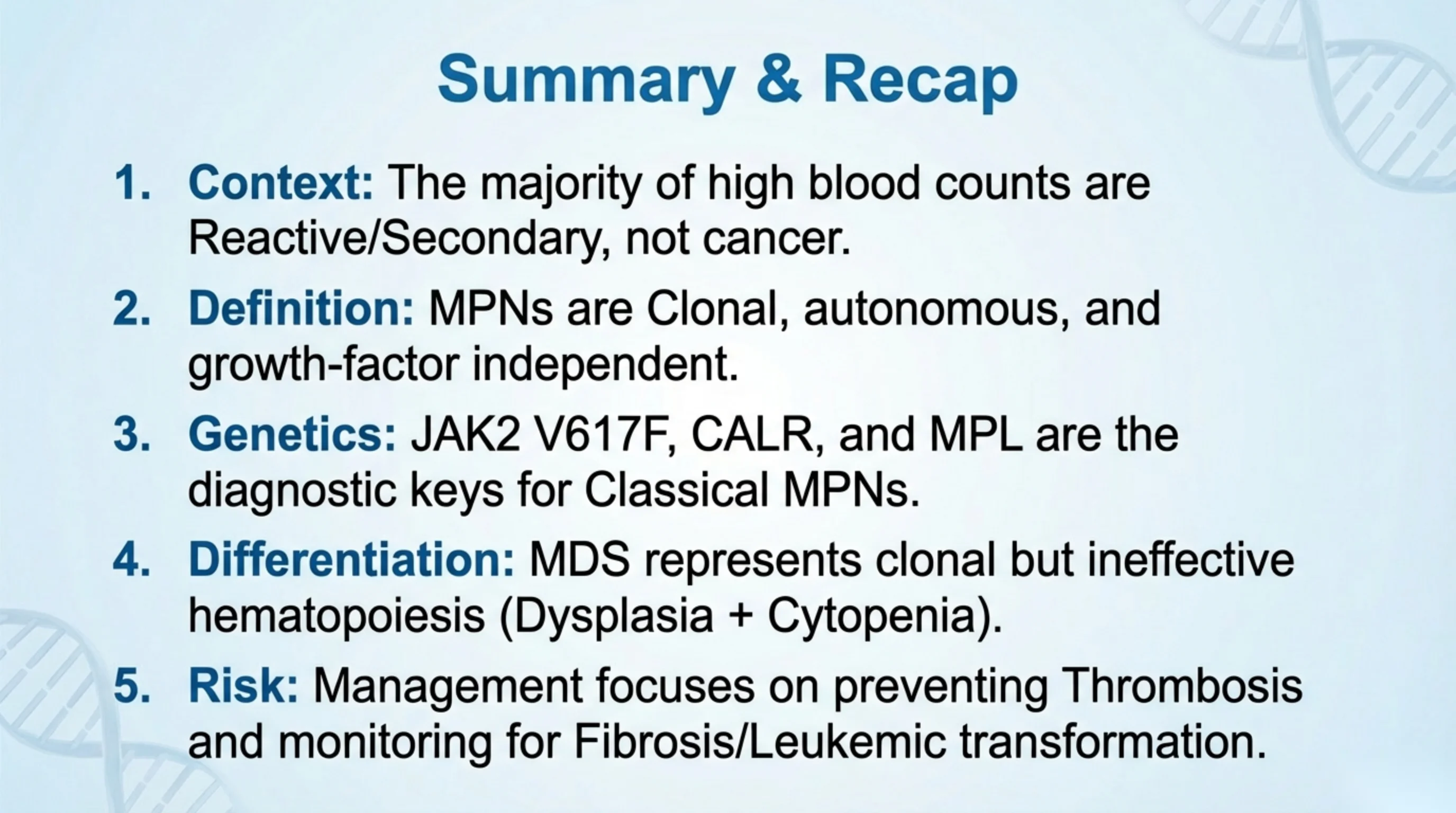
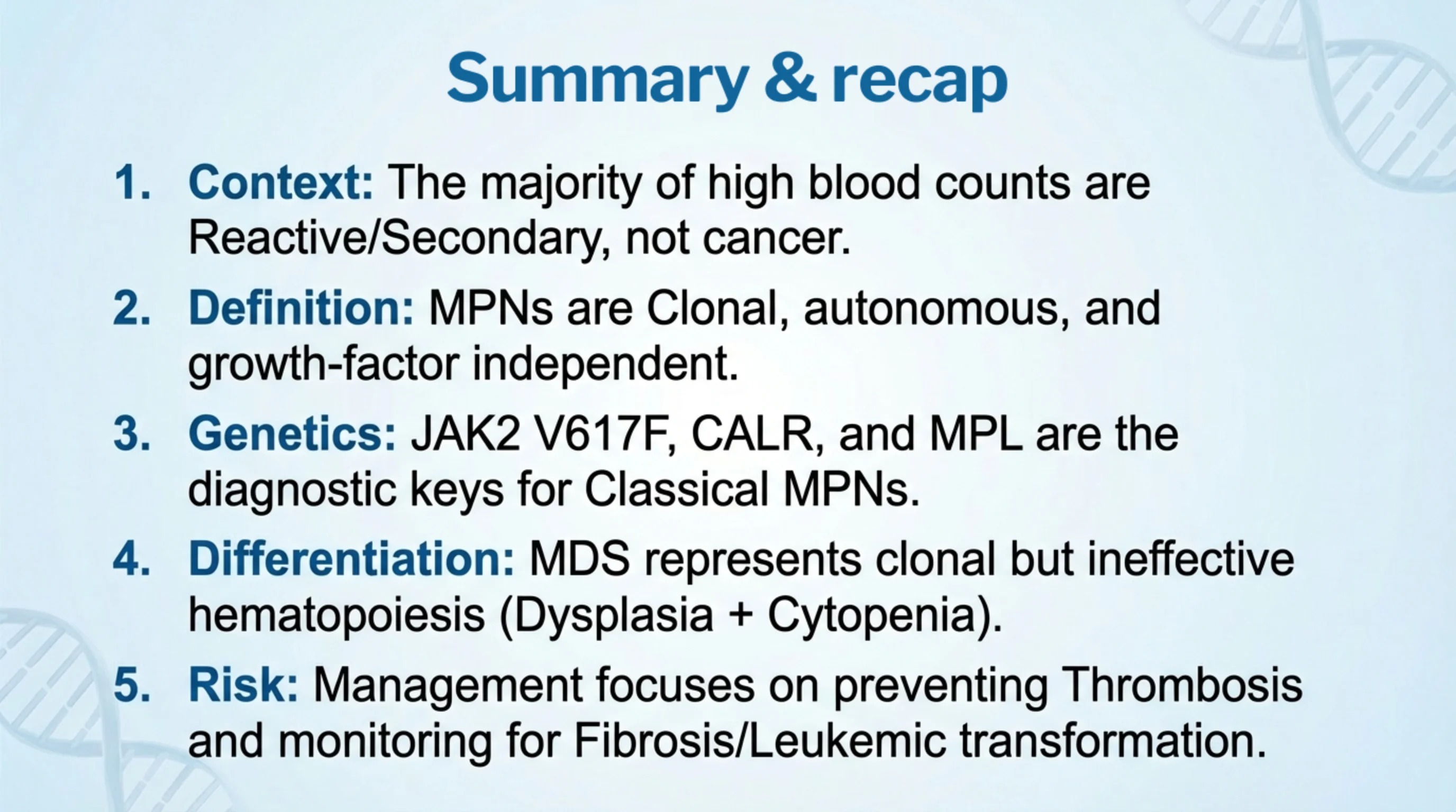
Context. The majority of high blood counts are reactive or secondary, not cancer. The first question in front of any raised count is what the marrow is responding to.
Definition. Myeloproliferative neoplasms are clonal and autonomous. Their defining property is that the proliferation is independent of growth factors; the switch is stuck on.
Genetics. JAK2 V617F, CALR and MPL are the diagnostic keys to the classical, Philadelphia-negative neoplasms, and the Philadelphia chromosome defines CML. Finding one of them turns a suspicion into a diagnosis.
Differentiation. Myelodysplastic syndromes are the counterpoint: clonal but ineffective hematopoiesis, presenting with dysplasia and cytopenia rather than cytosis.
Risk. Management focuses on preventing thrombosis, and on monitoring for the two ways the diseases evolve, fibrosis and leukemic transformation.

Questions and references
The lecture ends here. If you want to test whether the framework has taken, go back to the three cases and, for each, name the question that was asked at every step: reactive or clonal; which lineage; which mutation; what complication is the patient at risk of; and which quadrant of management applies.
Questions about the material can be sent to the address on the slide.