Lymphoma
From pathogenesis to management — classification, presentation, staging, and immunotherapy.
These slides are an outline only. Read the accompanying material with them: the notes under each slide below, and the Hematology Handbook chapters on Hodgkin lymphoma, non-Hodgkin lymphoma and T-cell lymphomas. Then work through the Internal Medicine Casebook and the Hematology Question Bank.
What this lecture is about
These slides are an outline only. Read the accompanying material with them: the notes under each slide on this page, and the Hematology Handbook chapters on Hodgkin lymphoma, non-Hodgkin lymphoma and T-cell lymphomas. Then work through the Internal Medicine Casebook and the Hematology Question Bank.
Lymphoma is a malignancy of lymphocytes, and it is the great mimic of clinical medicine: it can present as a lump in the neck, as a fever of unknown origin, as a bowel obstruction, as a rash, or as a neurological syndrome. This lecture is built to give you a framework for recognising it and a discipline for confirming it.
The subtitle names the four sections. Physiology first, because every lymphoma is a normal lymphocyte frozen at one stage of its development, and the classification only makes sense once you know those stages. Classification next, using the WHO system and the anatomy of the lymph node. Diagnosis third: the presentation, the examination, the emergencies, and a case that shows why the right biopsy matters. Immunotherapy last, because the treatment of lymphoma is where antibodies against surface markers first changed the practice of oncology.
The cell on the title slide is a lymphocyte, its surface covered in the projections that carry the receptors you will meet on the next slide.
The defenders: three kinds of lymphocyte
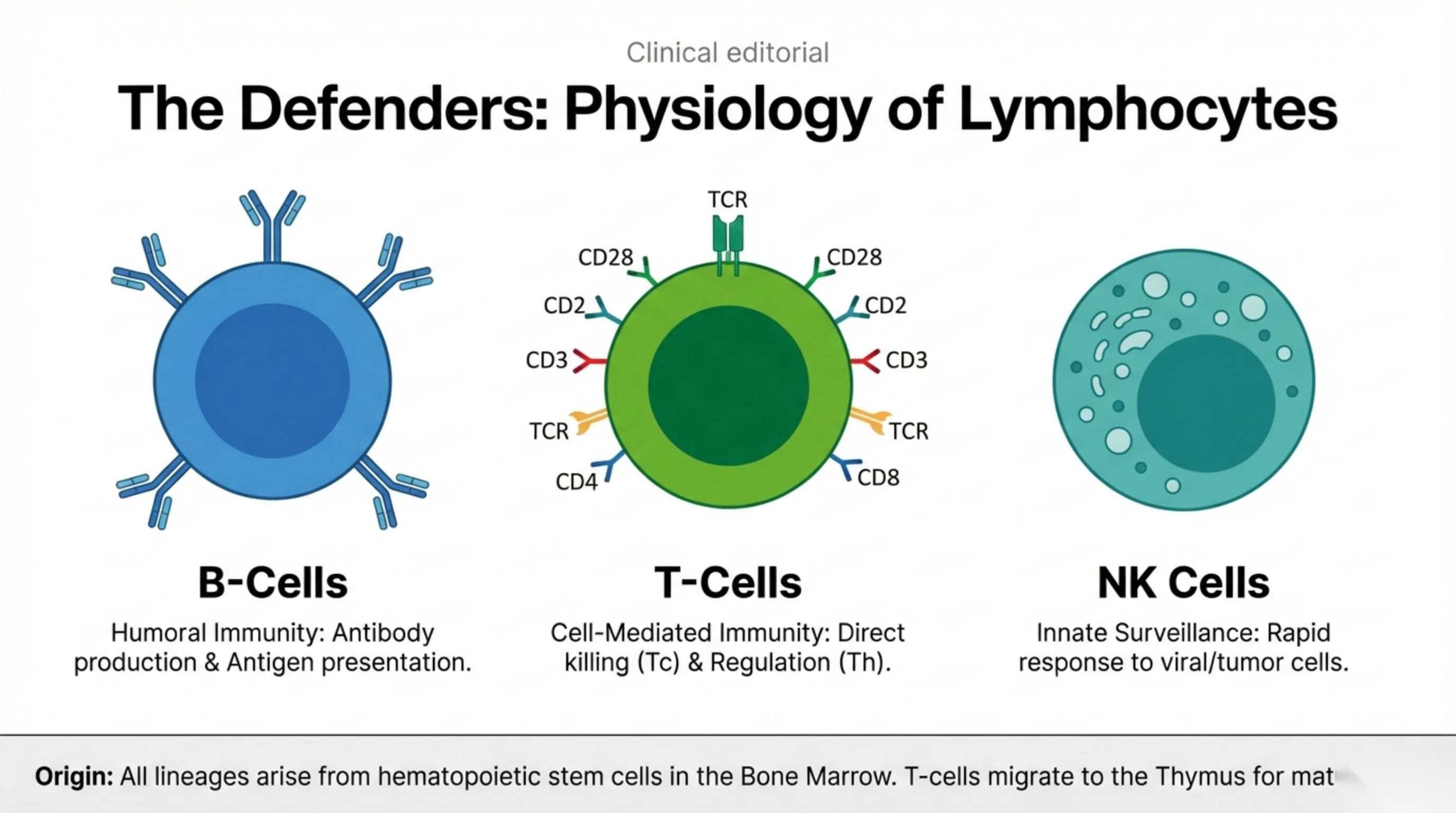
The immune system's adaptive arm is built from three lymphocyte populations, and lymphomas are classified by which of them the malignant cell resembles. B cells are responsible for humoral immunity: they make antibody and they present antigen to T cells. T cells provide cell-mediated immunity, in two flavours: cytotoxic T cells that kill infected or malignant cells directly, and helper T cells that regulate the response. NK cells, natural killers, belong to innate immunity and mount a rapid response to virus-infected and tumour cells without needing prior sensitisation.
Look at the surface markers drawn on the T cell: the T-cell receptor, CD3, CD4 and CD8, CD2 and CD28. These CD molecules are the labels by which a pathologist identifies a lymphocyte's lineage, using the technique of immunophenotyping. When a biopsy is reported as a T-cell or B-cell lymphoma, this is how the decision was made, and later in the lecture a treatment will be aimed at one of these markers.
All three lineages arise from hematopoietic stem cells in the bone marrow. B cells complete their maturation there; T cells migrate to the thymus to mature, which is where their name comes from.
The journey of a B cell: three compartments, three sets of markers
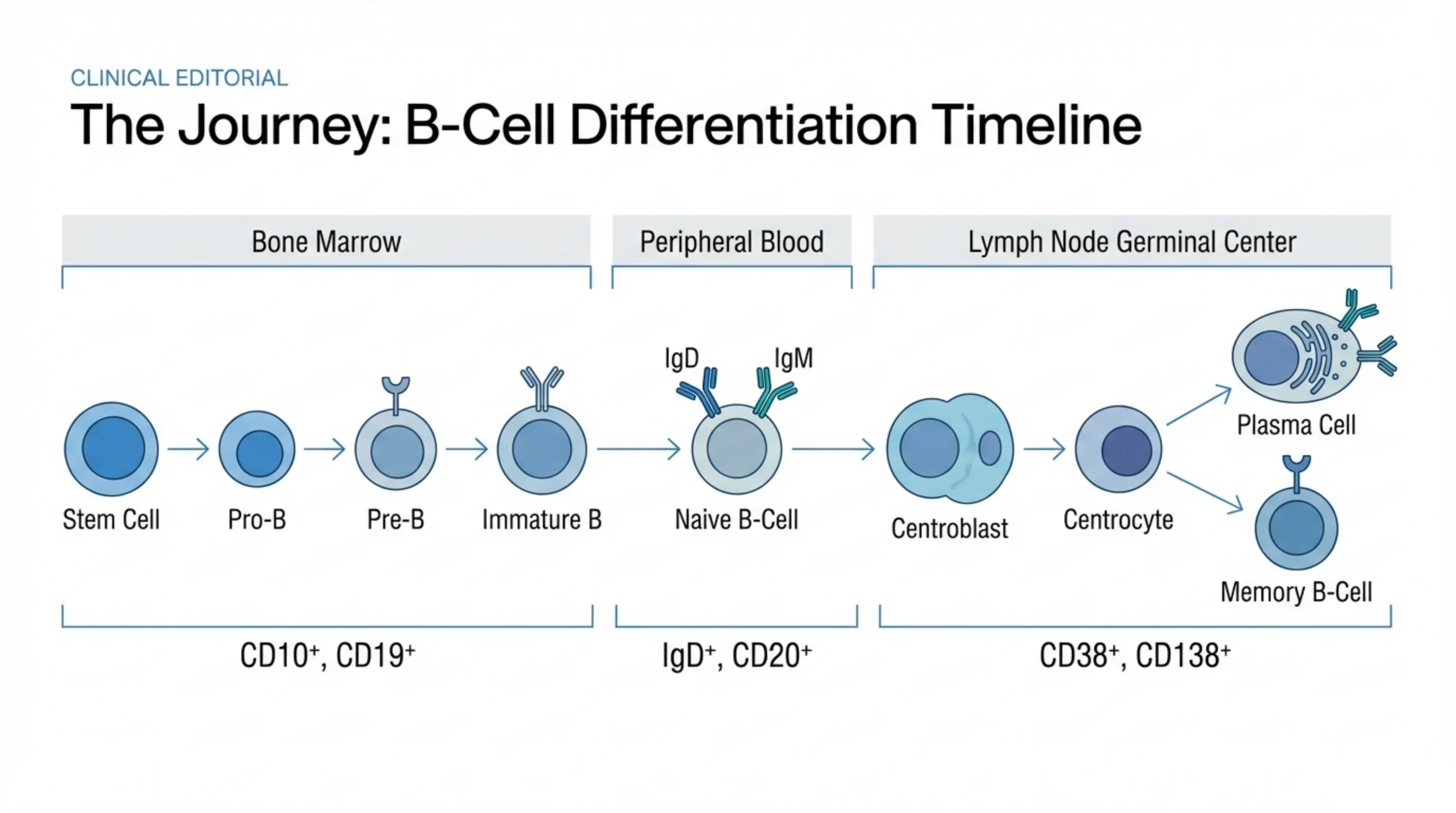
This slide is the key to the whole classification, so follow it from left to right. A B cell passes through three anatomical compartments during its life, and at each stage it wears a different set of surface markers.
In the bone marrow it develops from a stem cell through the pro-B and pre-B stages to an immature B cell, carrying CD10 and CD19. It then enters the peripheral blood as a naive B cell, displaying surface IgM and IgD and now expressing CD20. When it meets its antigen it enters the germinal centre of a lymph node, where it proliferates as a centroblast, matures into a centrocyte, and leaves as either a plasma cell, which secretes antibody and carries CD38 and CD138, or a long-lived memory B cell.
Every station on this timeline has a malignant counterpart. A tumour of the marrow stage is a lymphoblastic leukemia or lymphoma, the precursor neoplasm of slide 5. Tumours of the mature CD20-positive stages are the common B-cell lymphomas, and CD20 is the target of the antibody on slide 14. A tumour of the plasma cell, CD138 positive, is myeloma, which has its own lecture. Slide 6 will map the germinal centre stages onto specific lymphomas.
Pathogenesis: what turns a lymphocyte malignant
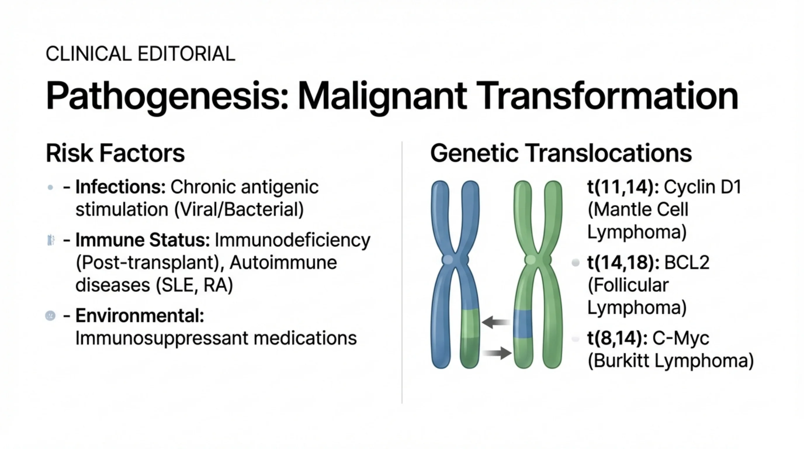
The left column lists the conditions under which lymphoma arises, and they share a theme: a lymphocyte population that is being driven to proliferate for a long time, or that has lost its normal supervision. Chronic infection, viral or bacterial, keeps lymphocytes dividing in response to persistent antigen. Immunodeficiency, for example after transplantation, removes the surveillance that would otherwise eliminate an abnormal clone, and autoimmune diseases such as lupus and rheumatoid arthritis provide both chronic stimulation and, through their treatment with immunosuppressant medications, reduced surveillance.
The right column names the genetic lesions, and three translocations are worth memorising because each defines a lymphoma. In t(11;14) the cyclin D1 gene is moved next to a highly active immunoglobulin gene and drives the cell cycle: this is mantle cell lymphoma. In t(14;18) the BCL2 gene is overexpressed; BCL2 blocks apoptosis, so the cells fail to die: this is follicular lymphoma. In t(8;14) the MYC oncogene is overexpressed and drives relentless proliferation: this is Burkitt lymphoma.
Notice that chromosome 14 appears in all three. It carries the immunoglobulin heavy chain locus, which is switched on at full power in every B cell, and whatever gene is translocated beside it is switched on with it. Each translocation gives a different defect, failing to stop dividing, failing to die, or dividing too fast, and the resulting lymphomas behave differently in the clinic as a result.
See it online.
- Mantle cell lymphoma, the t(11;14) lymphoma ASH Image Bank
- Follicular lymphoma, the t(14;18) lymphoma ASH Image Bank
- The starry-sky pattern of Burkitt lymphoma, the t(8;14) lymphoma ASH Image Bank
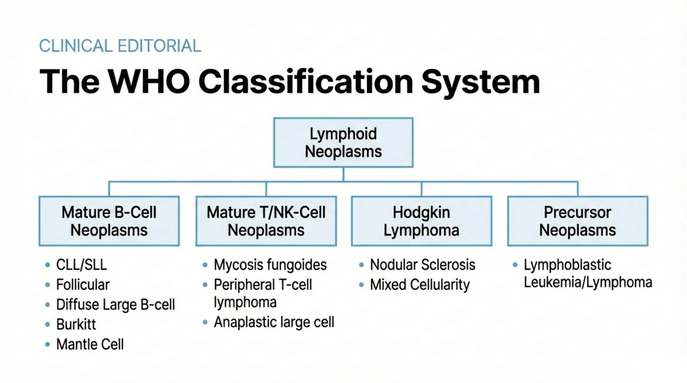
The WHO classification: four families
The WHO system classifies lymphoid neoplasms by the cell they derive from and its stage of maturity, not by where in the body they happen to appear. The diagram groups them into four families.
Mature B-cell neoplasms are the largest family and contain most of the lymphomas you will see: chronic lymphocytic leukemia and its tissue form small lymphocytic lymphoma, follicular lymphoma, diffuse large B-cell lymphoma, Burkitt lymphoma and mantle cell lymphoma. Three of these you have just met by their translocations. Mature T- and NK-cell neoplasms are less common and include mycosis fungoides, a lymphoma of the skin, peripheral T-cell lymphoma and anaplastic large cell lymphoma. Hodgkin lymphoma stands as its own family because of its distinctive biology, histology and treatment; nodular sclerosis and mixed cellularity are its two commonest subtypes. Precursor neoplasms are tumours of immature lymphoblasts, and the same disease is called lymphoblastic leukemia when it is in the marrow and blood and lymphoblastic lymphoma when it forms a mass.
You are not expected to know every entity. You are expected to know the four families, to be able to place the common named lymphomas within them, and to understand that the name on the pathology report determines the treatment. That is why the biopsy, on slide 12, has to be done properly.
Read next. Case 55 · The Chronic Leukaemias in the casebook, in its section on chronic lymphocytic leukaemia, follows the first entity on this slide in one patient: a well woman with a high lymphocyte count, diagnosed on blood flow cytometry by CD5, CD19 and CD23 together. It also teaches watch and wait as a treatment decision, and Richter transformation, where fast-growing nodes and a rising LDH call for an urgent biopsy.
See it online.
- Reed-Sternberg cells in nodular sclerosis Hodgkin lymphoma ASH Image Bank
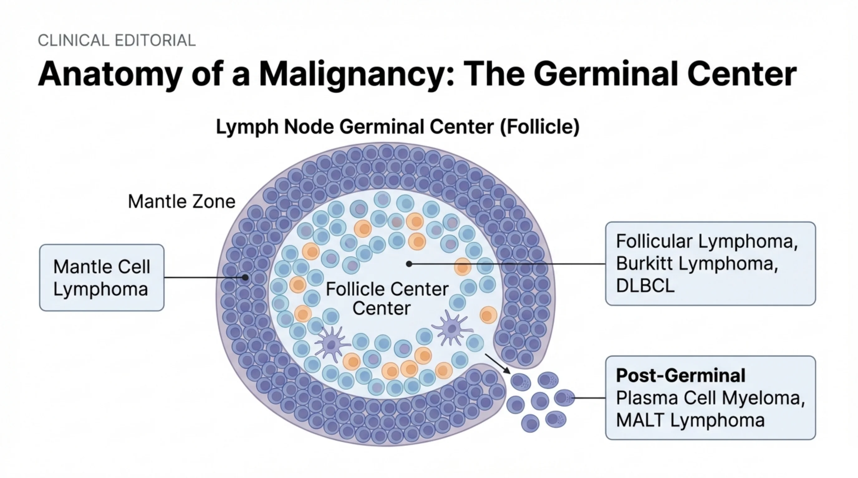
Anatomy of a malignancy: where in the follicle each lymphoma is born
The cross-section shows a single lymphoid follicle in a lymph node, and it is the anatomical version of the timeline on slide 3. The follicle has a pale follicle centre, the germinal centre where B cells that have met antigen proliferate and mature, surrounded by a darker mantle zone of naive B cells that have not yet been stimulated.
Each zone gives rise to the lymphomas that resemble its normal occupants. A tumour of mantle zone cells is mantle cell lymphoma. Tumours of germinal centre cells include follicular lymphoma, which recreates the follicle architecture, Burkitt lymphoma and many cases of diffuse large B-cell lymphoma. Cells that have completed the germinal centre reaction and are leaving it give rise to the post-germinal tumours: plasma cell myeloma, the tumour of the fully differentiated antibody-secreting cell, and MALT lymphoma, arising from the memory cells of mucosal lymphoid tissue.
The lesson is that a lymphoma is a snapshot of a normal B cell arrested at one station, and its behaviour is inherited from that station. Germinal centre cells are built to proliferate fast, and Burkitt lymphoma is one of the fastest-growing human tumours. Memory cells are built to persist quietly, and follicular and MALT lymphomas tend to be indolent.
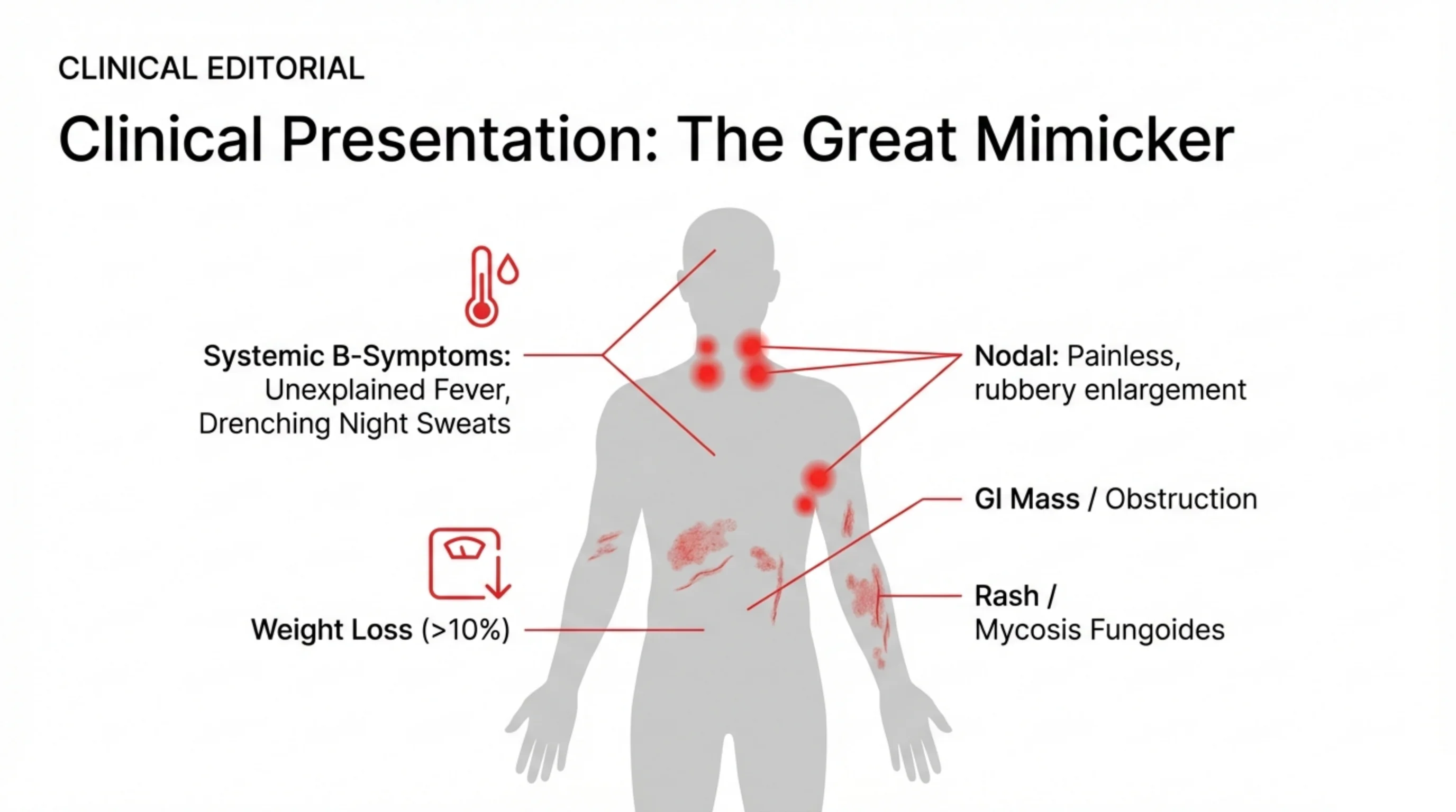
Clinical presentation: the great mimicker
Lymphoma has no single presentation, which is why the slide draws its manifestations across a whole body. The commonest is the painless, rubbery lymph node: enlarged, firm but not hard, mobile and not tender, unlike the tender nodes of infection. But many patients never notice a node.
The B symptoms are systemic and are recorded in staging because they carry prognostic weight: unexplained fever, drenching night sweats that soak the bedclothes, and loss of more than 10 percent of body weight. Ask about them specifically, because patients rarely volunteer sweats.
Extranodal disease presents in the organ involved. Lymphoma of the gut can present as an abdominal mass or as an obstruction. Lymphoma of the skin, mycosis fungoides, presents as a chronic rash that may be treated for years as eczema or psoriasis before a biopsy is taken. Add to these the paraneoplastic syndromes on slide 9 and you see why a lymphoma should be on the differential of almost any persistent, unexplained illness.
See it online.
- Mycosis fungoides on a skin biopsy ASH Image Bank
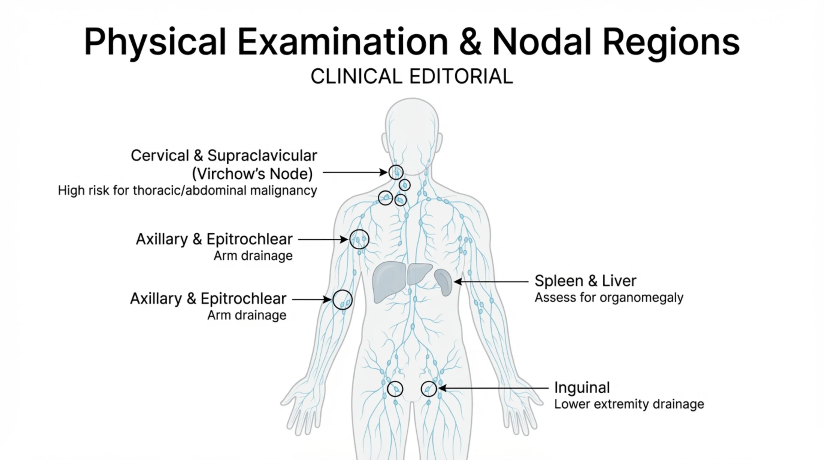
Examination: every station, and the two organs
The lymphatic system is drawn over the body to remind you that examination for lymphoma is a systematic tour of the nodal stations, not a glance at the neck. Each region drains a territory, so the site of an enlarged node points to where a primary process might be.
The cervical and supraclavicular nodes are the most important. A supraclavicular node, especially the left-sided Virchow's node, drains the thoracic duct and carries a high risk of malignancy in the chest or abdomen; it should never be dismissed. The axillary and epitrochlear nodes drain the arm; an epitrochlear node above the elbow is unusual and worth noting. The inguinal nodes drain the lower limb and are so often mildly enlarged from minor infections of the leg that they carry the least weight.
Two organs complete the examination. Palpate for the spleen and liver, because lymphoma commonly involves both, and organomegaly may be the only physical sign when the disease is internal, as it was in the case on slide 11. Describe every node you find by site, size, consistency, tenderness and mobility.
Paraneoplastic syndromes: when the lymphoma announces itself from a distance
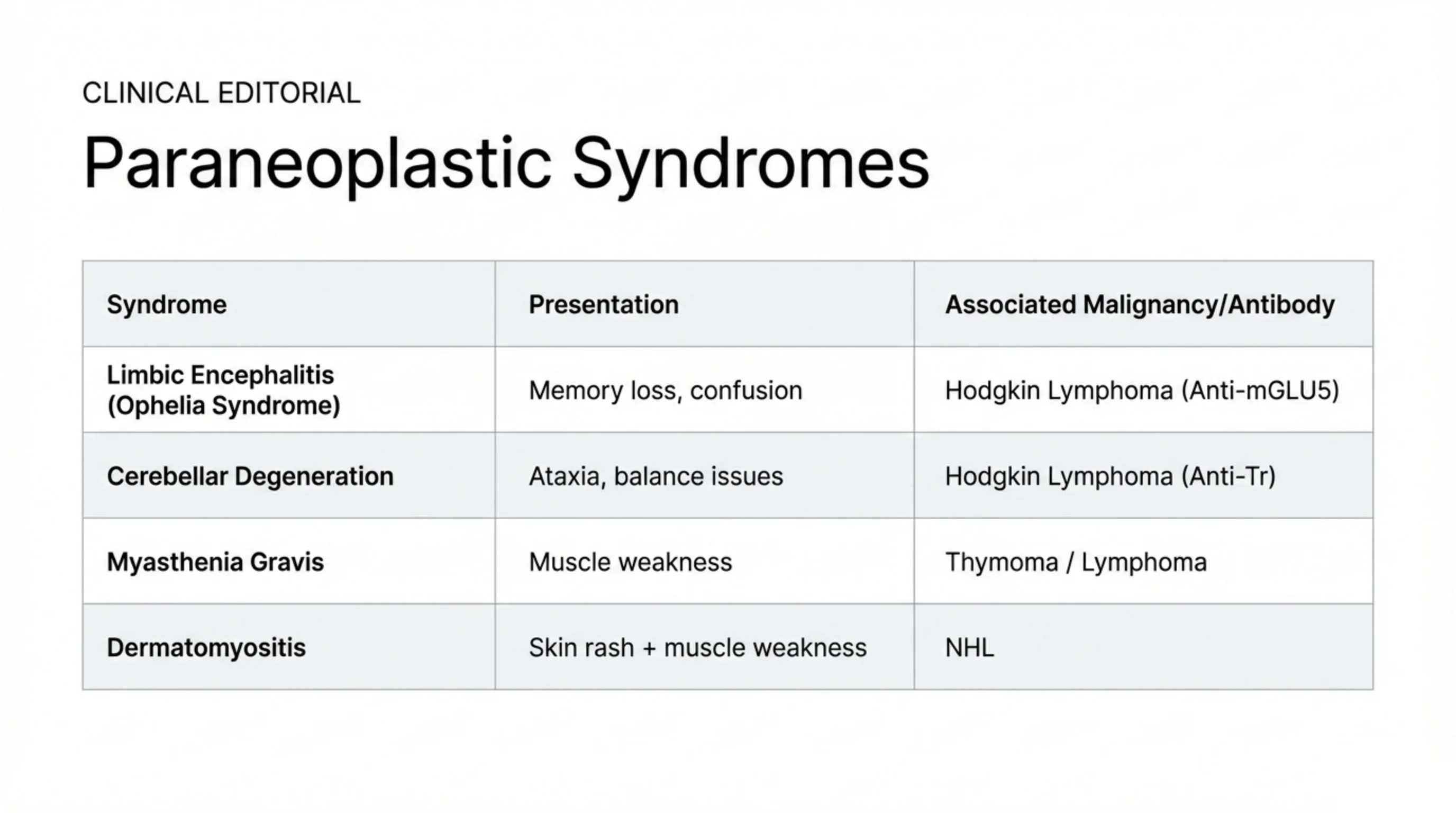
A paraneoplastic syndrome is an illness caused not by the tumour's bulk but by the immune response it provokes, typically an antibody that cross-reacts with normal tissue. The table lists four that are associated with lymphoma, and each may appear before the lymphoma itself is evident.
Two are neurological and both are linked to Hodgkin lymphoma. Limbic encephalitis presents with memory loss and confusion; in association with Hodgkin lymphoma it is called Ophelia syndrome and is mediated by antibodies against the mGluR5 receptor. Cerebellar degeneration presents with ataxia and loss of balance, mediated by the anti-Tr antibody. Myasthenia gravis, with fatigable muscle weakness, is classically associated with thymoma but also occurs with lymphoma. Dermatomyositis, a rash with proximal muscle weakness, is associated with non-Hodgkin lymphoma among other cancers.
The practical message is that an unexplained subacute neurological or dermatological syndrome in an adult should prompt a search for an occult malignancy, and lymphoma is high on that list. Treating the lymphoma is usually the only effective treatment of the syndrome.
Tumor lysis syndrome: the emergency of a tumour that dies too fast
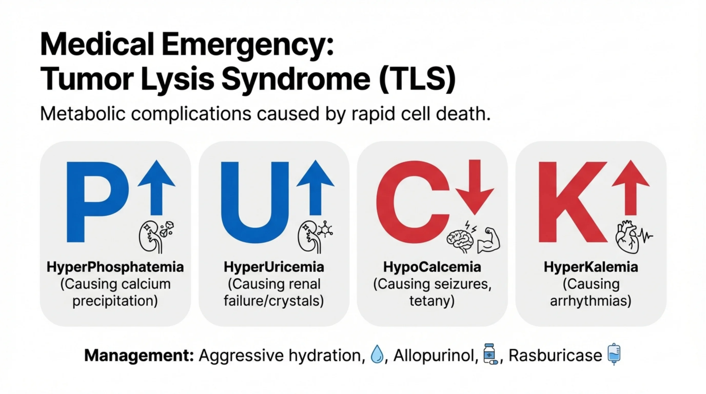
Lymphoma cells that divide rapidly also die rapidly, especially in the first days after chemotherapy begins, and when a large mass of cells breaks open at once their contents overwhelm the kidneys. The result is tumor lysis syndrome, and the slide organises its four derangements with the mnemonic PUCK.
P: hyperphosphatemia. Dying cells release phosphate, which binds calcium and precipitates as calcium phosphate in the tissues and renal tubules. U: hyperuricemia. Nucleic acids from the broken cells are metabolised to uric acid, which crystallises in the tubules and causes renal failure. C: hypocalcemia, the one arrow pointing down, because the calcium has been consumed by the phosphate; it causes tetany and seizures. K: hyperkalemia. Potassium, the main intracellular cation, floods the plasma and causes arrhythmias, which is the immediately fatal part of the syndrome.
Management is mostly prevention in patients known to be at risk. Aggressive intravenous hydration keeps the tubules flushed. Allopurinol blocks the enzyme that produces uric acid, preventing further accumulation. Rasburicase breaks down uric acid that has already formed and is used in high-risk patients. Electrolytes are monitored closely and potassium is treated as it rises. The same syndrome appears in the acute leukemia lecture; it is a hazard of any rapidly proliferating hematological malignancy.
Case study: a 22-year-old woman with a positive ANA
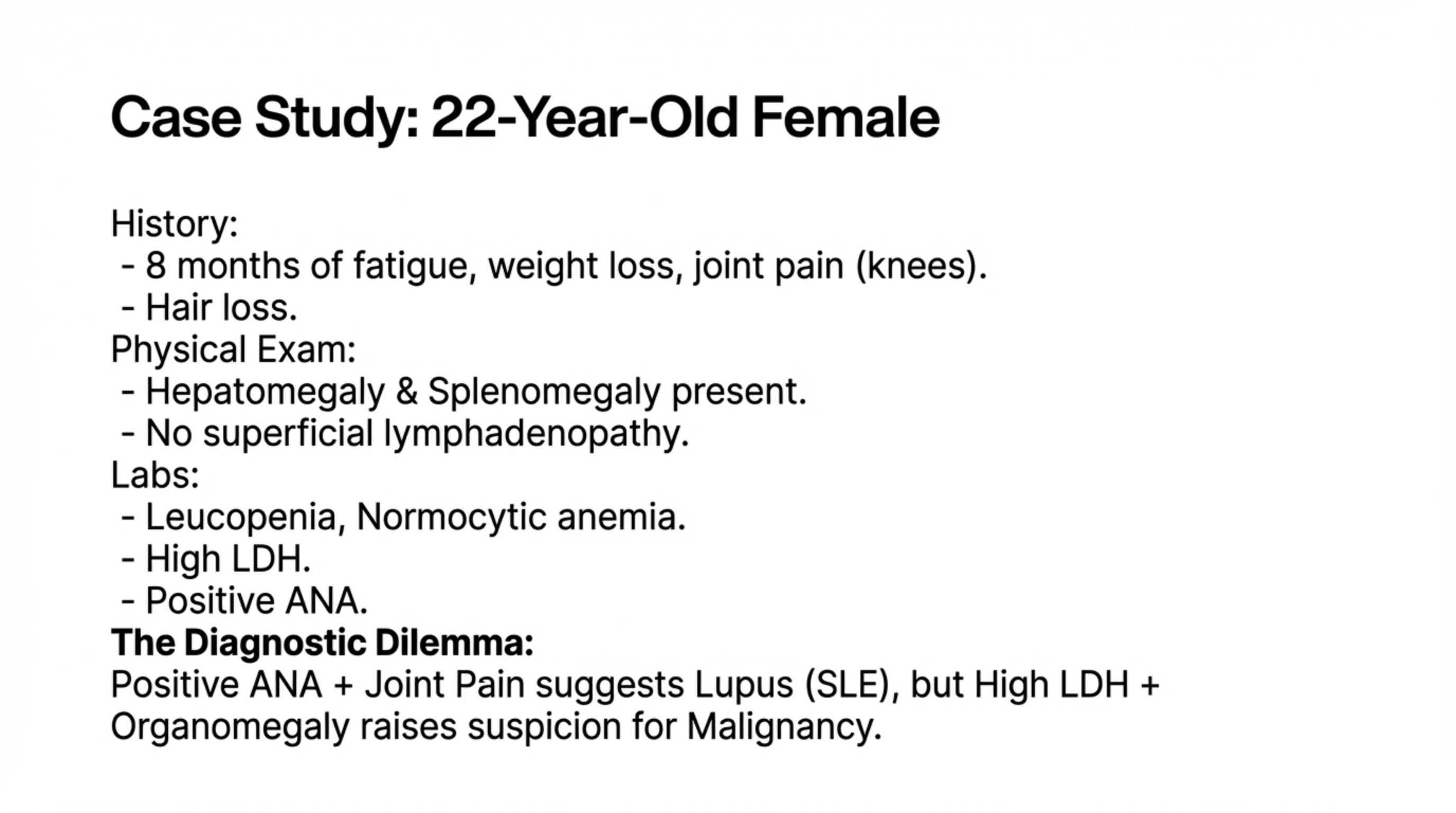
The case is chosen because it is a trap. A young woman has eight months of fatigue, weight loss and pain in the knees, and she has been losing her hair. On examination the liver and spleen are enlarged, but there are no palpable lymph nodes. Her blood shows a low white cell count, a normocytic anemia, a high lactate dehydrogenase and a positive antinuclear antibody.
Read the story one way and it is systemic lupus erythematosus: a young woman, joint pain, hair loss, cytopenias, and a positive ANA. Most clinicians would reach for that diagnosis. But two findings do not fit comfortably. Organomegaly of this degree is unusual in lupus, and a high LDH signals rapid cell turnover, which is the signature of a proliferating tumour rather than an autoimmune disease.
The dilemma teaches two things. First, a positive ANA is not specific: it occurs in healthy people, in infections and in malignancy, and it should confirm a clinical picture rather than create one. Second, when a patient has weight loss, organomegaly, cytopenias and a raised LDH, malignancy must be excluded with tissue, however plausible the alternative. The next slide is about how that tissue should be obtained.
Tissue is the issue: why the wrong biopsy gives the wrong answer
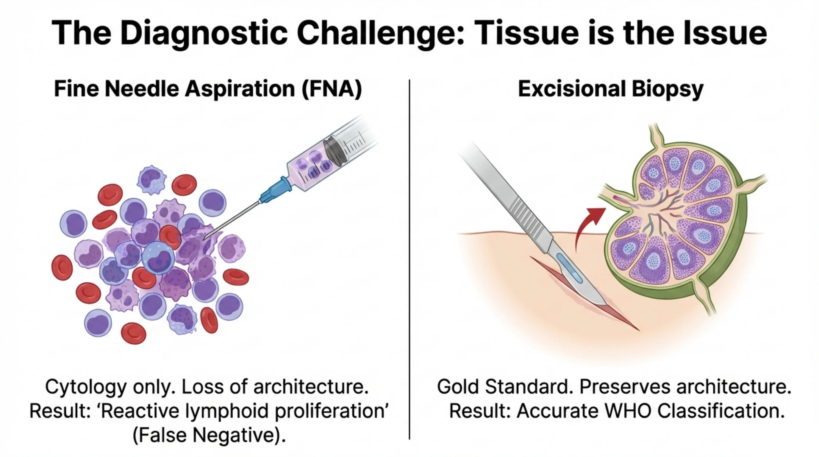
The diagnosis of lymphoma is made under a microscope, and the slide contrasts the two ways of getting the tissue there. The contrast is the single most important practical lesson in the lecture.
Fine needle aspiration draws cells out of a node through a needle. It yields cytology, individual cells in suspension, and it destroys the one thing the pathologist most needs: the architecture of the node. Whether the follicles are preserved, effaced or replaced by sheets of cells, whether the pattern is nodular or diffuse, is what separates a reactive node from a lymphoma and one lymphoma from another. Without it the report often reads "reactive lymphoid proliferation", which in a patient who actually has lymphoma is a false negative, and a dangerous one because it reassures.
Excisional biopsy, removing the whole node intact, is the gold standard. It preserves the architecture, gives enough tissue for immunophenotyping and genetic studies, and allows an accurate WHO classification, which is what the treatment depends on. When lymphoma is a serious possibility, ask for an excisional biopsy of the most accessible abnormal node, and do not accept a negative needle aspirate as an answer.
Read next. Case 56 · Lymphoma in the casebook is this slide in one patient: a young woman with a rubbery node in the neck and night sweats. Its most important investigation is the one on this slide, an excision biopsy and not a needle aspirate. It adds a second rule: do not give steroids before the biopsy, because they can kill lymphoma cells and leave the histology impossible to read.
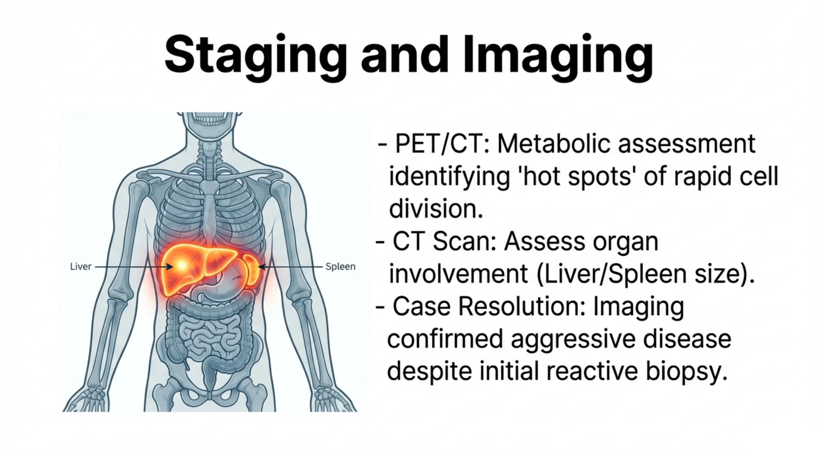
Staging and imaging, and how the case resolved
Once lymphoma is diagnosed, treatment depends on how far it has spread, and staging is done by imaging. PET/CT combines a metabolic scan with anatomical CT: lymphoma cells take up glucose avidly because they are dividing quickly, so active disease lights up as hot spots wherever it is, including in nodes of normal size and in organs that look normal on CT alone. Conventional CT measures the size of nodes and the involvement of organs, in particular the liver and spleen highlighted on the illustration.
The case from slide 11 resolves here. Imaging showed aggressive, widespread disease despite the initial biopsy having been reported as reactive. That sequence, a reassuring needle biopsy followed by imaging that revealed the truth, is exactly the failure mode the previous slide warned about. When the clinical picture and the biopsy disagree, believe the clinical picture and obtain better tissue.
Imaging also gives a baseline. The same scan repeated after treatment shows whether the hot spots have gone, which is how response is judged.
See it online.
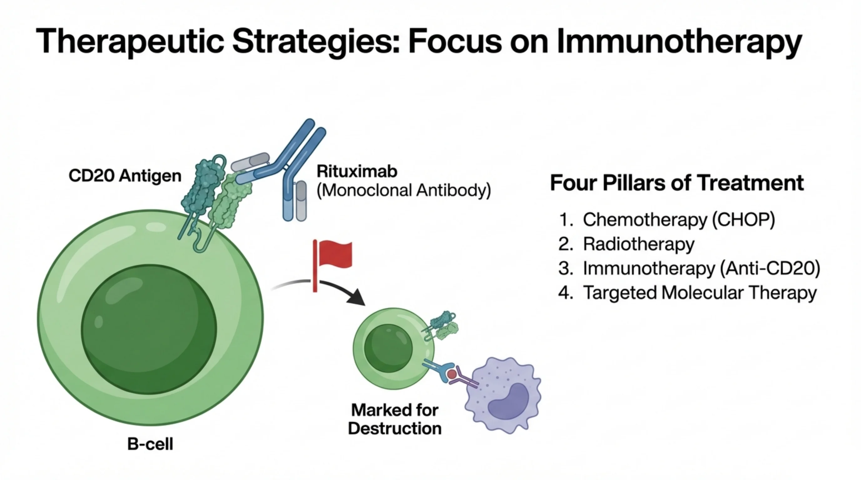
Treatment: four pillars, and the antibody that changed lymphoma
The diagram shows the idea that transformed the treatment of B-cell lymphoma. Rituximab is a monoclonal antibody engineered to bind CD20, the surface marker you saw appear on the naive B cell on slide 3. Once bound, it flags the cell for destruction by the patient's own immune effector cells and complement. Because CD20 is present on mature B cells but not on stem cells, precursors or plasma cells, the antibody removes the malignant B cells and their normal counterparts while sparing everything else, and normal B cells regenerate afterwards from the CD20-negative precursors.
Rituximab is one of four pillars. Chemotherapy, most often the CHOP combination, remains the backbone for aggressive lymphomas. Radiotherapy treats localised disease and bulky sites. Immunotherapy with anti-CD20 antibody is added to chemotherapy for CD20-positive B-cell lymphomas. Targeted molecular therapy attacks the specific signalling pathways that particular lymphomas depend on.
Which pillars are used, and in what combination, depends entirely on the histological subtype, which closes the loop back to the biopsy. A lymphoma that has been classified correctly can be treated precisely; one that has not cannot.
See it online.
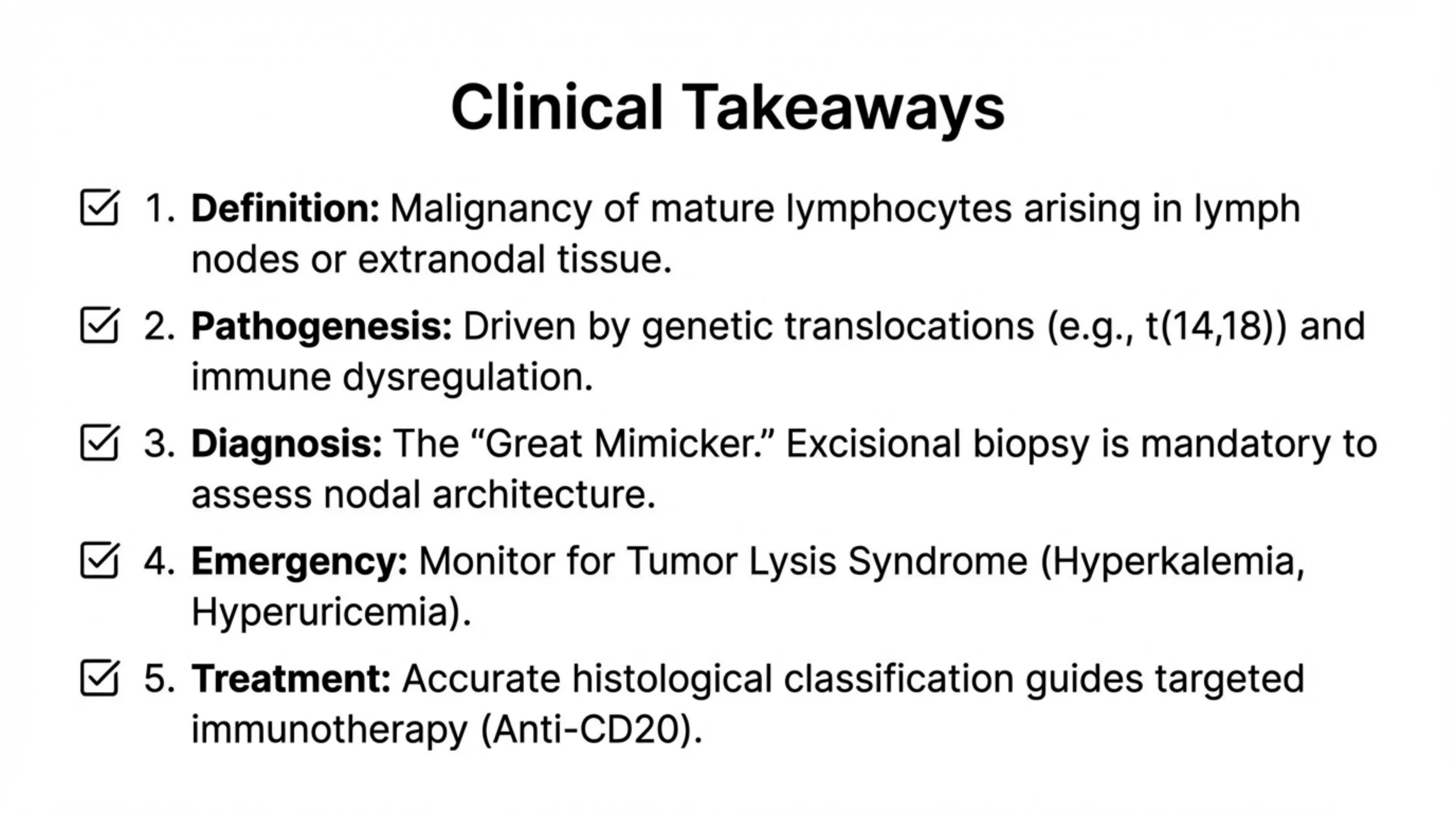
Five things to take away
Definition. Lymphoma is a malignancy of mature lymphocytes arising in lymph nodes or in extranodal tissue. It is classified by the normal cell and developmental stage it resembles, and the WHO system groups it into four families.
Pathogenesis. It is driven by genetic translocations, of which t(14;18) and BCL2 in follicular lymphoma is the archetype, arising on a background of chronic antigenic stimulation or immune dysregulation.
Diagnosis. Lymphoma is the great mimicker, presenting through nodes, B symptoms, organs, skin or paraneoplastic syndromes. Excisional biopsy is mandatory, because only an intact node shows the architecture on which classification depends.
Emergency. Watch for tumor lysis syndrome, above all the hyperkalemia and hyperuricemia, in any patient with a rapidly proliferating lymphoma, especially in the first days of treatment.
Treatment. Accurate histological classification is what makes targeted immunotherapy possible; anti-CD20 antibody is the model, and it works because the pathologist first identified the marker on the cell.