Acute Leukemia
From origin to emergency — pathogenesis, diagnosis, and the oncologic emergencies.
These slides are an outline only. Read the accompanying material with them: the notes under each slide below, and the Hematology Handbook. Then work through the Internal Medicine Casebook and the Hematology Question Bank.
What this lecture is about
These slides are an outline only. Read the accompanying material with them: the notes under each slide on this page, and the Hematology Handbook. Then work through the Internal Medicine Casebook and the Hematology Question Bank.
Acute leukemia is the disease that every doctor fears missing, because it can kill within days of the first symptom and yet is often curable if it is recognised in time. This lecture is built to make sure you recognise it.
It runs in four movements. First, normal blood production, because leukemia only makes sense as a failure of that process. Second, the two genetic faults that turn a normal marrow into a "broken factory" that churns out immature cells called blasts. Third, diagnosis, which depends far more on the pattern of the full blood count and the peripheral smear than on the white cell count that most people look at first. Fourth, the emergencies: the handful of complications that kill leukemia patients early, and what to do about each of them in the first hour.
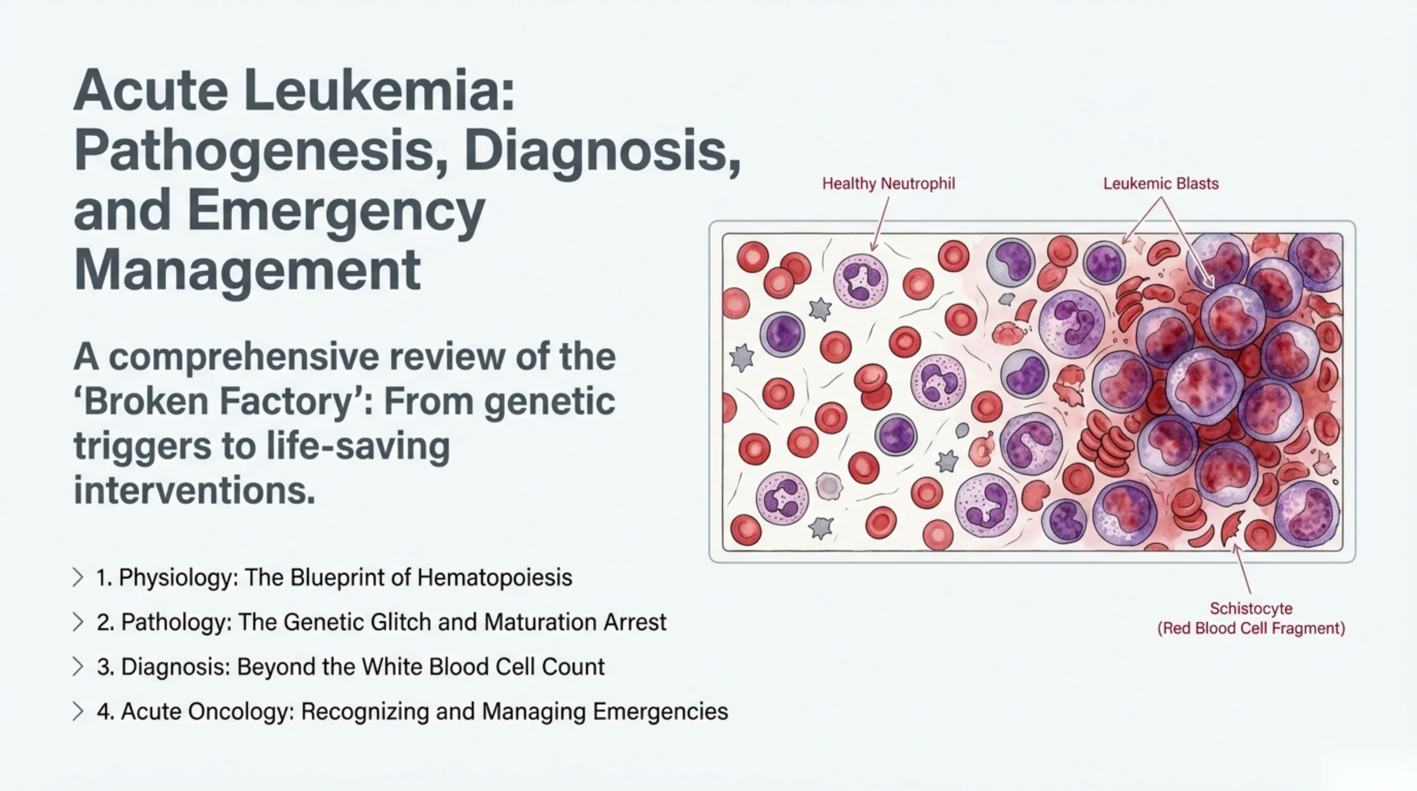
The picture on the title slide sets the scene: a healthy neutrophil, a cluster of leukemic blasts, and a fragmented red cell. By the end you should be able to explain why all three appear on the same smear.
Normal blood production: one stem cell, every blood cell
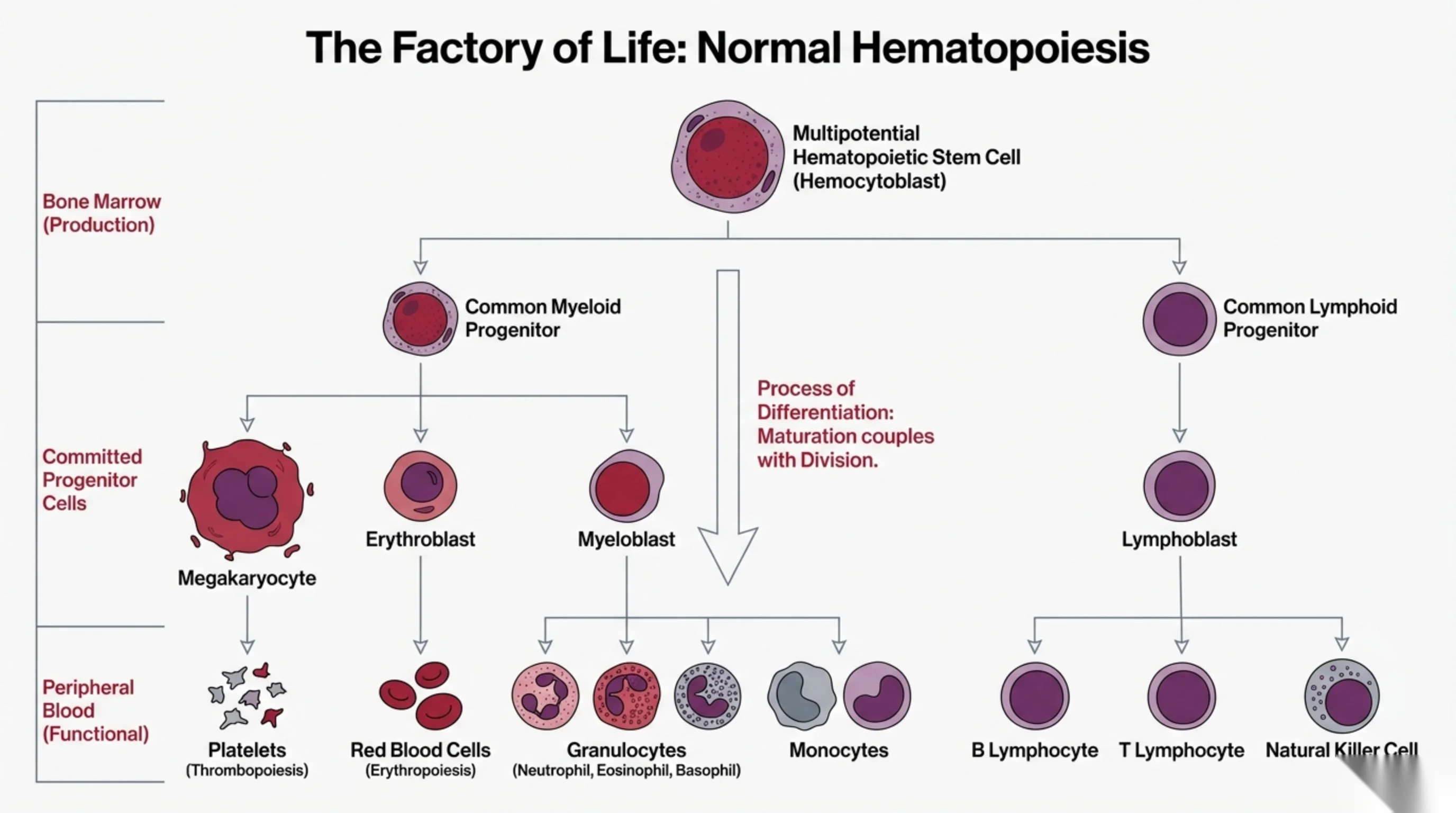
All the cells in the blood descend from a single kind of cell in the bone marrow, the multipotential hematopoietic stem cell. It can copy itself indefinitely, and it can commit to one of two great branches.
The myeloid branch produces most of the blood: the megakaryocyte, which fragments into platelets; the erythroblast, which matures into the red cell; and the myeloblast, which becomes the granulocytes (neutrophils, eosinophils, basophils) and the monocytes. The lymphoid branch produces the lymphoblast, which becomes B lymphocytes, T lymphocytes and natural killer cells.
The slide is arranged in three tiers because there are three stages to the journey: the stem cells and early progenitors in the marrow, the committed progenitors that have chosen a lineage, and the mature functional cells released into the blood. The key phrase is on the central arrow: maturation is coupled with division. Each time a developing cell divides, it also takes a step toward its final form, so by the time it has divided enough to make useful numbers, it is fully mature and ready to work. Leukemia is what happens when division and maturation are uncoupled.
Two words from this slide will be used constantly: a blast is the earliest recognisable cell of a lineage (myeloblast, lymphoblast), and the lineage a blast belongs to, myeloid or lymphoid, is what separates acute myeloid leukemia (AML) from acute lymphoblastic leukemia (ALL).
What a healthy marrow looks like, and what leukemia does to it
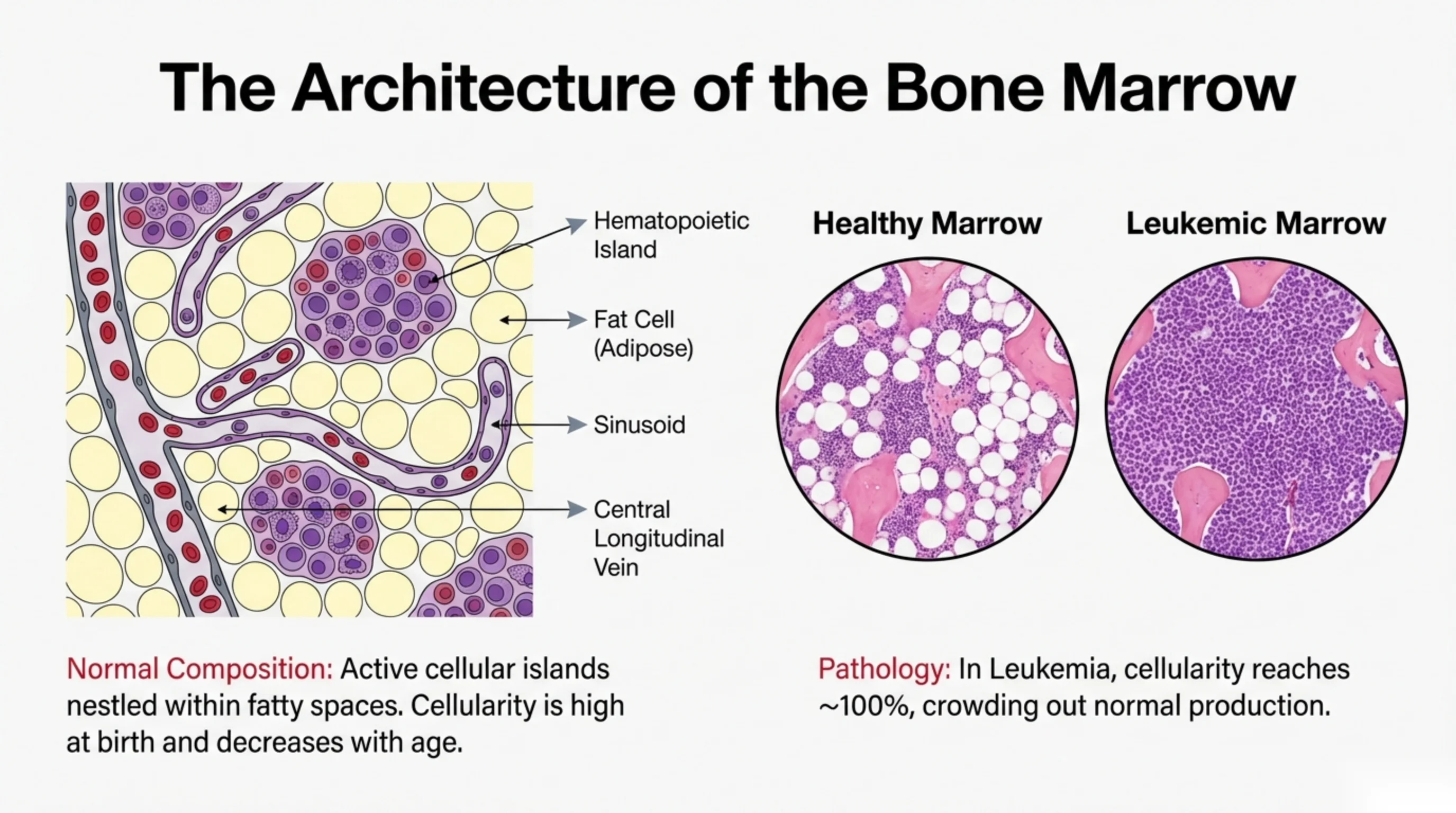
Bone marrow is not a solid mass of cells. It is organised into islands of blood-forming cells set among fat cells, threaded by thin-walled blood channels called sinusoids that drain into a central vein. Mature cells squeeze through the sinusoid walls to enter the circulation; immature cells cannot, which is why blasts are normally confined to the marrow.
The proportion of the marrow that is cellular rather than fatty is called cellularity, and it changes with age. A newborn's marrow is almost entirely cellular; by old age about half of it has been replaced by fat. A pathologist reading a biopsy therefore judges cellularity against the patient's age.
The two micrographs show the contrast that matters. In healthy marrow the cellular islands are separated by clear fat spaces and contain a mixture of cell types at every stage of maturation. In leukemic marrow the fat has vanished and the cellularity approaches 100 percent: the space is packed edge to edge with a single population of identical blasts. There is simply no room left for the erythroblasts, megakaryocytes and maturing granulocytes that should be there. This physical crowding-out is the first reason the blood counts fall, and it is why the diagnosis of leukemia is confirmed in the marrow rather than in the blood.
See it online.
- Clusters of blasts on a bone marrow biopsy ASH Image Bank
The two mutations behind every acute leukemia
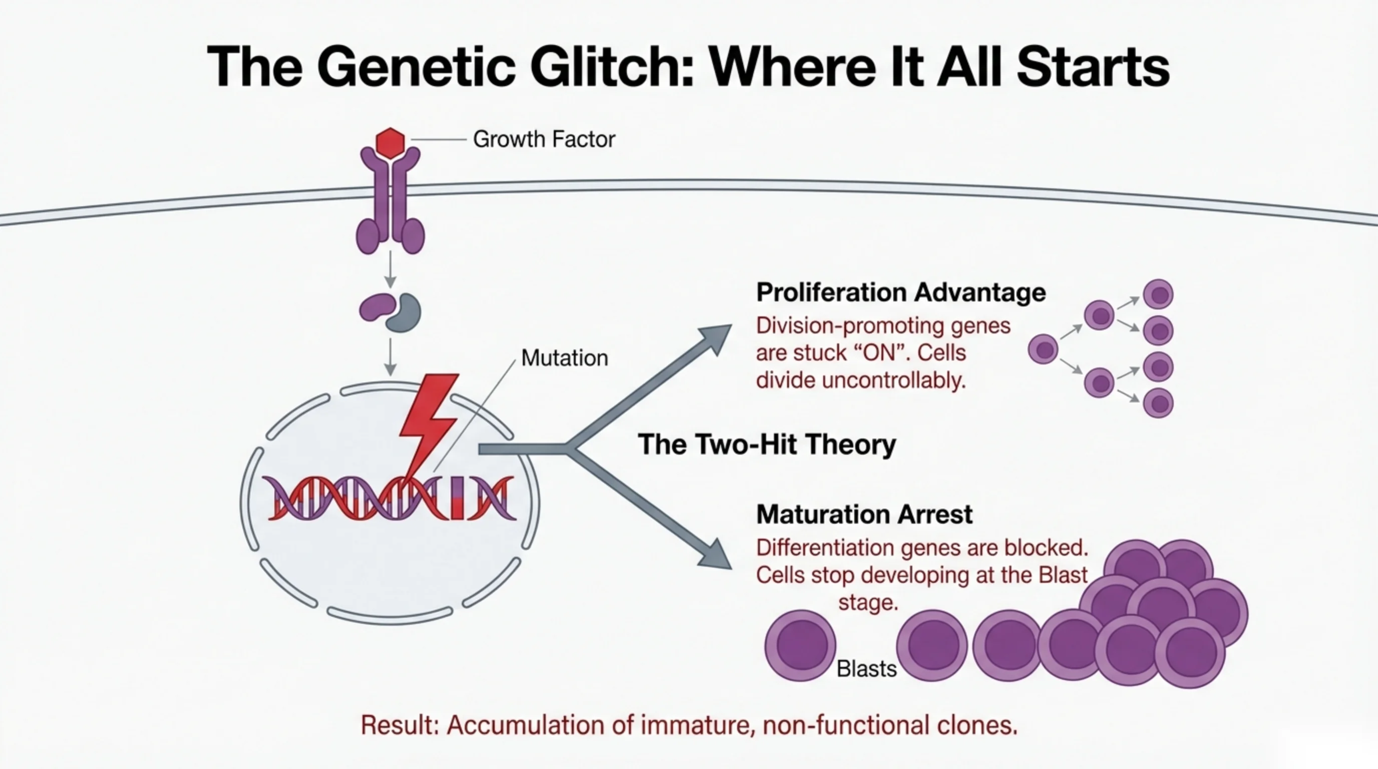
A normal blood cell divides only when told to. A growth factor in the plasma binds a receptor on the cell surface, the receptor sends a signal through the cytoplasm to the nucleus, and genes that control division and maturation switch on in the right order. Leukemia begins when a mutation in the DNA corrupts this control. Because a single mutation is rarely enough, this is called the two-hit theory: two different kinds of damage combine to make a leukemic cell.
The first hit gives a proliferation advantage. A mutation locks a division-promoting gene in the "on" position, so the cell divides whether or not any growth factor is present. On its own this produces too many cells, but they can still mature.
The second hit causes maturation arrest. A mutation blocks the genes that drive differentiation, so the cell can no longer take the next step toward becoming a neutrophil or a lymphocyte. It stays frozen at the blast stage.
Put the two together and you have a cell that divides without limit and never grows up. Its descendants are an ever-expanding clone of identical, immature, non-functional blasts. They do not fight infection, carry oxygen or stop bleeding; they only take up space. That combination, uncontrolled division plus frozen maturation, is the definition of acute leukemia, and the next slide shows what happens when only one of the two hits is present.
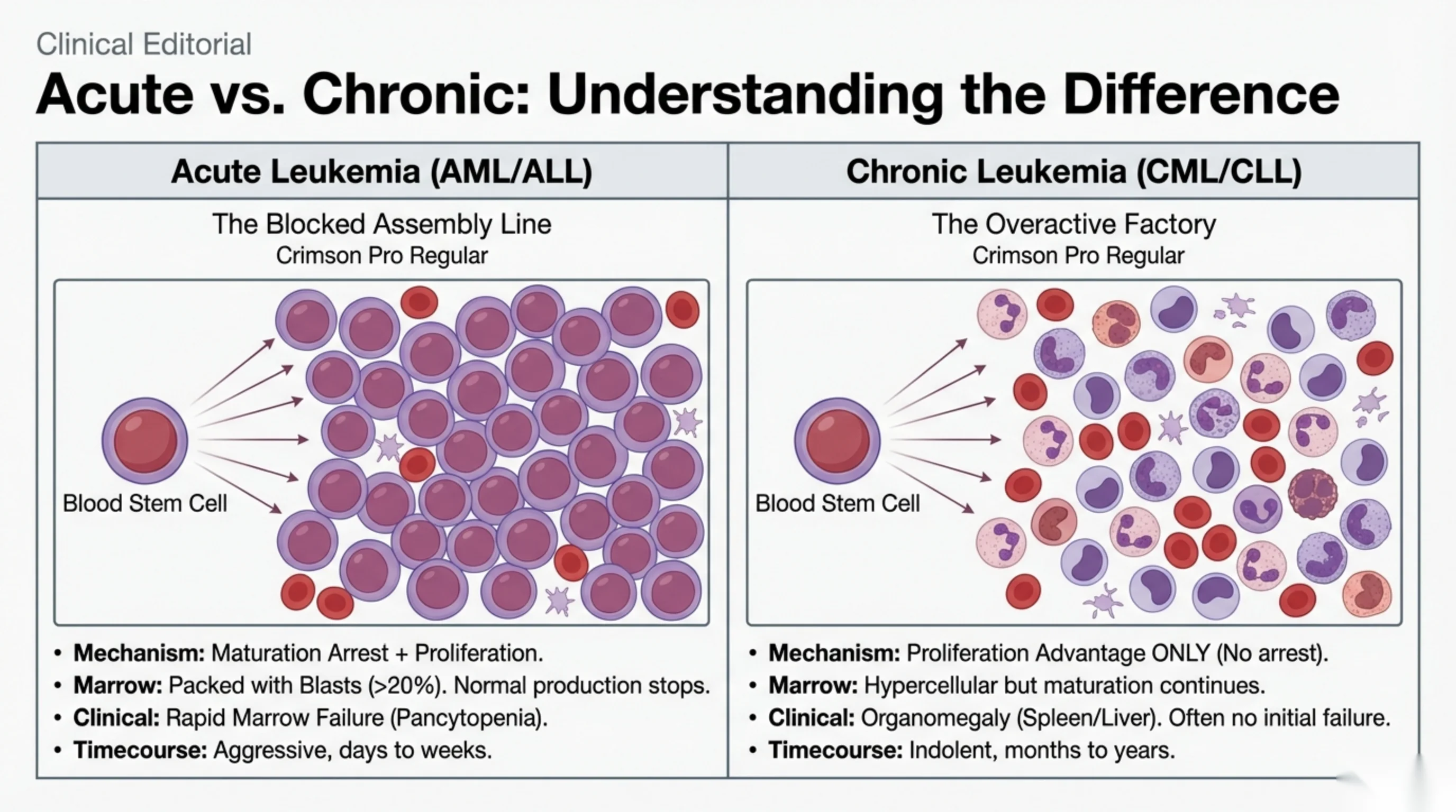
Acute versus chronic is a difference of mechanism, not just of speed
Students often think "acute" and "chronic" leukemia simply describe how fast the disease moves. They do, but the speed is a consequence of a deeper difference, and the slide's two factory pictures capture it.
Acute leukemia is the blocked assembly line. Both hits are present: proliferation and maturation arrest. The marrow fills with blasts, more than 20 percent of the cells, and because nothing matures, the production of normal red cells, neutrophils and platelets stops. The patient presents with marrow failure, that is, pancytopenia, and the disease is aggressive, unfolding over days to weeks. Untreated, it is fatal within months. The two acute leukemias are AML and ALL.
Chronic leukemia is the overactive factory. Only the proliferation hit is present. Cells divide too much, but they still mature, so the blood fills with excess mature cells: granulocytes in chronic myeloid leukemia (CML), lymphocytes in chronic lymphocytic leukemia (CLL). The marrow is hypercellular but still working, so there is usually no marrow failure at first. Instead the surplus cells pile up in the spleen and liver, and the patient presents with organomegaly, often found incidentally. The course is indolent, over months to years.
The practical consequence: an acutely ill patient with low counts and blasts on the smear has acute leukemia and needs admission today; a well patient with a high white count of mature cells and a big spleen probably has chronic leukemia and can be worked up in clinic. The rest of this lecture is about the first patient.
Read next. The other half of this slide is worked through in Case 55 · The Chronic Leukaemias in the casebook. A 46-year-old man has a white cell count of 168 × 109/L found on an insurance medical, a spleen 12 cm below the costal margin, and a film showing granulocytes at every stage of maturation with basophilia — the maturation that acute leukemia has lost. About half of these patients are found by accident on a routine count, exactly as this one was. The case also carries the chronic lymphocytic half, where the diagnosis is made on flow cytometry of the blood and no marrow examination is needed, and where the right management of early disease is to watch and wait — which the case insists you write down for the patient, because otherwise it is heard as neglect.
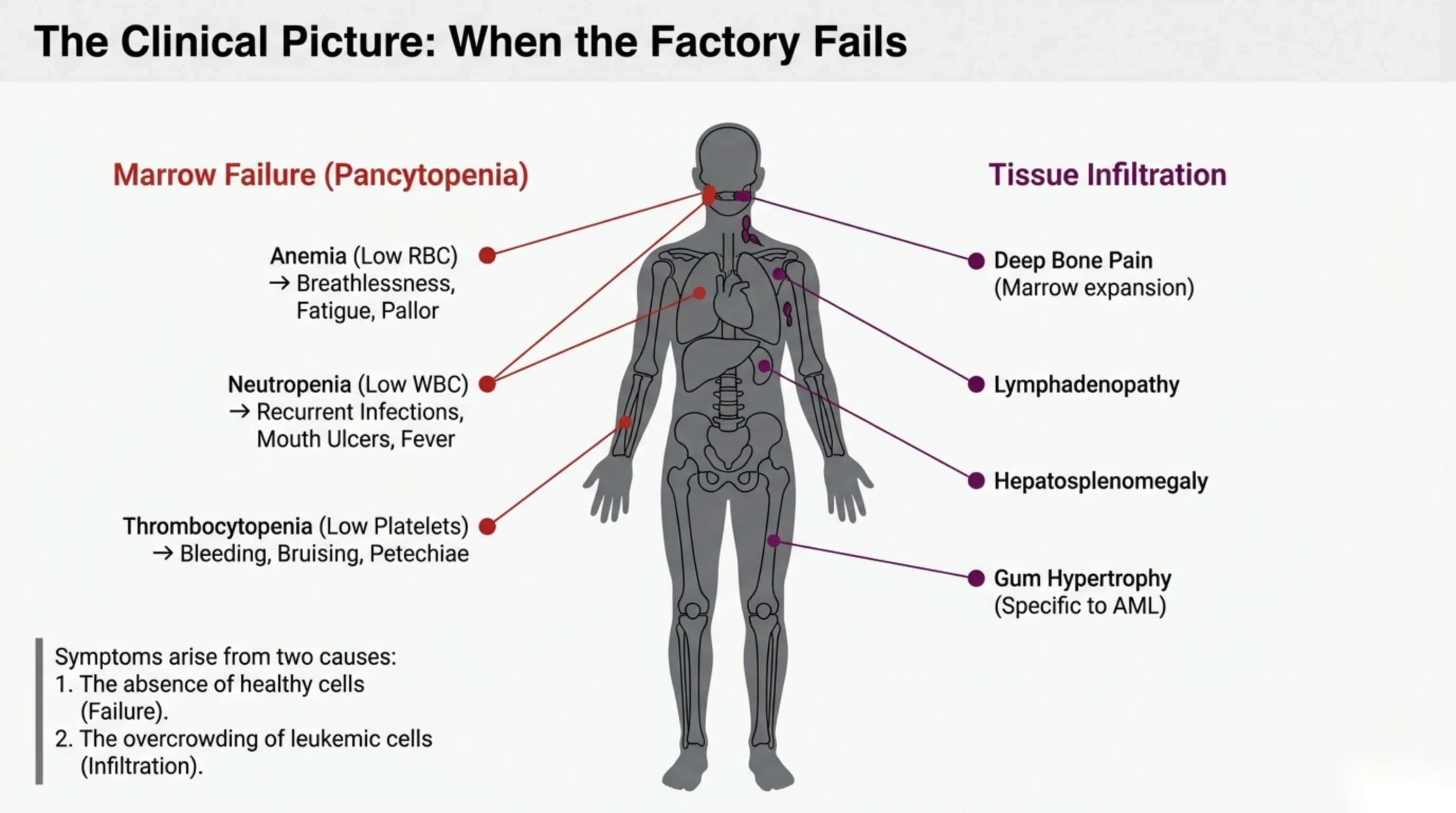
Two reasons a leukemia patient feels ill
Every symptom of acute leukemia comes from one of two processes, and sorting a patient's complaints into these two groups is a good way to organise the history.
Marrow failure. Because the blasts have crowded out normal production, all three cell lines fall, and each shortfall has its own signature. Too few red cells (anemia) causes fatigue, breathlessness and pallor. Too few neutrophils (neutropenia) removes the body's first defence against bacteria, so the patient has recurrent infections, painful mouth ulcers, and fever. Too few platelets (thrombocytopenia) causes bruising, bleeding from gums and nose, and petechiae, the pinpoint red spots of tiny skin bleeds that do not blanch on pressure. A patient with symptoms from all three lines at once, tired, feverish and bruising, is describing pancytopenia, and that is the classic presentation.
Tissue infiltration. The blasts do not stay in the marrow. Their sheer bulk stretches the marrow cavity and produces deep, aching bone pain, especially in children. They spill into the blood and lodge in the lymph nodes (lymphadenopathy), the liver and spleen (hepatosplenomegaly), and, characteristically in the monocytic forms of AML, the gums, which swell and bleed (gum hypertrophy). They can also reach the skin, the testes and the meninges.
Notice that none of these symptoms is specific. Fatigue, fever and bruising are common complaints. What should alert you is the combination, and the next slide shows how the blood count confirms it.
Read next. Open Case 54 · Acute Leukaemia in the casebook: a 24-year-old man with three weeks of fatigue and breathlessness, bruising without injury, gums that bleed when he brushes his teeth, and a week of fever and sore throat that two courses of antibiotics have not touched. The case reads that presentation back as the single process this slide describes, the marrow replaced and all three lineages failing at once, and it adds the parts of the examination the slide does not cover: fundoscopy for retinal haemorrhage, a deliberate search for infection in the mouth, the perineum, the skin folds and between the toes, and the rule that you do not perform a rectal examination in profound neutropenia, because it risks bacteraemia and a perianal abscess.
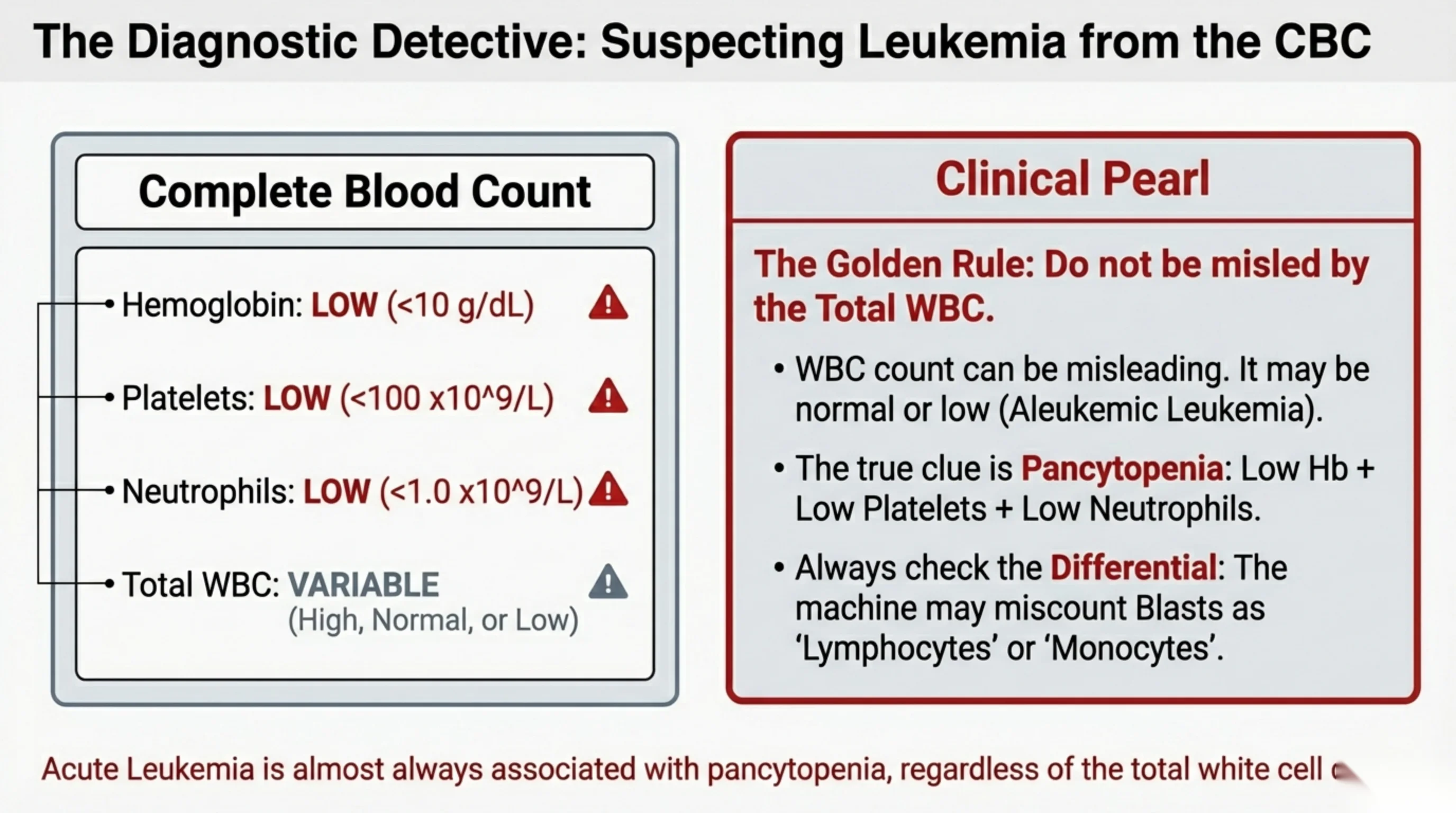
Reading the blood count: the white cell count is the least useful number
Most people, asked to check a blood count for leukemia, look at the total white cell count. This is the mistake the slide is designed to correct.
The total white cell count in acute leukemia is variable. It may be very high, because blasts have flooded the blood. It may be normal. It may be low, because the blasts are still confined to the marrow and the normal white cells have been suppressed; this presentation is called aleukemic leukemia, and it is precisely the one that gets missed. A normal or low white count therefore never excludes leukemia.
The reliable clue is pancytopenia: a low hemoglobin (on the slide, below 10 g/dL), a low platelet count (below 100 × 10⁹/L) and a low neutrophil count (below 1.0 × 10⁹/L) together. When all three lines are down, something has replaced or destroyed the marrow, and acute leukemia is at the top of the list. The neutrophil count, in particular, is almost always low.
The second rule is to always look at the differential, not just the total. The automated analyser sorts cells by size and granularity, and it has no category for blasts. It will file them as "lymphocytes" or "monocytes", so a report showing an unexplained lymphocytosis or monocytosis in a pancytopenic patient may in fact be showing you blasts. The analyser usually flags such samples for review; the flag is your cue to ask the laboratory for a smear and to look at it yourself.
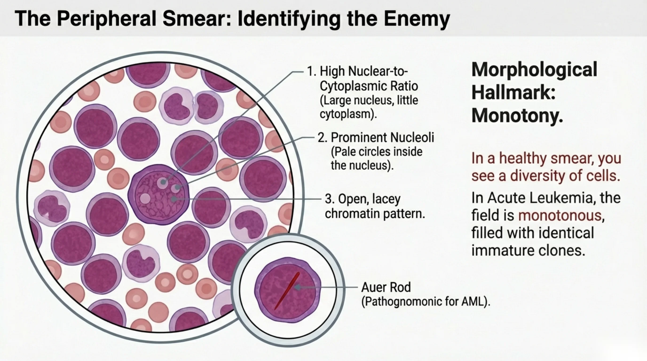
Recognising a blast on the smear
The peripheral blood smear is where leukemia is first seen, and a blast has three features that, once learned, are hard to miss.
- A high nuclear-to-cytoplasmic ratio. The nucleus fills almost the whole cell, leaving only a thin rim of cytoplasm. A mature lymphocyte also has a large nucleus, but its chromatin is dense and dark; the blast's is not.
- Prominent nucleoli. One or more pale, round structures are visible inside the nucleus. Nucleoli are where ribosomes are assembled, and a cell that shows them is a cell preparing to divide. Mature blood cells never display them.
- Open, lacy chromatin. The nuclear material looks fine and evenly dispersed, like a net, rather than clumped. This is the chromatin of an actively transcribing, immature cell.
One additional finding settles the lineage on the spot. An Auer rod is a needle-shaped pink or red streak in the cytoplasm, formed from fused granules. It occurs only in myeloid blasts, so a single Auer rod makes the diagnosis of AML rather than ALL, and many Auer rods bundled together suggest the promyelocytic variant that carries a special bleeding risk (slide 13).
Step back from the individual cell and there is a second, more subtle sign: monotony. A normal smear is a crowd of different cells at different stages. A leukemic smear is a field of identical clones, the same size, the same nucleus, the same blank cytoplasm, repeated across the slide. When every cell looks like every other cell, you are looking at a clone, and a clone is a malignancy.
See it online.
- A myeloblast on a blood film: large nucleus, fine chromatin, prominent nucleoli ASH Image Bank
- An Auer rod in a blast ASH Image Bank
What real blasts look like
The previous slide drew a blast. These are photographs of real ones, from bone marrow smears in the atlas of the Armed Forces Institute of Pathology.
Left, acute myeloid leukemia. Check the three features from the previous slide on any one of the large cells. The nucleus fills most of the cell. The chromatin is fine and open, not clumped. Pale nucleoli show inside several of the nuclei. Then look at the field as a whole. It is made of one kind of cell. The atlas notes that these blasts vary in size and in how much cytoplasm they have, so monotony does not mean identical copies. It means that the mixture of cells at every stage of maturity, which a healthy marrow shows, is gone. That is why this form is called AML without maturation.
Right, acute lymphoblastic leukemia. The atlas describes these lymphoblasts as having very little cytoplasm, an irregular outline to the nucleus, and some variation in size and in how dense the chromatin is. Some have small nucleoli.
Compare the two panels, then keep in mind the point of the slide after next. A blast under the microscope can look much the same whether it is myeloid or lymphoid, and the two are treated differently, so the lineage is confirmed by flow cytometry, not by eye.
See it online.
- Myeloblasts in acute myeloid leukemia, full size Wikimedia Commons · Public domain · AFIP
- Lymphoblasts in acute lymphoblastic leukemia, full size Wikimedia Commons · Public domain · AFIP
- Lymphoblasts in the blood in acute lymphoblastic leukemia Wikimedia Commons · CC BY-SA 4.0 · SpicyMilkBoy
Auer rods, one and many
The inset on slide 8 drew an Auer rod. These are real ones, enlarged from bone marrow smears.
The first two panels are two myeloblasts, each with a single rod. In the first it lies at the upper left of the cell; in the second, at the lower right, in the thin rim of cytoplasm. Look for a thin, straight, pink-red streak. It is made of fused granules and occurs only in myeloid blasts, so one rod like these makes the diagnosis AML rather than ALL.
The third panel is a faggot cell from acute promyelocytic leukemia. Its cytoplasm holds many rods lying across each other, like a bundle of sticks, which is where the name comes from. This is what slide 8 means by many Auer rods bundled together, and it points to the promyelocytic variant with its special bleeding risk (slide 13). The atlas notes that this smear was taken after chemotherapy, so the cells around it are normal neutrophil precursors.
See it online.
- Two myeloblasts with Auer rods, full field Wikimedia Commons · Public domain · AFIP
- A faggot cell in acute promyelocytic leukemia, full field Wikimedia Commons · Public domain · AFIP
- Blasts with Auer rods beside maturing myeloid cells Wikimedia Commons · Public domain · AFIP
- Several blasts with Auer rods in a marrow aspirate Wikimedia Commons · CC BY-SA 3.0 · VashiDonsk
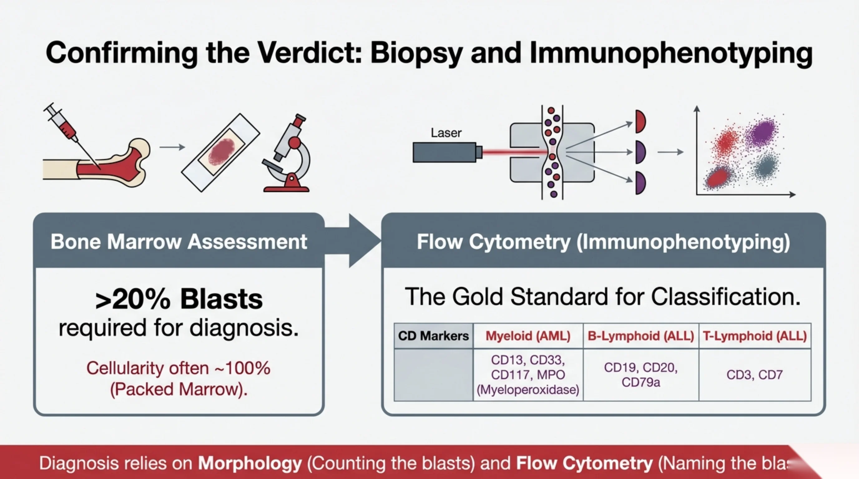
Confirming the diagnosis: count the blasts, then name them
The smear raises the suspicion; the bone marrow confirms it. Confirmation has two steps, and they answer two different questions.
Morphology answers "how many?" A bone marrow aspirate and biopsy are taken, usually from the back of the pelvis, and the cells are counted under the microscope. The diagnostic threshold for acute leukemia is more than 20 percent blasts among the nucleated marrow cells. Below that figure, the picture is classified as a myelodysplastic syndrome or a related condition rather than acute leukemia. The biopsy also shows the cellularity, which in leukemia approaches 100 percent, a packed marrow with no fat spaces left.
Flow cytometry answers "what kind?" A blast under the microscope looks much the same whether it is myeloid or lymphoid, yet the treatments for AML and ALL are entirely different, so the lineage must be established. Flow cytometry, also called immunophenotyping, does this by tagging the cells with fluorescent antibodies against surface and internal proteins called CD markers, then passing them one by one through a laser that reads which tags are present. Each lineage carries its own set: myeloid blasts express CD13, CD33, CD117 and the enzyme myeloperoxidase; B-lymphoid blasts express CD19, CD20 and CD79a; T-lymphoid blasts express CD3 and CD7. Flow cytometry is the gold standard for classification and returns a result within hours.
In practice a third step follows: cytogenetic and molecular testing of the same marrow sample, which does not change the diagnosis but, as slide 15 explains, largely decides the prognosis and the choice of consolidation treatment.
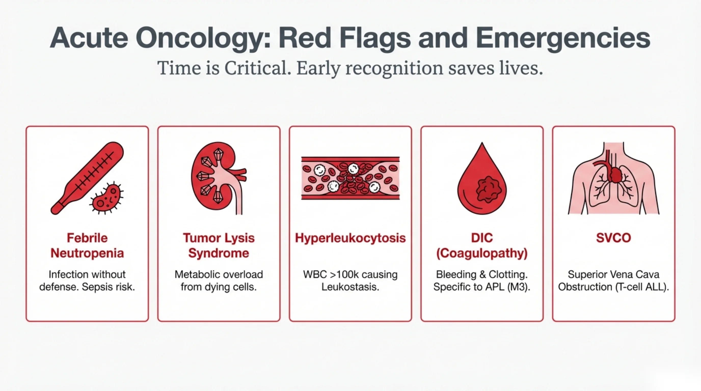
Five ways acute leukemia kills in the first days
Most deaths in the first weeks of acute leukemia are not caused by the leukemia itself but by one of a small number of complications, all of which are preventable or treatable if they are anticipated. This slide is the list to keep in your head; the next two slides explain each in detail.
- Febrile neutropenia. A fever in a patient with no neutrophils. There is nothing to contain a bacterial infection, and sepsis can develop in hours.
- Tumor lysis syndrome. When large numbers of blasts die at once, before or after chemotherapy starts, they release their contents into the blood faster than the kidneys can clear them.
- Hyperleukocytosis. A white cell count above 100 × 10⁹/L makes the blood viscous and blasts clog small vessels, a state called leukostasis.
- Disseminated intravascular coagulation (DIC). Widespread activation of clotting that consumes platelets and clotting factors and causes bleeding. It is particularly associated with acute promyelocytic leukemia, the M3 subtype of AML.
- Superior vena cava obstruction (SVCO). A mass of leukemic lymph nodes in the chest, typical of T-cell ALL, compresses the great vein returning blood from the head and arms.
The common thread is that time is the treatment. Each of these is recognised from the bedside and the first set of bloods, and each has an intervention that works when started early. Acute oncology is the discipline of looking for them before they announce themselves.
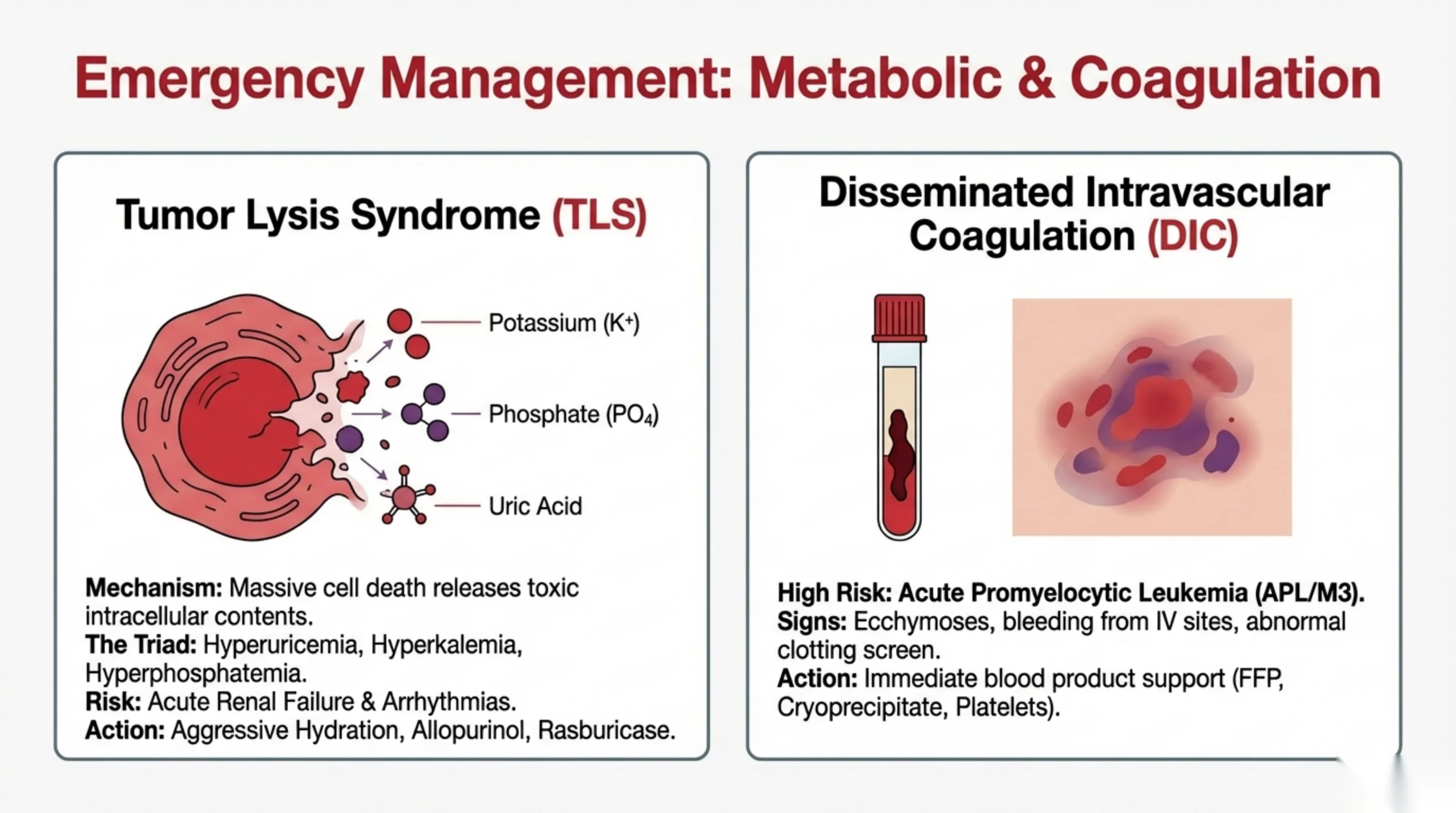
Tumor lysis and DIC: when the chemistry and the clotting go wrong
Tumor lysis syndrome. Every cell is a bag of potassium, phosphate and nucleic acid. When a large mass of leukemic cells dies, spontaneously in a rapidly growing leukemia or, more often, in the first days of chemotherapy, those contents flood the plasma. Potassium rises, and hyperkalemia can stop the heart. Phosphate rises, binds calcium, and the calcium phosphate crystallises in the kidney tubules. Nucleic acids are broken down to uric acid, which also crystallises in the tubules. The result is the triad of hyperuricemia, hyperkalemia and hyperphosphatemia, and its two dangers are acute renal failure and cardiac arrhythmia.
The management is mostly prevention. Aggressive intravenous hydration keeps the urine flowing and dilutes the crystals. Allopurinol blocks the enzyme that makes uric acid, so it stops new uric acid forming. Rasburicase goes further: it is an enzyme that breaks down uric acid already present, and it is used in high-risk patients or when the uric acid is already high. Potassium and phosphate are watched closely and treated as they rise.
Disseminated intravascular coagulation. Leukemic cells, above all the promyelocytes of acute promyelocytic leukemia (APL, the M3 subtype), release substances that activate the clotting cascade throughout the circulation. Tiny clots form everywhere, consuming platelets and clotting factors, and the patient then bleeds because there is nothing left to clot with. Look for spreading bruises, oozing from cannula sites, and a clotting screen showing prolonged times, low fibrinogen and low platelets. The treatment is immediate replacement of what has been consumed: fresh frozen plasma for clotting factors, cryoprecipitate for fibrinogen, and platelets. In APL specifically, starting the targeted drug all-trans retinoic acid at the first suspicion, before the diagnosis is even confirmed, is what stops the bleeding at its source, and it is why APL is treated as an emergency on the day of presentation.
Read next. The tumour lysis row of the emergencies table in Case 54 · Acute Leukaemia in the casebook carries a rule this slide does not. Rasburicase is contraindicated in glucose-6-phosphate dehydrogenase deficiency, and the case is explicit that the status is checked before the drug is given. It is an easy step to skip under pressure, and it matters more here than in most places, because G6PD deficiency is common in this population. The case sets all four emergencies out together, each with the action it demands and the time it has to be done in.
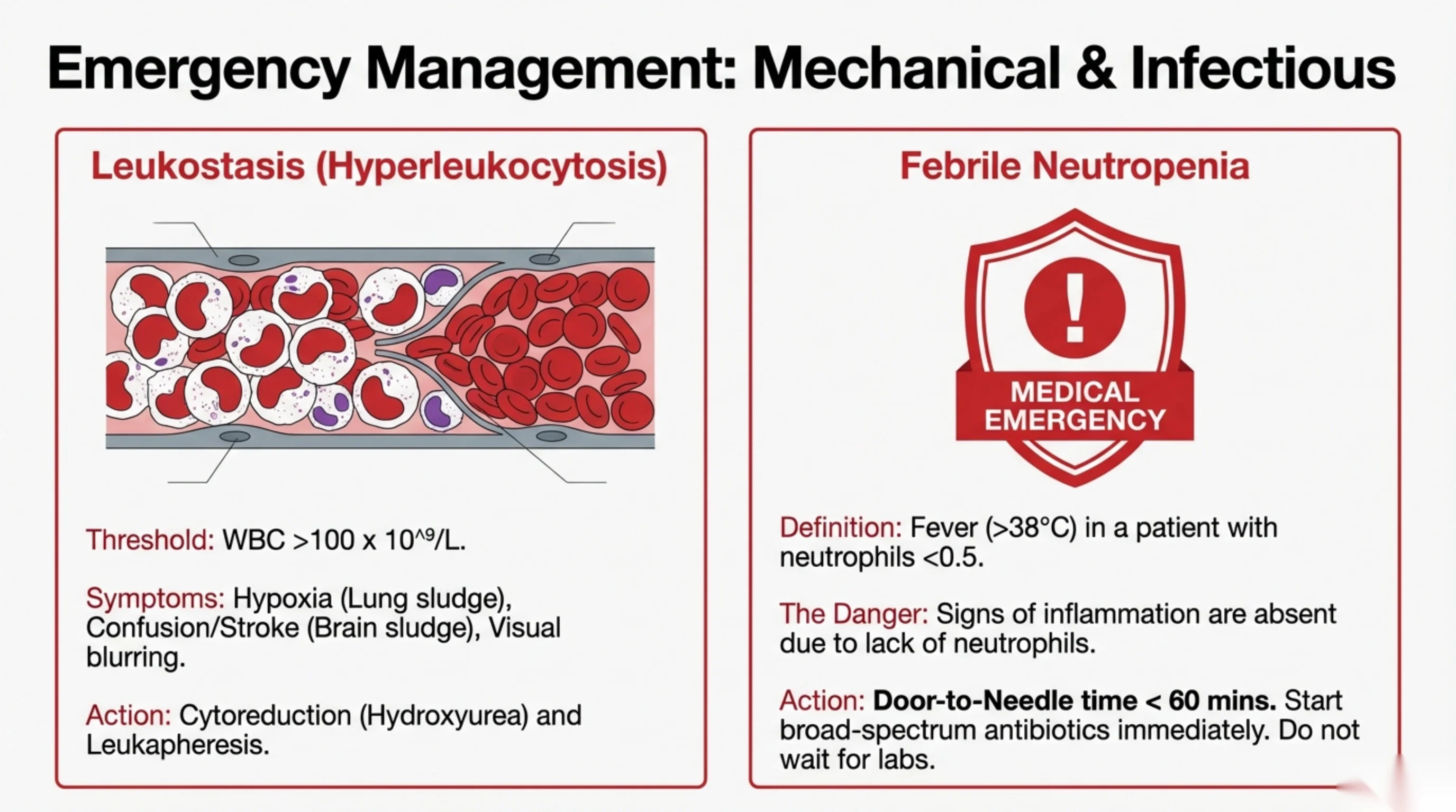
Leukostasis and febrile neutropenia: the sludge and the silent infection
Leukostasis. When the white cell count climbs above 100 × 10⁹/L, the blood itself changes. Blasts are larger and stiffer than normal white cells, and in that number they thicken the blood and plug the smallest vessels. The organs with the finest capillary beds suffer first. In the lungs the sludge causes hypoxia and breathlessness; in the brain it causes confusion, headache and stroke; in the retina it causes blurred vision. The treatment is to bring the count down quickly: hydroxyurea, an oral drug that stops cell division, for cytoreduction, and in severe cases leukapheresis, in which the patient's blood is run through a machine that removes the white cells and returns the rest. One caution follows from the physics: a red cell transfusion given before the count is reduced raises the viscosity further and can precipitate a stroke, so transfuse cautiously in hyperleukocytosis.
Febrile neutropenia. Defined as a temperature above 38 °C in a patient whose neutrophil count is below 0.5 × 10⁹/L, this is the commonest emergency in leukemia and the one most often mishandled. The danger is that neutrophils are what produce the signs of infection: the pus, the redness, the swelling, the shadow on the chest film. Without neutrophils, a patient can have a life-threatening bacterial infection and show nothing except the fever, and sometimes not even that. So the fever must be treated as sepsis until proven otherwise.
The rule is a door-to-needle time of under 60 minutes: blood cultures are taken and broad-spectrum intravenous antibiotics are running within an hour of the patient arriving, without waiting for the blood count, the cultures or any imaging to come back. Every hour of delay measurably increases mortality. If you learn one number from this lecture, make it that one.
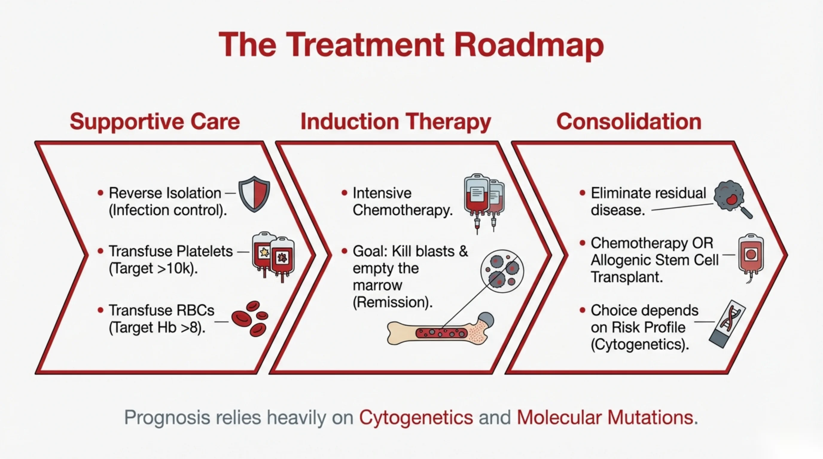
How acute leukemia is treated
Treatment runs on three tracks, one of which begins before the diagnosis is even confirmed.
Supportive care keeps the patient alive while the disease is attacked. Reverse isolation, a single room with strict hand hygiene, protects the neutropenic patient from the infections other people carry. Platelets are transfused to keep the count above 10 × 10⁹/L, the level below which spontaneous bleeding becomes likely, and higher if the patient is bleeding or febrile. Red cells are transfused to keep the hemoglobin above 8 g/dL. Infections are treated early, and the emergencies of the previous slides are anticipated.
Induction therapy is the first course of intensive chemotherapy. Its goal is remission: to kill the blasts and empty the marrow so that normal stem cells can regrow. The patient's counts fall to nothing for two to three weeks before recovering, and it is during this period that supportive care matters most. Remission means the blasts have fallen below the diagnostic threshold and normal blood production has returned; it does not mean cure, because leukemic cells invisible to the microscope remain.
Consolidation is aimed at those residual cells. It is either further cycles of chemotherapy or, for patients whose disease is likely to return, an allogeneic stem cell transplant, in which the patient's marrow is replaced with a donor's after high-dose treatment. The choice depends on the risk profile, and that profile is read mainly from the cytogenetics and molecular mutations of the original blasts. Some chromosomal abnormalities predict an excellent response to chemotherapy alone; others predict relapse and point to transplant. This is why the genetic tests on the marrow sample, though they do not change the diagnosis, largely determine the treatment plan and the patient's prospects.
Read next. The management section of Case 54 · Acute Leukaemia in the casebook carries three things this slide does not, and all three belong to the first day rather than the first cycle. Fertility preservation is discussed and arranged before the first dose of chemotherapy — sperm banking, or oocyte or ovarian tissue cryopreservation — because once treatment starts the opportunity has usually gone, and the patient in the case is 24. The patient and his siblings are tissue-typed early, since a transplant decision may follow. And the virology, the echocardiogram before anthracyclines and the dental assessment are all done before treatment starts, not after.
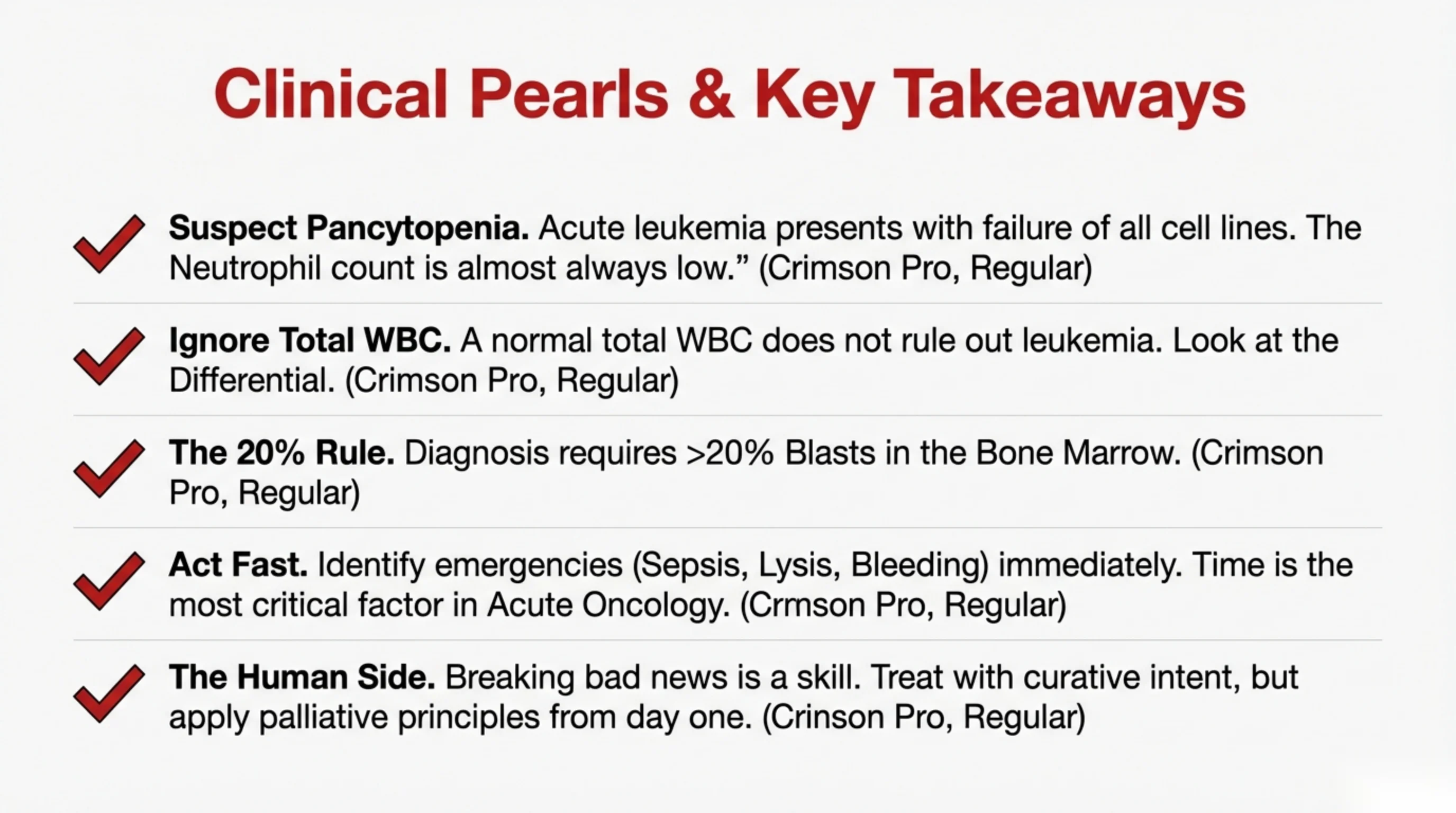
Five things to take away
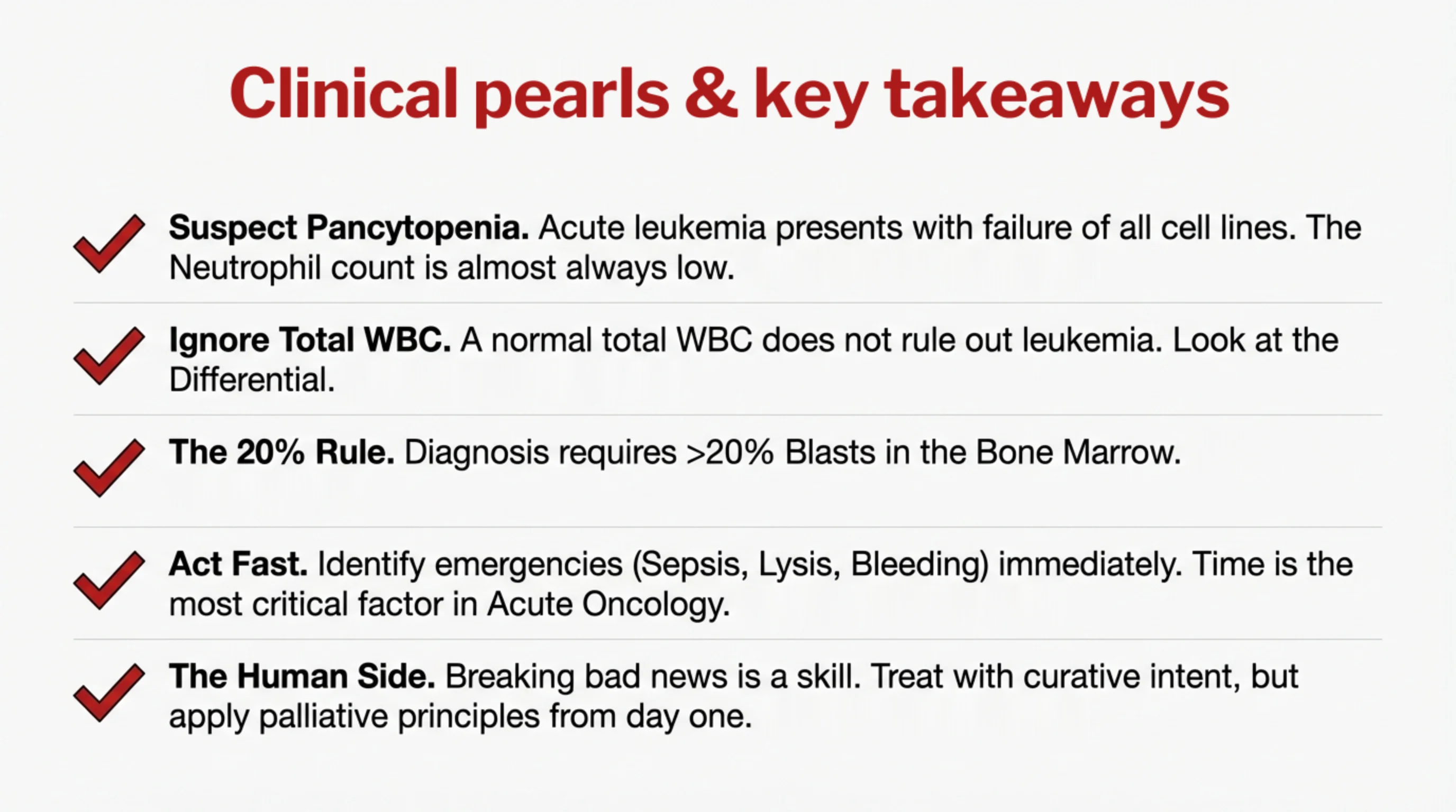
- Suspect pancytopenia. Acute leukemia presents as failure of all three cell lines. Whatever else the blood count shows, the neutrophil count is almost always low. A tired, feverish, bruising patient with three low counts has leukemia until the marrow says otherwise.
- Ignore the total white cell count. It may be high, normal or low, and a normal value excludes nothing. Read the differential, and remember the analyser may have labelled blasts as lymphocytes or monocytes. Ask for the smear.
- The 20 percent rule. The diagnosis requires more than 20 percent blasts in the bone marrow. Morphology counts them, flow cytometry names their lineage, and cytogenetics sets the prognosis.
- Act fast. Sepsis in the neutropenic patient, tumor lysis, bleeding from DIC, leukostasis and SVC obstruction are identified at the bedside and treated within the hour. Time is the single most important variable in acute oncology.
- The human side. A diagnosis of acute leukemia is delivered to a frightened person, often young, and how it is delivered will be remembered for the rest of their life. Breaking bad news is a skill to be learned and practised. Treat with curative intent, but bring the principles of palliative care, control of symptoms, honesty, attention to the family, in from the first day, not the last.
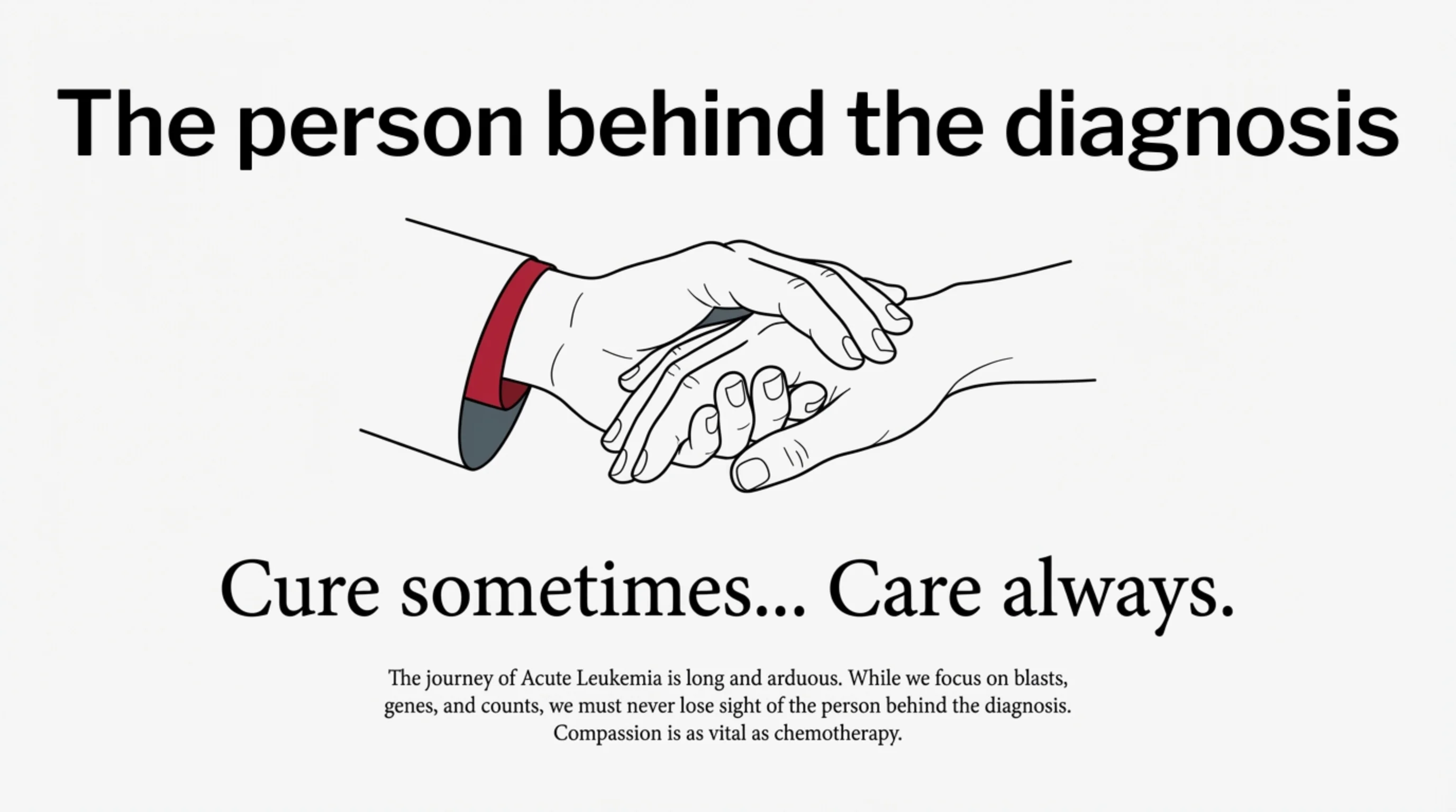
Cure sometimes, care always
The phrase adapts an old motto of medicine: to cure sometimes, to relieve often, to comfort always. It belongs at the end of this lecture because acute leukemia stretches both halves of it further than almost any other disease.
The cure is real. A young adult with acute lymphoblastic leukemia, or an older patient with a favourable-risk AML, has a genuine chance of being alive and well years later, and that chance did not exist two generations ago. But the road there is long: months of intensive chemotherapy, weeks at a time in isolation, repeated transfusions, infections, and for some a transplant with its own risks. And for the patients whose disease does not respond, or returns, the road ends differently.
Through all of it, the person on the bed will experience the disease as fatigue, fear, boredom, pain, separation from family, and a constant stream of numbers they do not understand. The clinician who explains those numbers, who sits down, who says what is known and what is not, and who does not disappear when the news is bad, is practising medicine as fully as the one who chooses the chemotherapy. Compassion is not a supplement to the treatment of leukemia. It is part of the treatment.