Disorders of Hemostasis
A clinical approach to bleeding — primary vs secondary, quantitative vs qualitative, congenital vs acquired.
These slides are an outline only. Read the accompanying material with them: the notes under each slide below, and chapters 20 to 25 of the Hematology Handbook, from Principles of Hemostasis to Hemophilia. Then work through the Internal Medicine Casebook and the Hematology Question Bank.
What this lecture is about
These slides are an outline only. Read the accompanying material with them: the notes under each slide on this page, and chapters 20 to 25 of the Hematology Handbook, from Principles of Hemostasis to Hemophilia. Then work through the Internal Medicine Casebook and the Hematology Question Bank.
This is the clinical lecture that follows the physiology of hemostasis. It is about the patient who bleeds, and it has one organising idea: bleeding disorders can be sorted along three axes, and once you have placed a patient on each axis you know what to test and usually what is wrong.
The three axes are on the title slide. Primary versus secondary hemostasis: is the failure in the platelet plug or in the fibrin clot? Quantitative versus qualitative: is the component missing, or present but not working? Congenital versus acquired: was the patient born with it, or did something happen? The lecture builds the approach around these, works through a case of a 14-year-old girl whose bleeding disorder had been hiding in plain sight, and ends with the laboratory logic of the PT, the aPTT and the mixing study.
Primary versus secondary hemostasis: two kinds of bleeding
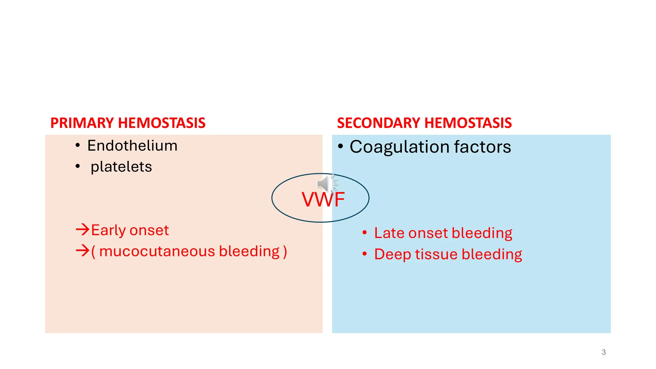
The first axis is the most useful at the bedside, because the two kinds of failure produce two recognisably different patterns of bleeding. Primary hemostasis is the platelet plug, made by the endothelium and the platelets. When it fails, bleeding starts early, immediately after the injury, because nothing plugs the hole, and it is mucocutaneous: bruises, petechiae, nosebleeds, gum bleeding, heavy periods, bleeding from the gut. Secondary hemostasis is the fibrin clot, made by the coagulation factors. When it fails, the platelet plug forms and bleeding stops at first, then starts again late, hours later, because the plug was never stabilised, and it is deep: into joints, into muscles, into the retroperitoneum.
The molecule circled between the two columns is von Willebrand factor, and it is circled because it belongs to both. It glues platelets to the wound, a primary function, and it carries factor VIII in the plasma, protecting it from destruction, a secondary function. Von Willebrand disease, the commonest inherited bleeding disorder and the diagnosis in this lecture's case, therefore presents as a primary disorder but can, when severe, behave like a secondary one too.
Read next. Case 53 · The Bleeding Patient in the casebook opens with this slide as a table: the mucocutaneous pattern of the platelets, the vessel wall and von Willebrand factor on one side, the deep-tissue pattern of the coagulation factors on the other, and the diagnoses to think of under each.
See it online.
- Petechiae on the legs Wikimedia Commons · CC BY-SA 3.0 · DrFO.Jr.Tn
Congenital versus acquired, quantity versus quality
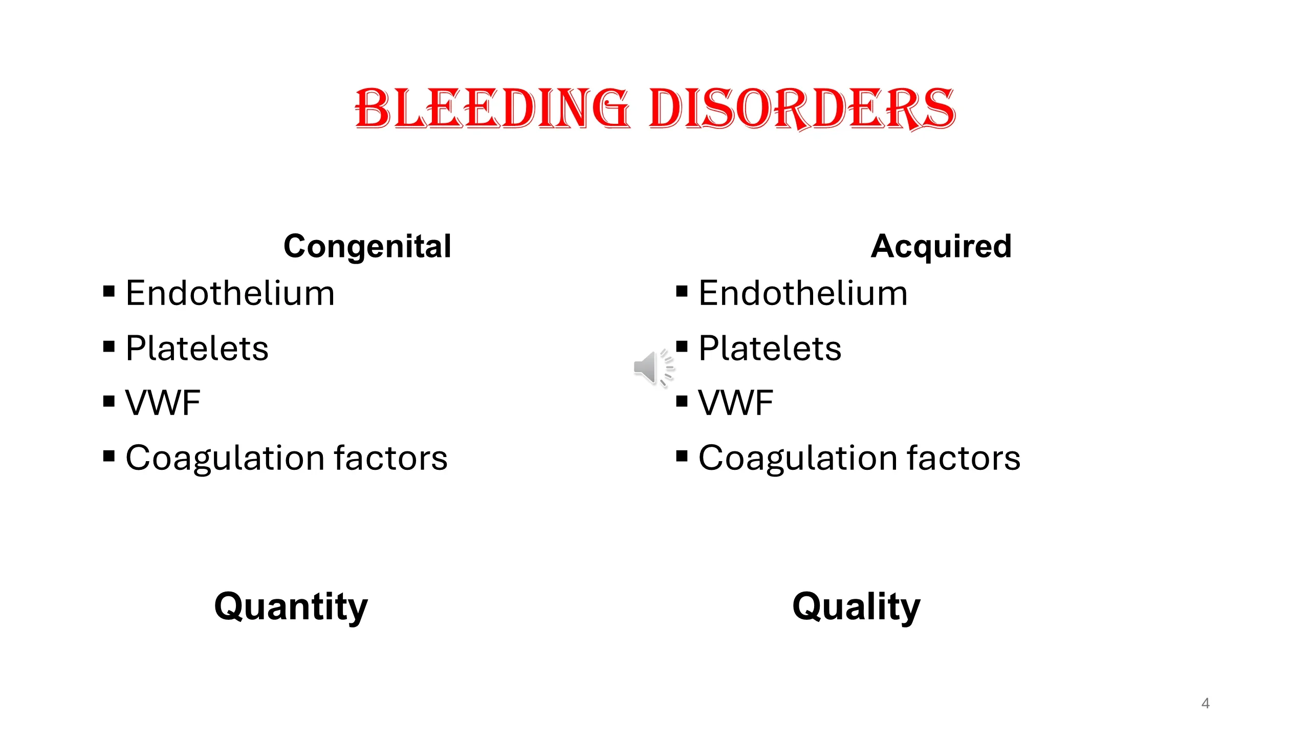
The other two axes are drawn as a grid. Any of the four components of hemostasis, the endothelium, the platelets, von Willebrand factor and the coagulation factors, can fail because of a defect the patient was born with, congenital, or because of something that happened later, acquired. And each can fail in two ways: there can be too little of it, a defect of quantity, or there can be a normal amount that does not work, a defect of quality.
Examples make the grid concrete. Thrombocytopenia is a quantitative platelet disorder; a platelet function defect caused by aspirin is a qualitative one, and acquired. Hemophilia is a congenital quantitative deficiency of factor VIII or IX; an acquired inhibitor against factor VIII is an acquired disorder in which the factor is present but neutralised. Von Willebrand disease type 1 is a quantitative deficiency and type 2 a qualitative one, as slides 19 to 21 will show.
The reason the grid matters is that the laboratory tests answer its questions in order. A count tells you quantity; a function test tells you quality; the history and family history tell you congenital from acquired.
The approach: suspect, then confirm
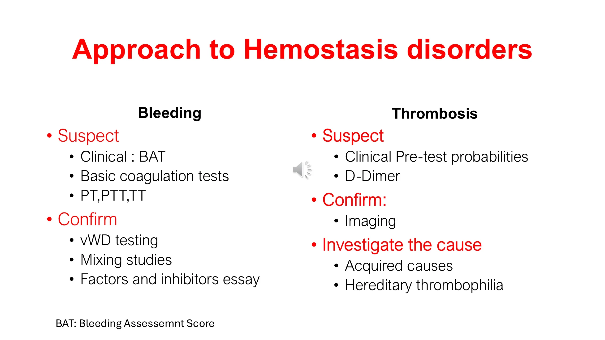
The slide sets the approach to bleeding beside the approach to thrombosis, because the two have the same shape: first suspect, using clinical assessment and screening tests, then confirm, with specific tests, then find the cause.
For bleeding, suspicion is raised clinically, and the lecture will use a structured tool for this, the bleeding assessment tool or BAT. It is supported by the basic coagulation tests: the prothrombin time, the partial thromboplastin time and the thrombin time. Confirmation then depends on what the pattern suggests: testing for von Willebrand disease, mixing studies to separate a deficiency from an inhibitor, and assays of individual factors and their inhibitors.
For thrombosis, suspicion is expressed as a clinical pretest probability, supported by the D-dimer; confirmation is by imaging; and the cause is then sought among acquired causes and the hereditary thrombophilias. That half of the slide belongs to the thrombosis seminar. Keep the parallel in mind: in both, the tests are interpreted in the light of the clinical probability, never on their own.
Local bleeding or a bleeding disorder? The pattern decides
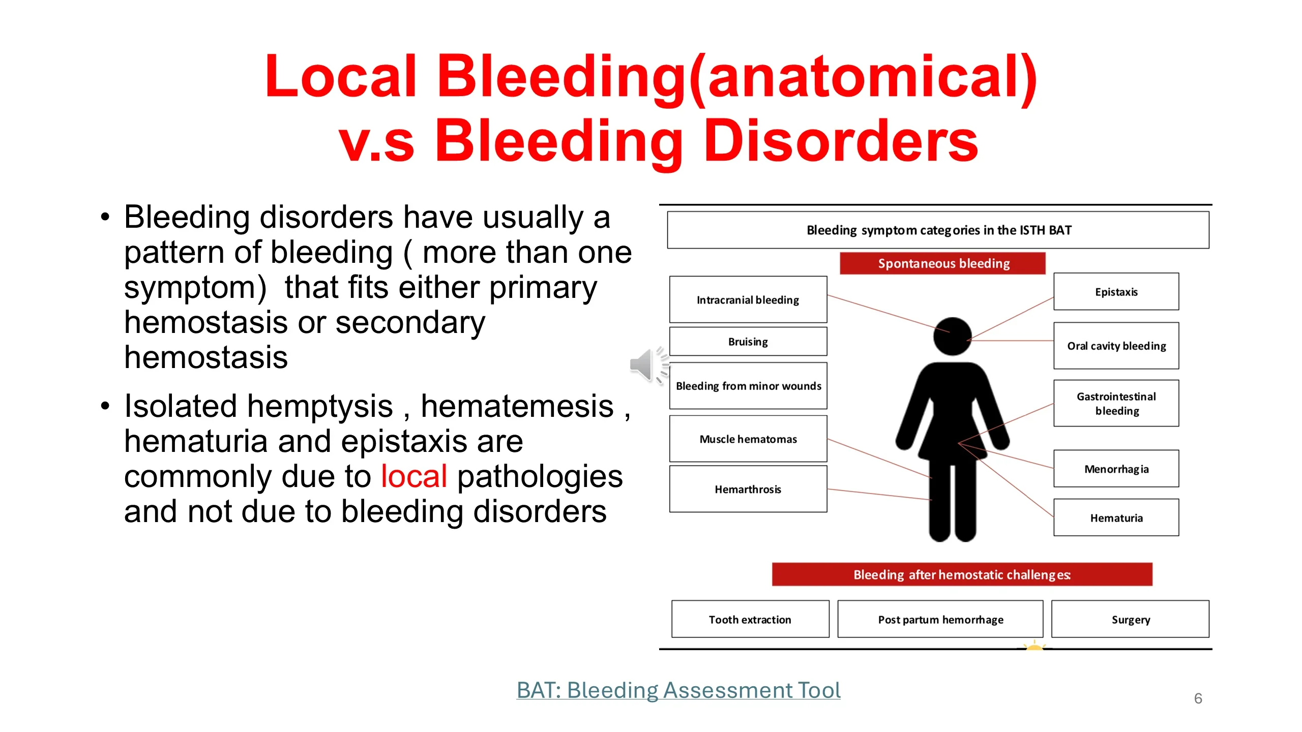
Most patients who bleed do not have a bleeding disorder. They have a local, anatomical cause: an ulcer, a polyp, a tumour, a vessel in the nose. The slide gives the rule for telling the two apart. A bleeding disorder produces a pattern, more than one kind of bleeding, and the pattern fits either primary or secondary hemostasis. A single isolated symptom, hemoptysis, hematemesis, hematuria or epistaxis on its own, is usually local and should be investigated as such, with endoscopy or imaging, not with a coagulation screen.
The body diagram lists the bleeding symptoms that the ISTH bleeding assessment tool scores, and they fall into two groups. Spontaneous bleeding: bruising, bleeding from minor wounds, epistaxis, bleeding in the mouth, gastrointestinal bleeding, menorrhagia, hematuria, muscle hematomas, hemarthrosis and intracranial bleeding. And bleeding after hemostatic challenges: tooth extraction, surgery and childbirth. The tool asks about each and scores its severity, and the total is what turns a vague impression of easy bruising into a number that can be compared with a threshold.
See it online.
- Hemarthrosis: the knee joint of a haemophiliac, a watercolour from 1881 Wikimedia Commons · CC BY 4.0 · Thomas Godart, via Wellcome Collection
- Bleeding in the mouth: petechiae and purpura on the lip in immune thrombocytopenia Wikimedia Commons · CC BY-SA 3.0 · Mdscottis
Two steps, in this order
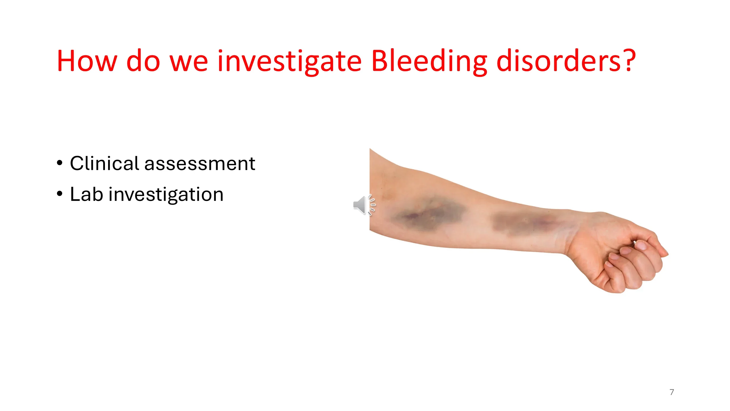
The investigation of a suspected bleeding disorder has two components, clinical assessment and laboratory investigation, and the order is not negotiable. The history comes first, because it determines which tests to send and how to interpret them, and because a normal coagulation screen does not exclude a bleeding disorder: von Willebrand disease and platelet function defects can both hide behind normal screening tests. The next four slides are the history.
The photograph shows what patients usually bring: bruises on the forearm. Bruising is common, and most of it is innocent. The skill is in the questions that follow.
See it online.
- A large bruise (ecchymosis) on the back of the hand ASH Image Bank
Taking the bleeding history: what kind of bleeding?
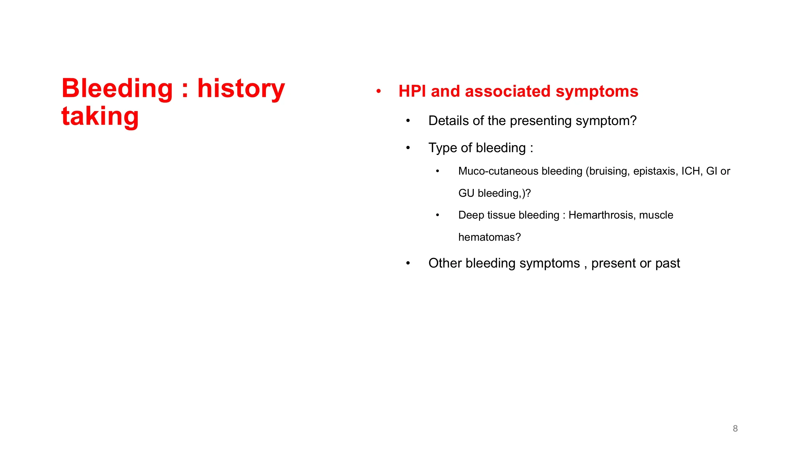
Begin with the presenting symptom and its details, then ask the question that places the patient on the first axis: what type of bleeding is this? Mucocutaneous bleeding, bruising, nosebleeds, bleeding from the gums, gastrointestinal or genitourinary bleeding and, at its most serious, intracranial hemorrhage, points to primary hemostasis, that is, to platelets or von Willebrand factor. Deep tissue bleeding, into joints and muscles, points to secondary hemostasis, that is, to the coagulation factors.
Then widen the net. A bleeding disorder produces a pattern, so ask about every other bleeding symptom, present and past, using the list from the bleeding assessment tool. Patients who have lived with a mild disorder all their lives often regard their bleeding as normal, and will not mention it unless asked directly.
Features that suggest a bleeding disorder
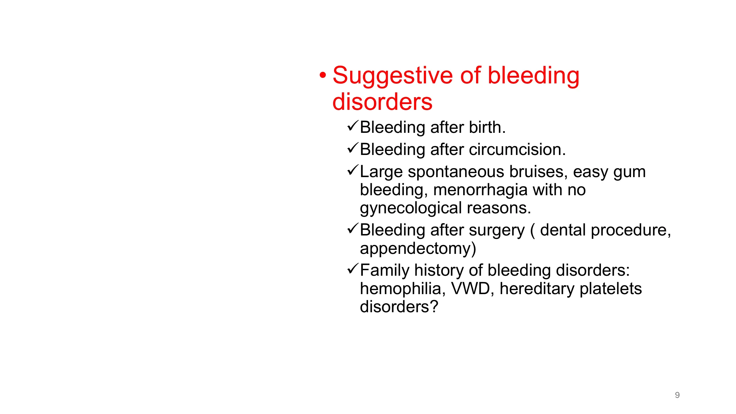
The most informative parts of a bleeding history are the hemostatic challenges the patient has already been through, because each is a natural test that the patient either passed or failed. Bleeding after birth, from the umbilical stump or into the scalp, and bleeding after circumcision, are the earliest challenges and point to a congenital disorder. Bleeding after dental extraction or after operations such as appendectomy, particularly if it required a return to theatre or a transfusion, is the challenge most adults have faced. A patient who has had surgery or extractions without trouble is unlikely to have a significant congenital disorder.
Spontaneous symptoms count when they are out of proportion: large bruises without injury, gums that bleed easily, and menorrhagia for which no gynecological cause has been found. The last is the one most often missed, and it is the subject of the case that follows.
Finally, ask about the family. Hemophilia, von Willebrand disease and the hereditary platelet disorders run in families, and a relative with a known diagnosis, or a story of a relative who bled badly after surgery, may be the most important fact in the history.
Medications, illnesses and drugs: the acquired causes
In adults, acquired bleeding disorders are commoner than congenital ones, and this slide is the checklist for them. A new medication may have caused a drug-induced thrombocytopenia. A recent viral illness, or a recent infection treated with antibiotics, may have done the same by an immune mechanism. Recent travel to an endemic area raises malaria and the viral hemorrhagic fevers.
Then the medical illnesses that disturb hemostasis. Chronic kidney disease impairs platelet function. Chronic liver disease reduces the synthesis of coagulation factors and lowers the platelet count. Cancer can cause consumption of factors and platelets. And pregnancy and the postpartum period are one of the settings in which acquired hemophilia appears, an autoantibody against factor VIII in a woman who has never bled before.
Last, and most often the answer, the drugs that are meant to do this: antiplatelet agents and anticoagulants. Ask for them by name, and include over-the-counter aspirin and the herbal preparations that patients do not count as medicines.
Read next. The focused history in Case 53 · The Bleeding Patient runs the same checklist on a real patient. It adds malabsorption, autoimmune disease, HIV and recent infection or vaccination to the list, and it asks about fever, weight loss, night sweats and bone pain, which point away from immune thrombocytopenia and towards disease of the marrow.
The hematology system review: is this bleeding, or is it marrow failure?
One question in the history changes the urgency of everything. Does the patient have fatigue or other symptoms of anemia, or fever? If mucocutaneous bleeding and petechiae are accompanied by the symptoms of anemia and by infections or fever, which are the symptoms of neutropenia, then all three cell lines are probably low. That is pancytopenia, and it means the problem is not a bleeding disorder but a failing bone marrow, from leukemia, aplastic anemia, or infiltration.
The bleeding in that patient is a symptom of the thrombocytopenia, and the thrombocytopenia is a symptom of the marrow. The blood count and film, not the coagulation screen, are the tests that matter, and the patient needs to be seen the same day. This is the link between this lecture and the acute leukemia lecture, where the same triad of tiredness, fever and bruising was the classic presentation.
See it online.
Case 1: a 14-year-old girl with fatigue and a hemoglobin of 9.7
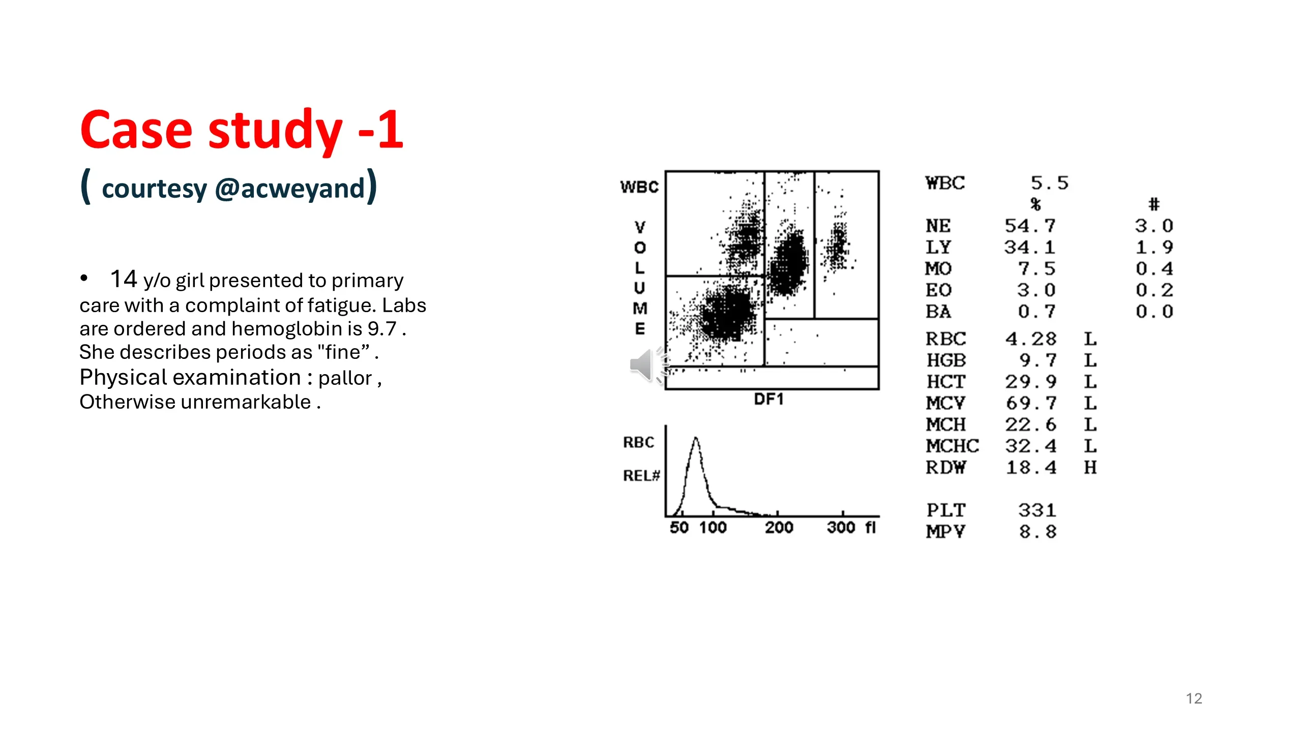
A 14-year-old girl comes to primary care tired. Her hemoglobin is 9.7 g/dL, she is pale, and the rest of the examination is normal. Asked about her periods, she says they are fine.
Read the blood count in the slide's table before reading on. The white cell count and differential are normal. The platelet count, 331, is normal. The red cell indices are all low: hemoglobin 9.7, hematocrit 29.9, mean cell volume 69.7, mean cell hemoglobin 22.6, and the red cell distribution width is high at 18.4. This is a microcytic, hypochromic anemia with marked variation in cell size, and the analyser's red cell histogram, shifted to the left of the normal range, shows the same thing graphically. The pattern is that of iron deficiency.
The next question is where the iron went. A 14-year-old girl who eats normally loses iron in one way above all others, through menstruation, and the answer "my periods are fine" should not close that line of enquiry. It should open it.
Case 1, continued: what "fine" turned out to mean
Asked for details rather than an adjective, the patient describes periods that last three to four weeks and require a change of pad or tampon between every class at school. That is not a normal period by any definition; it is heavy menstrual bleeding severe enough to explain her iron deficiency on its own.
The key point on the slide is one of the most practically important in the lecture. Many adolescent girls, and many adult women, do not know what a normal period is. They have nothing to compare their own with, their mothers and sisters may bleed the same way, and so they report heavy bleeding as normal. Never accept "fine" or "normal" as an answer about menstruation. Ask how many days, how many products, how often they are changed, whether there are clots, and whether bleeding ever soaks through clothing or bedding. The next slide gives the numbers to compare the answers against.
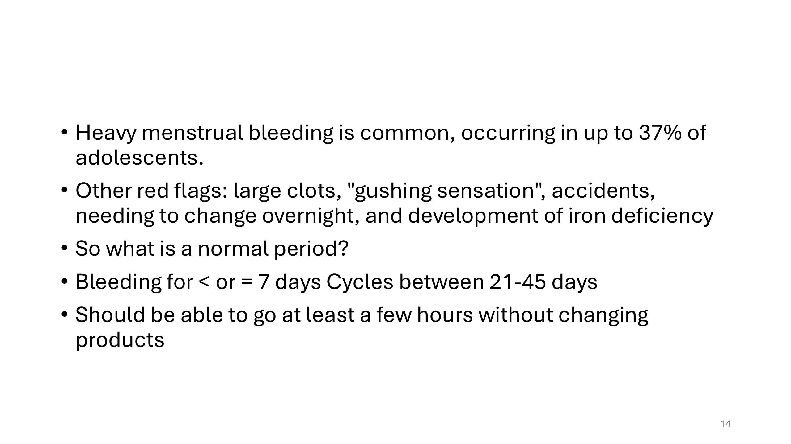
Heavy menstrual bleeding: how common, what to ask, and what is normal
Heavy menstrual bleeding is not rare. It occurs in up to 37 percent of adolescents, which means that a bleeding disorder presenting this way is easy to lose among the many girls who bleed heavily for other reasons. The red flags that mark the heavier end are on the slide: passing large clots, a sensation of gushing, accidents in which blood soaks through clothing, needing to change protection during the night, and the development of iron deficiency, which is the flag our patient raised.
Against these, the definition of a normal period: bleeding lasts seven days or fewer; cycles are between 21 and 45 days apart; and the woman can go at least a few hours without changing products. Put the case patient's history beside these criteria and every one is broken. The lesson is to have the criteria in your head so that the history can be measured against them in the consultation, not afterwards.
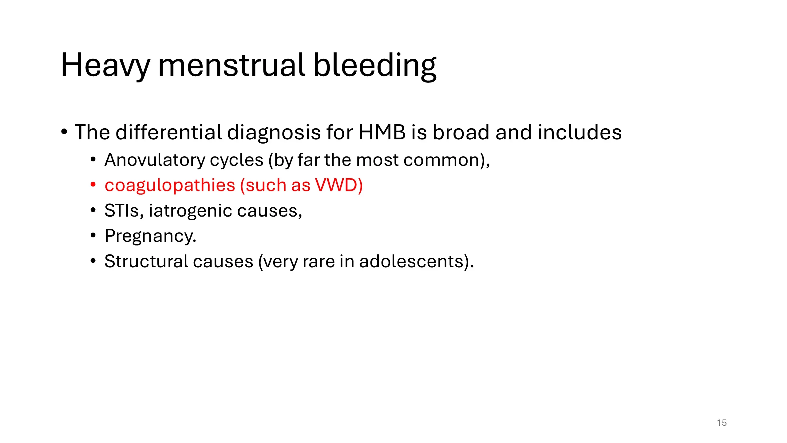
The differential diagnosis of heavy menstrual bleeding
Heavy menstrual bleeding has a broad differential, and the coagulopathies are highlighted in red because they are the part of it that this lecture is about, not because they are the commonest. In adolescents the commonest cause by far is anovulatory cycles: in the first years after menarche the hypothalamic-pituitary-ovarian axis is immature, cycles are irregular, and bleeding can be prolonged and heavy. Coagulopathies, above all von Willebrand disease, come next. Sexually transmitted infections, iatrogenic causes such as anticoagulants or hormonal treatments, and pregnancy must be considered. Structural causes, fibroids and polyps, which dominate the differential in older women, are very rare in adolescents.
The practical point is that heavy bleeding from the first period onwards, in a girl with other bleeding symptoms or a family history, should not be attributed to anovulation without asking the bleeding questions. That is what the next slide does.
Read next. One of the teaching points of Case 53 · The Bleeding Patient is this slide turned into a rule: ask every woman with heavy periods whether they have been heavy since menarche, because von Willebrand disease is badly under-diagnosed in women.
So what next? Ask the bleeding questions and score them
Having established that the periods are abnormal, the task is to find out whether the heavy bleeding is an isolated gynecological problem or one symptom of a systemic bleeding disorder. The history now focuses on bleeding elsewhere: nosebleeds, bleeding from the mouth and gums, bruising out of proportion to injury, and what happened after any surgery or dental extraction. It takes in the medical history, for the acquired causes on slide 9, and the family history, for the inherited ones.
And it is done with the bleeding assessment tool, not from memory. The BAT walks through every bleeding symptom from slide 5, scores each, and produces a total. An abnormal score is the justification for the laboratory testing that follows; a normal one makes a significant bleeding disorder unlikely and points back toward the gynecological causes.
The decision: this patient needs testing
The case reaches its hinge. The bleeding history is significant: heavy menstrual bleeding from menarche, severe enough to cause iron deficiency anemia, in a pattern that fits primary hemostasis. The bleeding assessment score is abnormal. The clinical assessment, the first of the two steps on slide 6, is complete, and its conclusion is that laboratory investigation is warranted.
The question the slide asks, how do we investigate bleeding disorders, is the second step, and the rest of the lecture answers it. The answer is organised, like everything else, along the primary-versus-secondary axis.
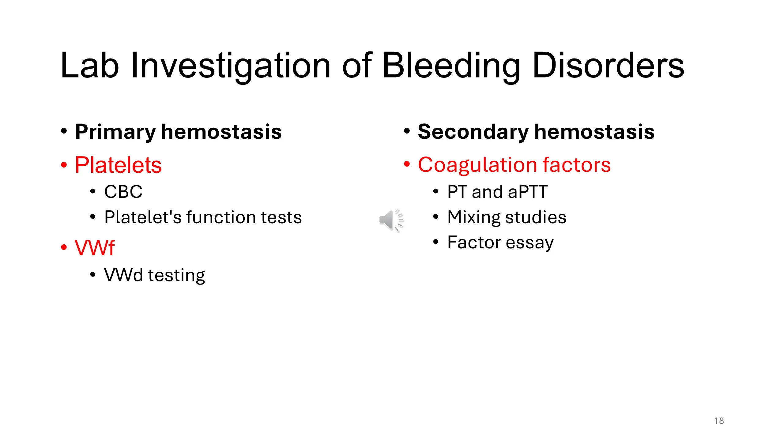
The laboratory investigation, sorted by axis
The tests for a bleeding disorder fall into two columns that correspond exactly to the two kinds of hemostasis on slide 2, and the history tells you which column to start in.
For primary hemostasis: the platelet count, which comes with the complete blood count; platelet function tests, if the count is normal but a qualitative platelet defect is suspected; and testing for von Willebrand disease, which means measuring von Willebrand factor antigen and activity together with factor VIII.
For secondary hemostasis: the screening tests, the prothrombin time and the activated partial thromboplastin time, which between them cover the whole coagulation cascade; mixing studies, when a screening test is prolonged, to decide whether the cause is a missing factor or an inhibitor; and assays of the individual factors to name the deficiency.
A patient with mucocutaneous bleeding is investigated down the left column first; a patient with joint and muscle bleeding down the right. Sending every test on every patient wastes resources and produces incidental abnormalities that then have to be explained.
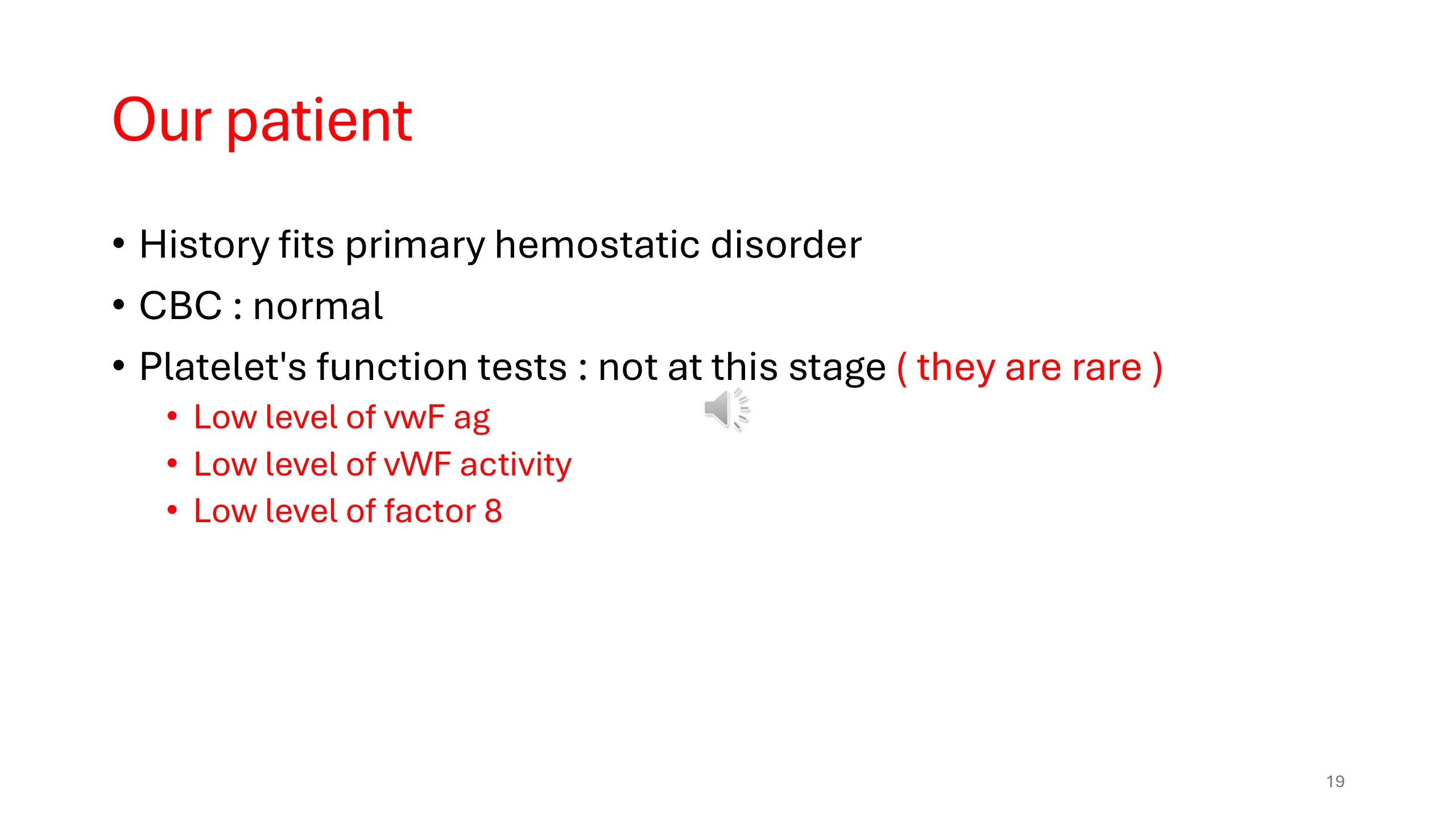
Our patient: the tests, and the answer
The history fits a disorder of primary hemostasis, so the investigation follows the left column of the previous slide. The complete blood count, apart from the iron deficiency anemia, is normal: in particular the platelet count is normal, so this is not a quantitative platelet disorder. Platelet function tests are not performed at this stage; the hereditary platelet function disorders are rare, and the commoner diagnosis should be sought first.
That diagnosis is found in the von Willebrand testing. The patient has a low level of von Willebrand factor antigen, so there is too little of the protein; a low von Willebrand factor activity, in proportion, so what there is works normally; and a low factor VIII. The last finding follows from the second function of von Willebrand factor on slide 2: it carries factor VIII in the plasma, and when the carrier is scarce the passenger is cleared faster. Low antigen with proportionately low activity is the pattern of a quantitative deficiency, which the classification on the next slide calls type 1 von Willebrand disease.
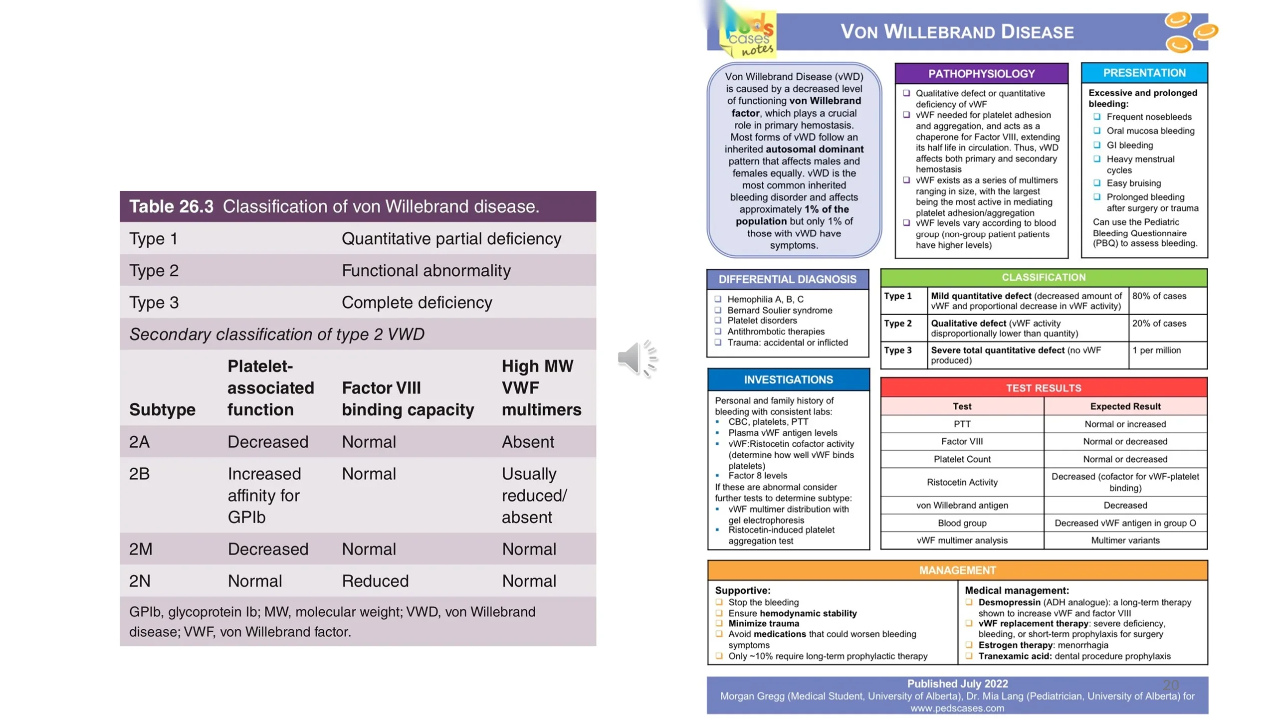
Classifying von Willebrand disease: the table and the summary card
Table 26.3 gives the classification in three lines. Type 1 is a partial quantitative deficiency: too little factor, but what there is works. Type 2 is a functional abnormality: the factor is present but defective. Type 3 is complete deficiency, with virtually no factor at all. Type 2 is subdivided by which function is defective, and the lower table lists the four subtypes by three properties: how well the factor binds platelets, how well it binds factor VIII, and whether the largest, most active multimers are present. Slide 21 turns those four rows into something memorable.
The PedsCases summary card fills in the clinical picture. Von Willebrand disease is the commonest inherited bleeding disorder, affecting about 1 percent of the population, though only about 1 percent of those have symptoms. It is usually autosomal dominant and affects males and females equally, which distinguishes it from hemophilia. Type 1 accounts for about 80 percent of cases and type 2 for about 20 percent; type 3 is rare, about one per million. Factor levels vary with blood group and are lower in group O, which has to be remembered when interpreting a borderline result.
The investigations are the ones our patient had: von Willebrand factor antigen, the ristocetin cofactor activity that measures platelet binding, and factor VIII, with multimer analysis and ristocetin-induced platelet aggregation to define the subtype when these are abnormal. Note that the PTT may be normal, so a normal screen does not exclude the disease. Management is on the card as well: desmopressin, which releases stored factor from the endothelium and works in most type 1 patients; von Willebrand factor concentrate for severe deficiency, bleeding or surgery; tranexamic acid for mucosal bleeding and dental work; and hormonal treatment for menorrhagia, which is how the case patient's presenting complaint would be controlled. Only about one in ten patients needs long-term prophylaxis.
Read next. The investigations in Case 53 · The Bleeding Patient say when to send these tests: mucocutaneous bleeding with a normal platelet count, especially with lifelong heavy periods, calls for von Willebrand factor antigen, von Willebrand factor activity and factor VIII.
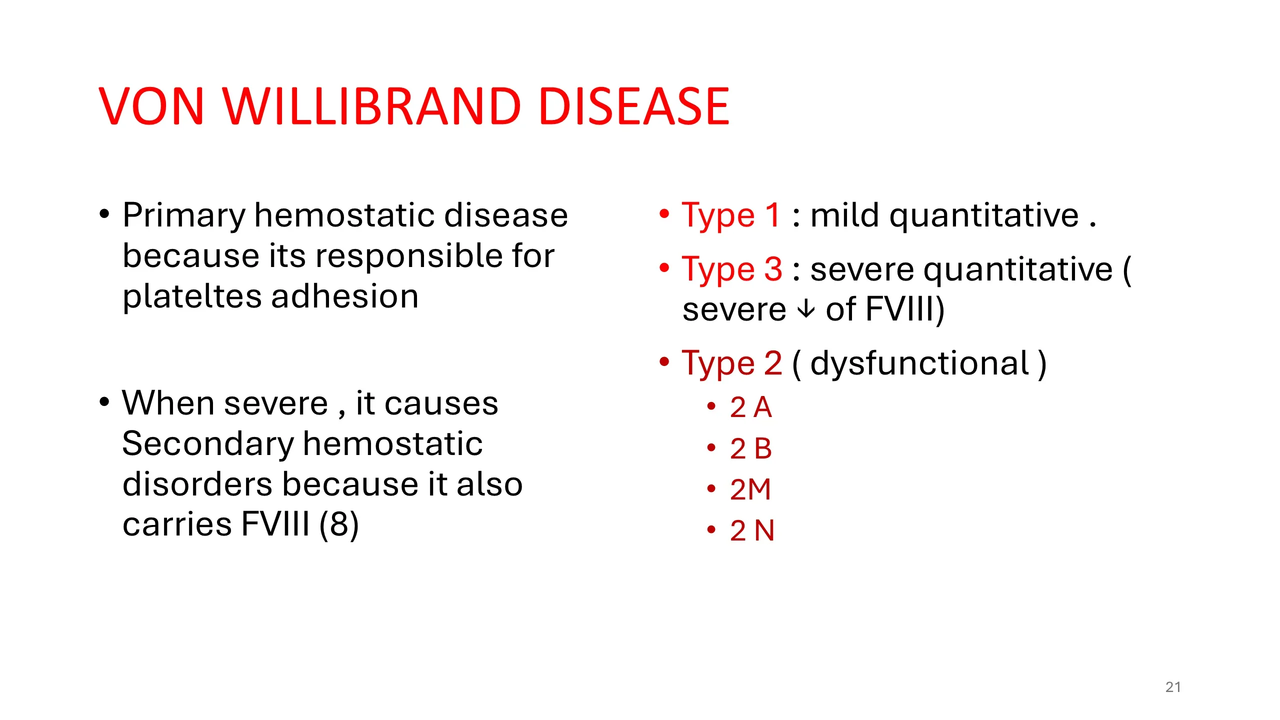
Von Willebrand disease: a primary disorder that can become a secondary one
This slide restates the point of the circled molecule on slide 2 with the classification attached. Von Willebrand disease is a disease of primary hemostasis, because von Willebrand factor is what allows platelets to adhere to the injured vessel wall; hence the mucocutaneous pattern of bleeding, the nosebleeds, the bruising and the menorrhagia. But because the factor also carries factor VIII, a severe deficiency drags factor VIII down with it, and the patient then also has a disorder of secondary hemostasis, with the joint and muscle bleeding of hemophilia.
The types map onto this. Type 1 is a mild quantitative deficiency, and factor VIII is modestly reduced, as in our patient. Type 3 is a severe quantitative deficiency, with a severe reduction of factor VIII, and these patients bleed like hemophiliacs. Type 2 is the dysfunctional group, with its four subtypes 2A, 2B, 2M and 2N, and the next slide gives a way to remember what each letter means.
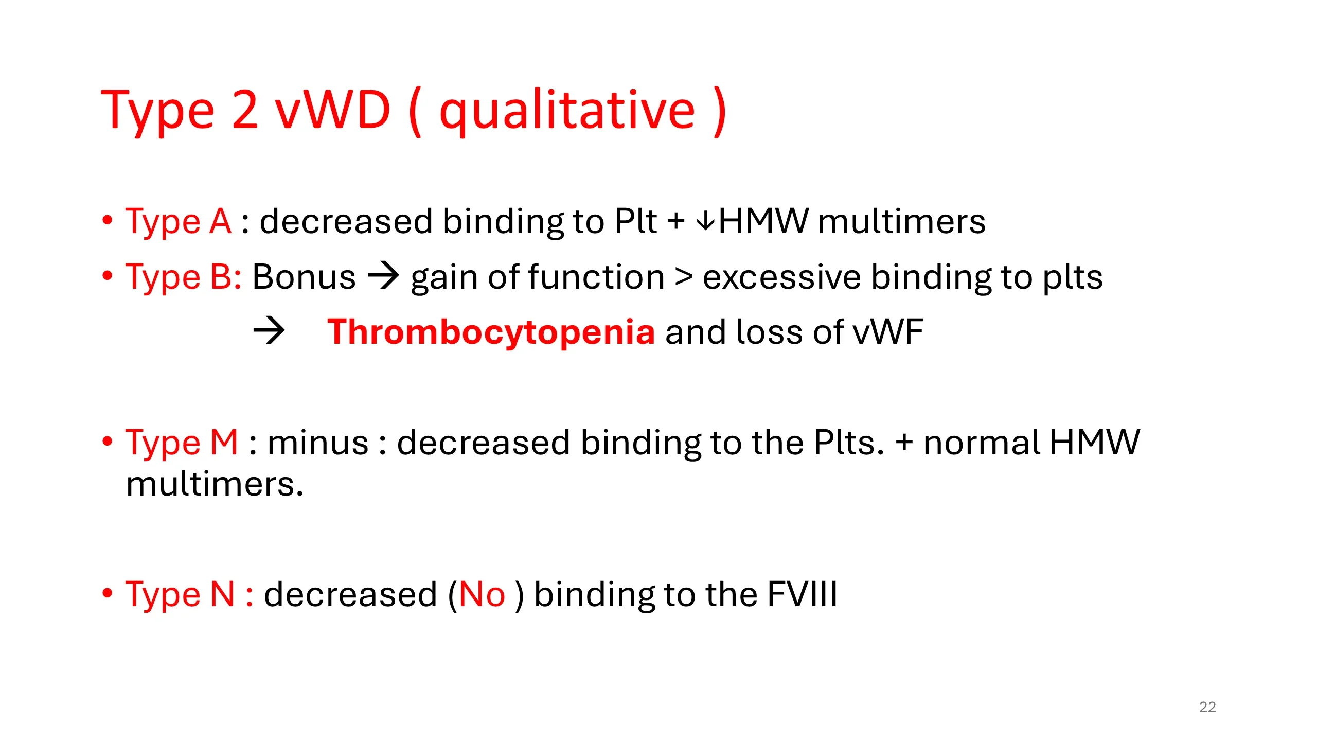
Type 2 von Willebrand disease: four letters, four defects
The four qualitative subtypes are hard to keep apart from the table alone, so the slide attaches a word to each letter.
2A: decreased binding to platelets, because the large, high-molecular-weight multimers that do most of the platelet binding are absent. 2B: think bonus. This is a gain-of-function mutation: the factor binds platelets too avidly, even in the circulation where it should not, and the platelet-factor complexes are cleared. The paradoxical result is a low platelet count and a loss of the largest multimers. 2M: think minus. Platelet binding is decreased, as in 2A, but the multimers are normal; the defect is in the binding site itself. 2N: think no binding to factor VIII. Platelet function is normal, but the factor cannot carry factor VIII, so the patient has a low factor VIII and looks exactly like a mild hemophilia A, except that the inheritance is autosomal and affects women.
The subtypes matter because they change treatment: what helps one can harm another, and the multimer and binding studies on slide 19 are how the subtype is established before treatment is chosen.
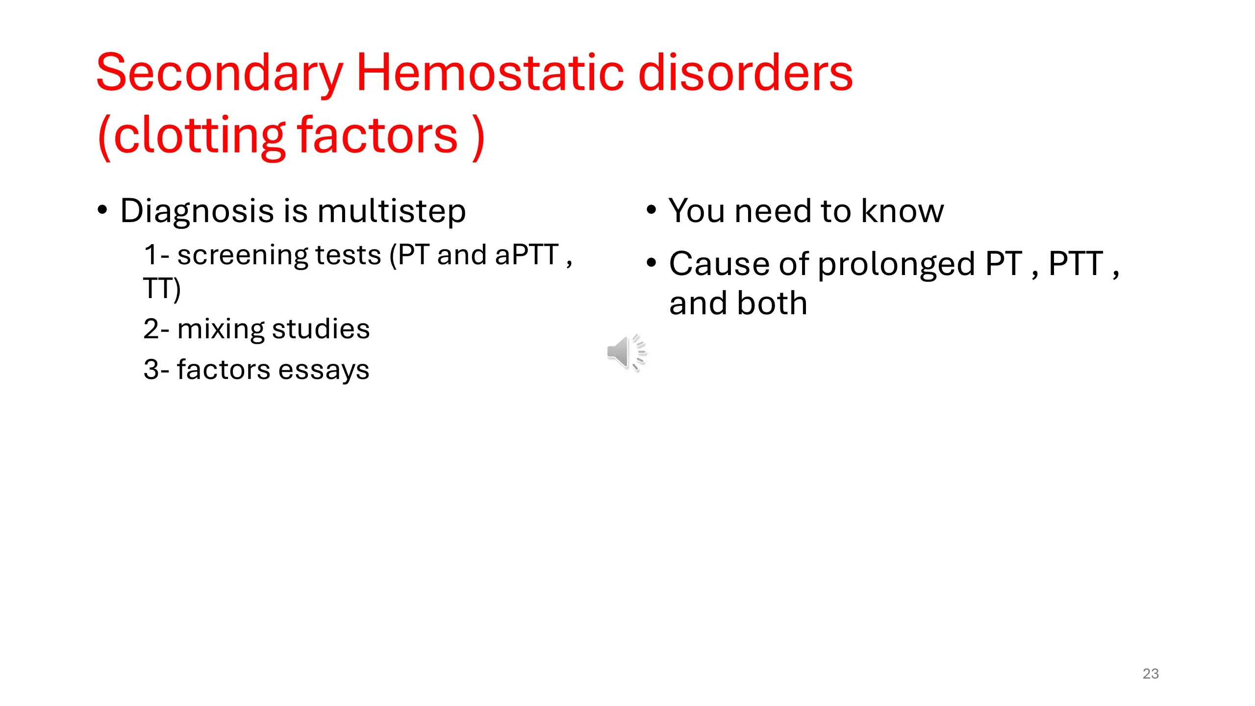
Secondary hemostatic disorders: a three-step diagnosis
The lecture now crosses to the right-hand column of slide 17, the disorders of the coagulation factors. Their diagnosis is multistep, and the steps are always taken in the same order. First, the screening tests: the PT, the aPTT and the thrombin time, which tell you that something in the cascade is deficient or inhibited and roughly where. Second, the mixing study, which tells you whether the prolonged test is caused by a missing factor or by an inhibitor. Third, the factor assays, which name the missing factor and measure how much is missing.
To use the first step you need to know one thing thoroughly, and the slide says so: the causes of a prolonged PT, of a prolonged PTT, and of both together. The slides that follow are built to teach exactly that.
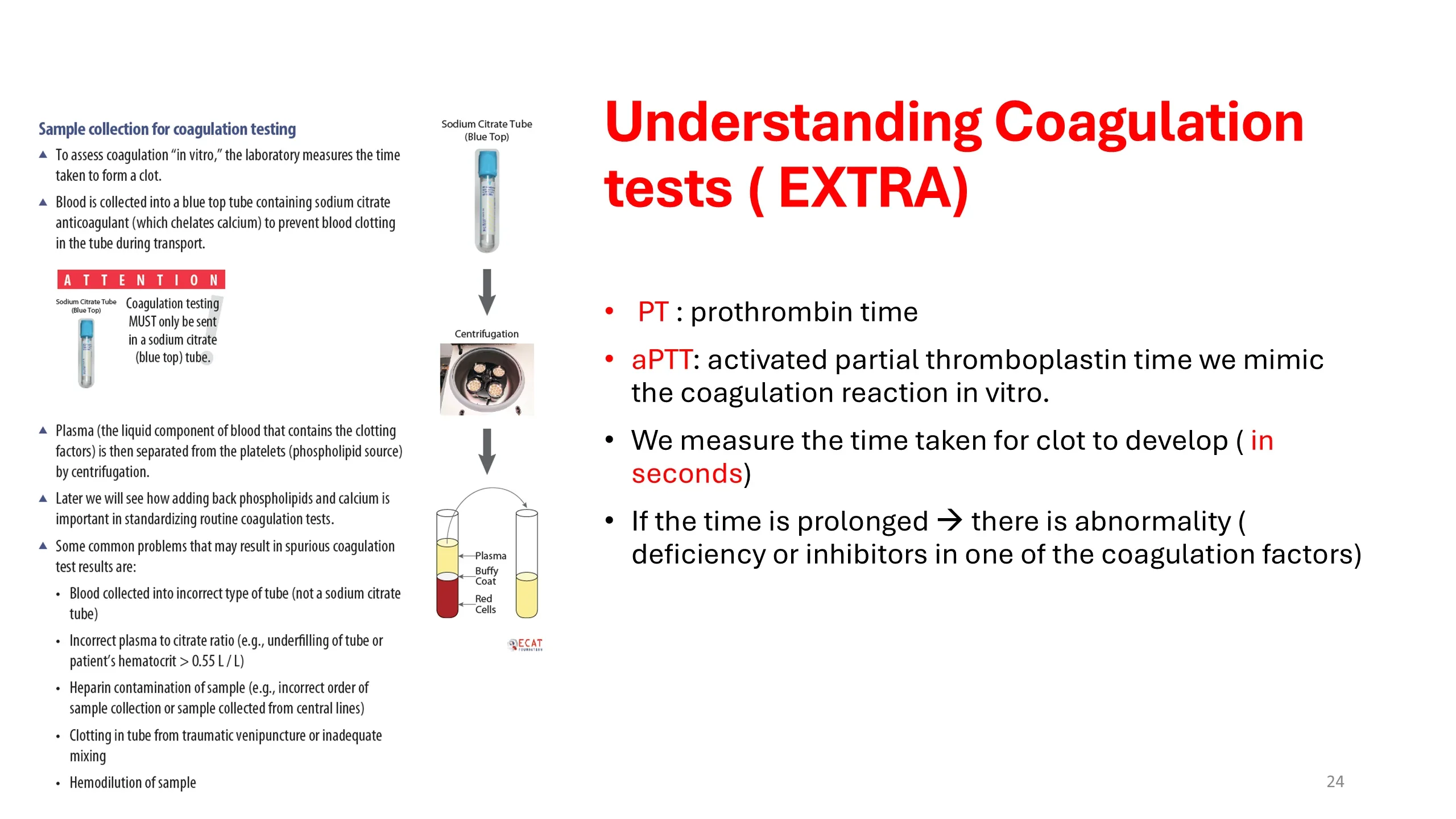
Understanding the coagulation tests: what the laboratory actually does
This slide is marked as extra material, but it explains why coagulation results can be wrong before they reach you, which every clinician should understand. A coagulation test measures, in seconds, how long a sample of plasma takes to clot when the reaction is started in a tube. The PT and the aPTT differ only in what is used to start it.
The sample must be collected into a blue-top tube containing sodium citrate. Citrate chelates calcium, and without calcium the cascade cannot run, so the blood stays liquid during transport. In the laboratory the tube is centrifuged, the plasma with its clotting factors is separated from the cells and platelets, and the test is performed by adding back phospholipid, to replace the platelet surface, and calcium, to release the brake. This is the only tube that works, and it must be correctly filled: the volume of citrate is calculated for a fixed volume of blood, so an underfilled tube, or a patient with a hematocrit above 0.55 whose blood contains less plasma, leaves too much citrate and falsely prolongs the times. Other spurious results come from heparin contamination, usually a sample drawn from a central line or collected in the wrong order, from a sample that clotted in the tube after a traumatic venepuncture, and from hemodilution.
The rule that follows: an unexpected prolonged result on a well patient is repeated on a fresh, correctly filled peripheral sample before anything else is done.
See it online.
- Fibrin strands and platelets on a film from a partly clotted sample Wikimedia Commons · CC BY-SA 4.0 · Spicy
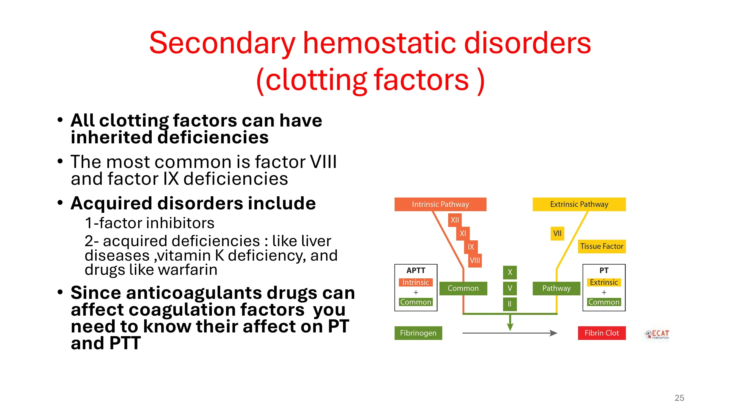
Disorders of the coagulation factors: inherited and acquired
Any of the coagulation factors can be deficient from birth, but two account for almost all inherited cases: factor VIII deficiency, hemophilia A, and factor IX deficiency, hemophilia B. Both are X-linked, both present with the deep bleeding of secondary hemostasis, and both prolong the aPTT while leaving the PT normal, because both factors sit in the intrinsic pathway, coloured on the cascade diagram.
The acquired disorders fall into two groups. Inhibitors are antibodies against a factor, most often factor VIII, arising in the settings listed on slide 9, the postpartum period, autoimmune disease, malignancy and old age. Acquired deficiencies arise when the factors are not made or are consumed: liver disease, vitamin K deficiency, and drugs such as warfarin that block vitamin K.
The last bullet is a demand rather than a fact. Anticoagulant drugs act on coagulation factors, so they change the PT and the aPTT, and you must know how each drug affects each test. A prolonged PT in a patient on warfarin is the intended effect; the same result in a patient on no drug is a diagnosis waiting to be made.
Read next. Case 53 · The Bleeding Patient ends with a table of the other bleeding disorders: von Willebrand disease, the hemophilias, disseminated intravascular coagulation, vitamin K deficiency and liver disease, and bleeding on anticoagulants, with the reversal agent for each drug.
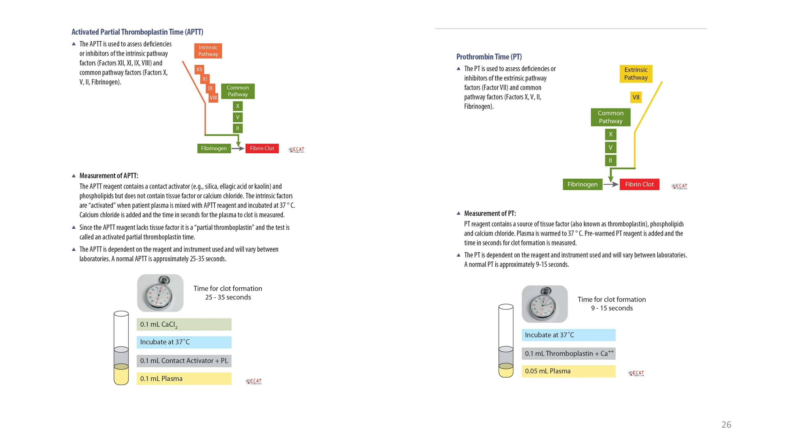
The aPTT and the PT: how each is measured and what each covers
The two screening tests are set side by side with their cascade diagrams and their reaction tubes, and the differences between them are entirely in the reagent.
The activated partial thromboplastin time covers the intrinsic pathway, factors XII, XI, IX and VIII, and the common pathway, X, V, II and fibrinogen. The reagent contains a contact activator, such as silica, kaolin or ellagic acid, and phospholipid, but no tissue factor and no calcium. Plasma and reagent are incubated at 37 degrees so that the contact factors are activated, calcium chloride is then added, and the clock runs until a clot forms. Because the reagent lacks tissue factor it is only a partial thromboplastin, which is where the name comes from. A normal aPTT is about 25 to 35 seconds, though the exact range depends on the reagent and instrument and varies between laboratories.
The prothrombin time covers the extrinsic pathway, factor VII, and the same common pathway. Its reagent contains tissue factor, the thromboplastin, together with phospholipid and calcium, all in one, so it is simply added to warmed plasma and the clot is timed. A normal PT is about 9 to 15 seconds, again laboratory-dependent. Notice that the PT is the shorter test: tissue factor drives coagulation faster than contact activation, which is the same lesson the cell-based model taught in the physiology lecture.
Because both tests share the common pathway, a defect there prolongs both; a defect confined to one arm prolongs only its own test. That is the logic of the next three slides.
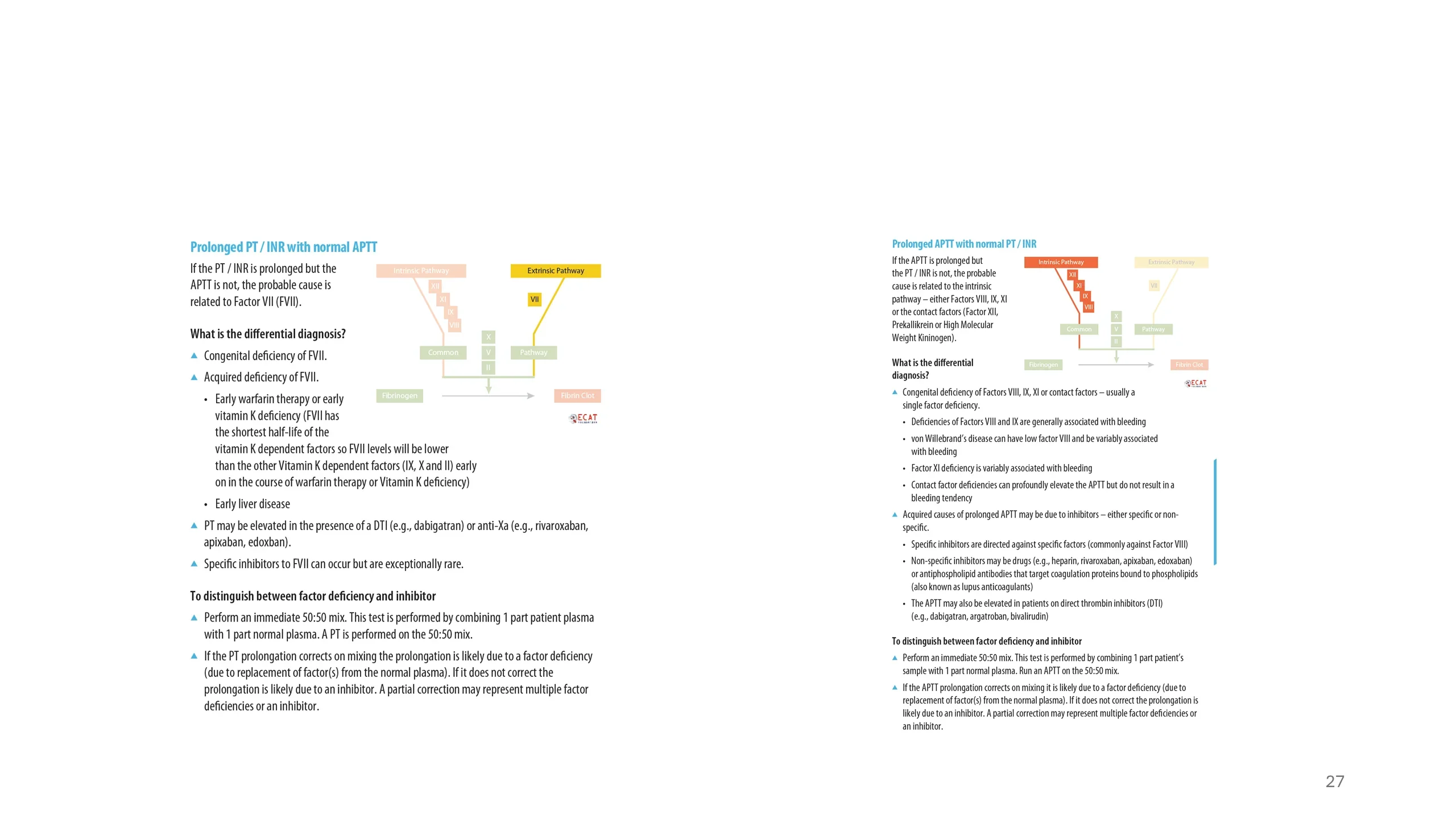
One test prolonged, the other normal: the two isolated patterns
A prolonged PT with a normal aPTT points to the only factor the PT tests that the aPTT does not: factor VII. Congenital factor VII deficiency exists but is rare. Far commoner is an early acquired deficiency: factor VII has the shortest half-life of the vitamin K-dependent factors, so in the first days of warfarin, or of vitamin K deficiency, or of liver disease, it falls before factors II, IX and X do and the PT lengthens alone. The direct oral anticoagulants can also raise the PT. A specific inhibitor of factor VII is exceptionally rare.
A prolonged aPTT with a normal PT points to the intrinsic pathway, and here the list is longer and the distinctions matter. Among the congenital deficiencies, factors VIII and IX cause bleeding, the hemophilias; von Willebrand disease can lower factor VIII and prolong the aPTT; factor XI deficiency is variably associated with bleeding; and deficiencies of the contact factors, XII, prekallikrein and high-molecular-weight kininogen, can prolong the aPTT dramatically yet cause no bleeding at all, for the reason the cell-based model gave. Among the acquired causes are inhibitors: specific ones, usually against factor VIII, and non-specific ones, which include heparin, the direct oral anticoagulants, the direct thrombin inhibitors, and the antiphospholipid antibodies known as lupus anticoagulants, which prolong the test in the tube but predispose to thrombosis in the patient.
For either pattern the next step is the same. Perform an immediate 50:50 mix of patient plasma with normal plasma and repeat the test. If the time corrects, the patient was missing a factor and the normal plasma supplied it. If it does not correct, something in the patient's plasma is inhibiting the reaction, and it inhibits the normal plasma too. A partial correction suggests either several deficiencies or an inhibitor.
Read next. The coagulation screen section of the casebook’s Interpretation chapter points to the table of these patterns in Case 53 and adds two traps: a lupus anticoagulant prolongs the aPTT in a patient who clots rather than bleeds, and in liver disease the INR does not measure the risk of bleeding.
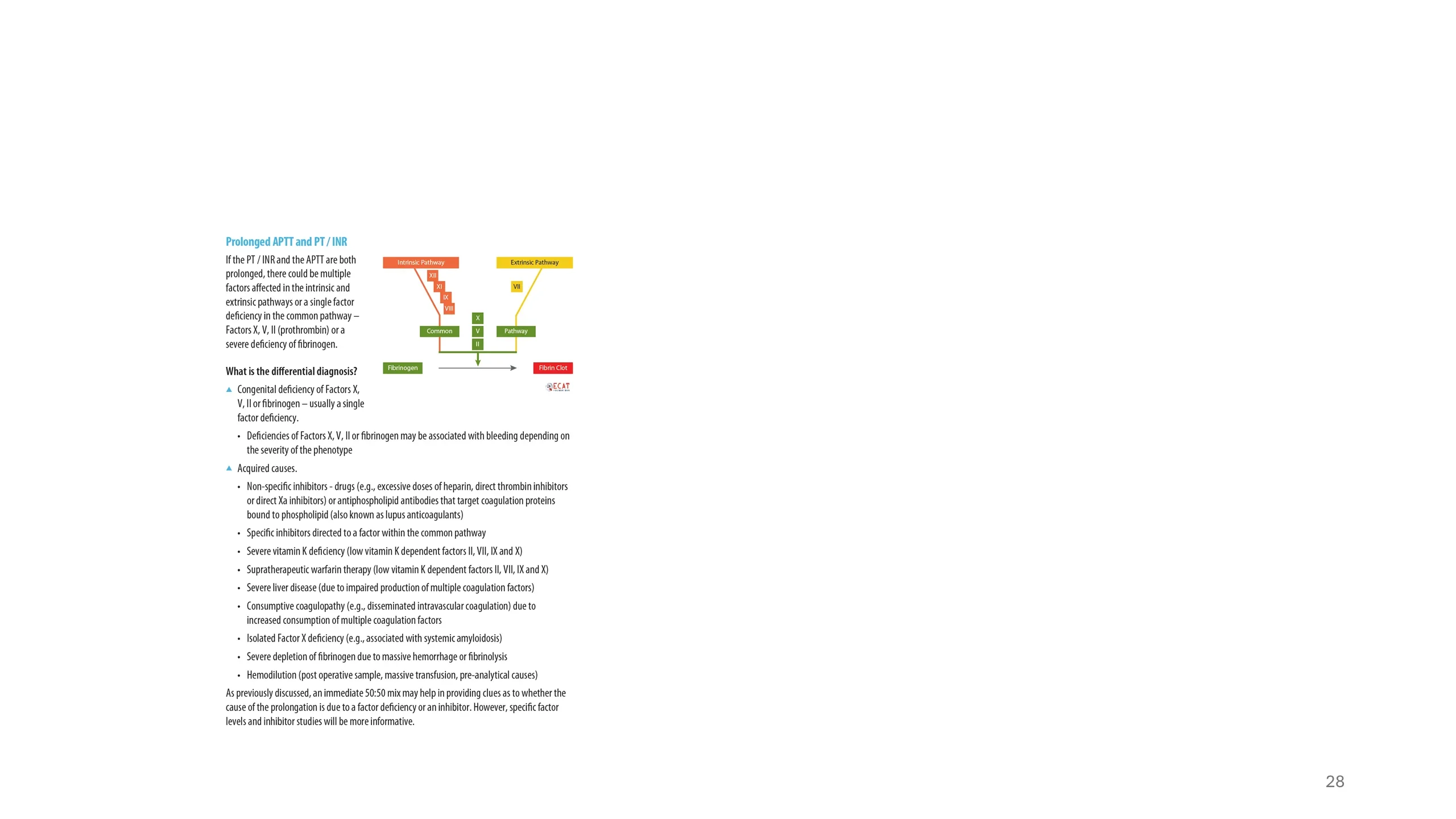
Both tests prolonged: the common pathway, or many factors at once
When the PT and the aPTT are both prolonged, either a single factor in the common pathway is deficient, X, V, II or fibrinogen, or several factors across both arms are affected together. The first is rare; the second is common, and its causes are the everyday business of hospital medicine.
The congenital causes are single deficiencies of X, V, II or fibrinogen, with bleeding that depends on the severity. The acquired list is where to concentrate. Severe vitamin K deficiency and supratherapeutic warfarin lower all four vitamin K-dependent factors, II, VII, IX and X, and so reach both arms. Severe liver disease impairs the synthesis of almost every factor. Disseminated intravascular coagulation consumes factors faster than they can be made. Massive hemorrhage and fibrinolysis deplete fibrinogen, and hemodilution, after massive transfusion or from a sample drawn near an infusion, dilutes everything. Excess heparin, the direct thrombin and factor Xa inhibitors, and lupus anticoagulants can prolong both tests, as can a specific inhibitor against a common pathway factor. One association worth remembering is isolated factor X deficiency in systemic amyloidosis, where the factor is adsorbed onto the amyloid.
A 50:50 mix helps here as elsewhere, but when many factors may be involved the mixing study gives only a clue; specific factor assays and inhibitor studies are what settle the diagnosis.
Read next. Case 51 · Acquired Haemolytic Anaemia and the Direct Antiglobulin Test in the casebook works through disseminated intravascular coagulation, one of the causes on this slide: both times prolonged, fibrinogen falling and D-dimer high. It sets it beside TTP and HUS, where the clotting screen is normal.
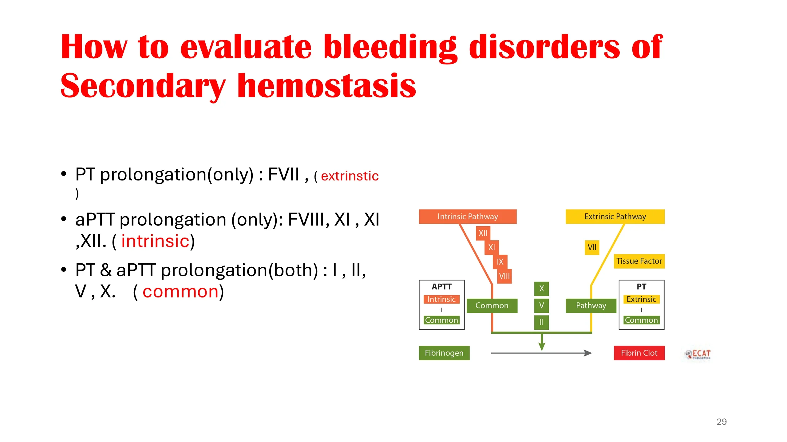
The map in three lines
This slide compresses the previous three into the form you should carry in your head, and the cascade diagram beside it is coloured to match.
PT prolonged alone: factor VII, the extrinsic pathway. aPTT prolonged alone: factors VIII, IX, XI and XII, the intrinsic pathway. Both prolonged: factors I, II, V and X, the common pathway, or multiple factors affected at once.
Read the diagram from the bottom up. The common pathway is shared, so anything wrong there shows in both tests. Above the junction the two arms separate, and each has its own test. When you receive a coagulation screen, place the abnormal result on this map first, then ask which of the causes on slides 26 and 27 fits the patient in front of you.
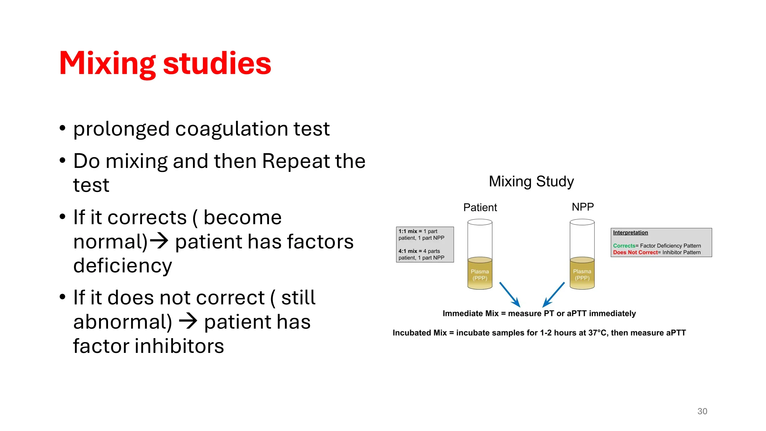
Mixing studies: deficiency or inhibitor?
A prolonged PT or aPTT tells you the cascade is not working. It does not tell you why, and the two possible reasons, a factor that is missing and a factor that is present but inhibited, have different causes and opposite treatments. The mixing study separates them, and its logic is simple enough to reason out rather than memorise.
Take the patient's plasma and mix it with normal pooled plasma, most often one part to one part, sometimes four parts patient to one part normal to make the test more sensitive to a weak inhibitor. Repeat the prolonged test on the mixture. Normal plasma contains every factor at a normal level, so if the patient's problem was a deficiency, the mixture now contains at least half the normal amount of the missing factor, which is enough to clot in normal time: the test corrects. If the patient's problem was an inhibitor, the inhibitor is still there and attacks the factors in the normal plasma as well: the test does not correct.
The diagram adds a refinement. Some inhibitors, notably antibodies against factor VIII, act slowly. An immediate mix corrects, and only after the mixture has been incubated for one to two hours at 37 degrees does the aPTT lengthen again. So the mixing study is read twice, immediately and after incubation, and a result that corrects at first and then fails is the signature of a time-dependent inhibitor.
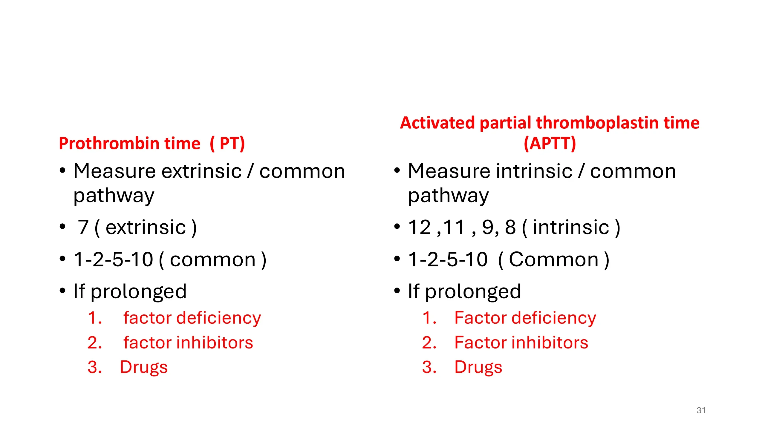
PT and aPTT, side by side
The two tests are summarised in parallel as a final revision aid. The prothrombin time measures the extrinsic and common pathways: factor 7, then factors 1, 2, 5 and 10. The activated partial thromboplastin time measures the intrinsic and common pathways: factors 12, 11, 9 and 8, then the same 1, 2, 5 and 10. The overlap is the common pathway; the difference is the arm.
When either is prolonged there are three possible explanations, and they are the same three for both tests: a factor deficiency, congenital or acquired; a factor inhibitor, specific or non-specific; or a drug, which is the commonest explanation of all in hospital patients. The mixing study separates the first from the second; the drug chart, read before any test is ordered, identifies the third.
A video to watch
The slide links to a video lecture on the approach to bleeding of unknown cause. It is worth watching after this lecture rather than before: it applies the same sequence, history and bleeding score, screening tests, mixing studies and specific assays, to patients whose diagnosis is not obvious, and it shows how often the answer lies in the history rather than the laboratory.
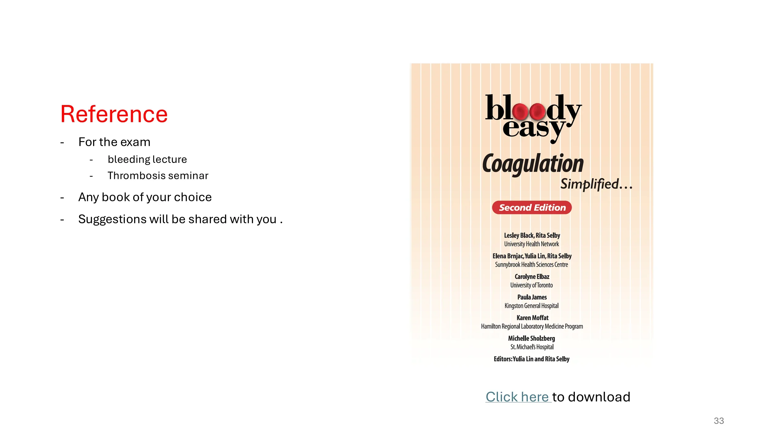
Reference and reading
For the examination, the material to master is this lecture on bleeding and the seminar on thrombosis, and any textbook of your choice; reading suggestions will be shared.
The book shown, Bloody Easy Coagulation Simplified, second edition, edited by Yulia Lin and Rita Selby with colleagues from the Toronto teaching hospitals, is recommended because it does what its title says. It covers the physiology, the laboratory tests and their interpretation, and the common bleeding and clotting disorders in a short, clinically organised format, and it is available to download free of charge from the link on the slide.