The Clinical Approach to Anemia
From pathophysiology to diagnosis — a three-part guide.
These slides are an outline only. Read the accompanying material with them: the notes under each slide below, and the Hematology Handbook. Then work through the Internal Medicine Casebook and the Hematology Question Bank.
Approach & Classification
What this lecture is about
These slides are an outline only. Read the accompanying material with them: the notes under each slide on this page, and the Hematology Handbook. Then work through the Internal Medicine Casebook and the Hematology Question Bank.
This is the first of three lectures on anemia. Before we can talk about hemolysis, thalassemia or sickle cell disease, we need a way of thinking about any patient whose hemoglobin is low. That is what Part 1 builds: a method, not a list of diseases.
The method has three moves. First, decide whether the patient is actually anemic for their physiology, not for a number printed on a lab form. Second, ask whether the bone marrow is trying to fix the problem (the kinetic question). Third, look at the size of the red cells (the morphologic question). Those two questions, taken together, shrink a very long differential diagnosis into a short one that the laboratory can then confirm.
Keep one idea in mind throughout: anemia is almost never a diagnosis in itself. It is a sign that something else is wrong, and the job of the clinician is to find that something.
Why the cutoff for anemia is not fixed
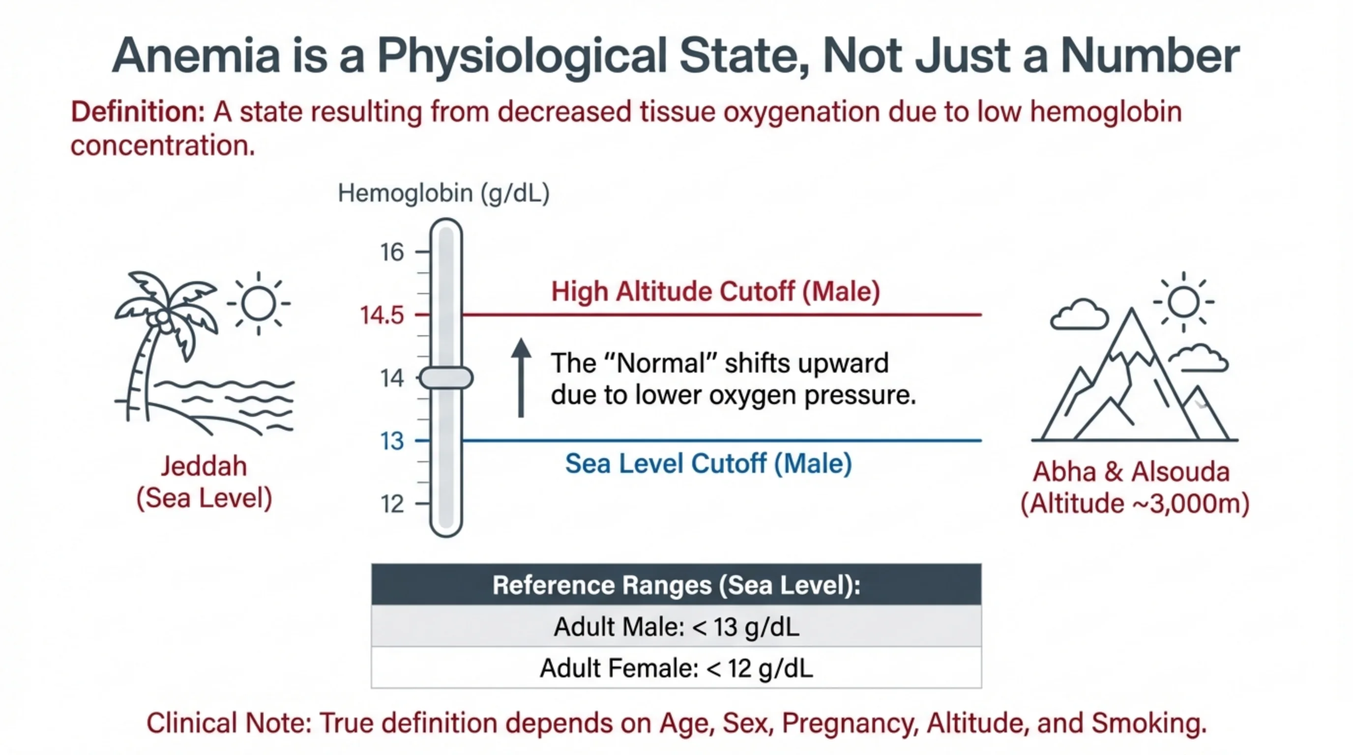
Anemia is defined by what hemoglobin does, not by what it measures. Hemoglobin carries oxygen to the tissues, so anemia is a state in which the tissues receive less oxygen than they need because there is too little hemoglobin to carry it. The number on the CBC is only a proxy for that state.
This matters because the "normal" range shifts with the body's oxygen needs. At sea level, as in Jeddah, the air holds plenty of oxygen and a man with a hemoglobin below 13 g/dL is anemic. In the highlands of ʿAsīr, the air pressure is lower, so each breath delivers less oxygen. The body compensates by making more red cells, and the whole normal range moves upward. The same man living at altitude may be anemic with a hemoglobin that would be perfectly normal in Jeddah; the slide shows the male cutoff rising to 14.5 g/dL. This is why a hospital in Abha cannot simply copy sea-level reference ranges.
The same principle explains the other adjustments. Women have a lower cutoff than men, and pregnant women lower still because plasma volume expands and dilutes the red cells. Smokers run higher hemoglobin because carbon monoxide occupies part of it. Age matters too. So when you read a hemoglobin, always ask: normal for whom, and where?
Think about probability before you think about tests
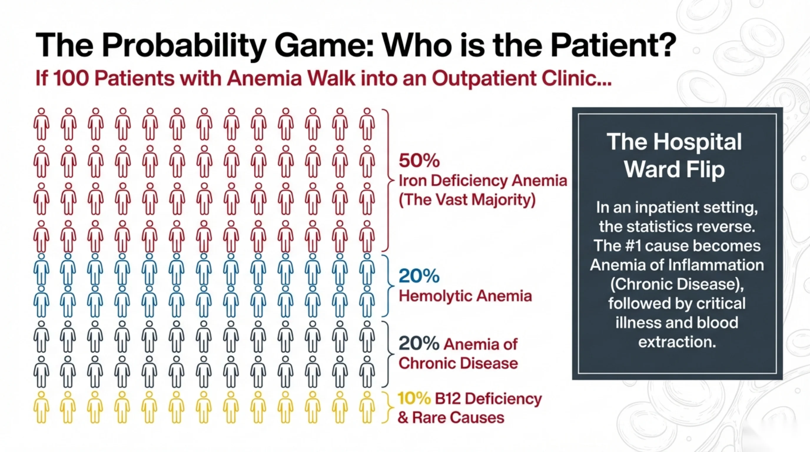
Good diagnosticians do not treat every anemic patient as an equal mystery. They start from what is likely and then look for evidence against it. This slide gives you a mental picture of the likely causes, and the key lesson is that the picture depends on where you meet the patient.
In an outpatient clinic, iron deficiency is by far the most common cause. Roughly half of all anemic patients walking through the door will turn out to be iron deficient, usually because they are losing blood slowly (heavy periods, a bleeding gut) or not absorbing enough iron. Hemolytic anemia and anemia of chronic disease each account for a smaller share, and B12 deficiency and the rare causes make up the remainder.
On a hospital ward, the picture flips. Inpatients are sick with something else, and that illness drives inflammation, which blocks the use of iron (you will see how on the hepcidin slide). Anemia of inflammation becomes the leading cause, followed by the effects of critical illness itself and, quite simply, the volume of blood we draw for tests every day.
The practical point: an anemic woman in clinic should make you think of blood loss and iron first; an anemic patient on the ward should make you think of their underlying illness first.
Red cells are made on a production line
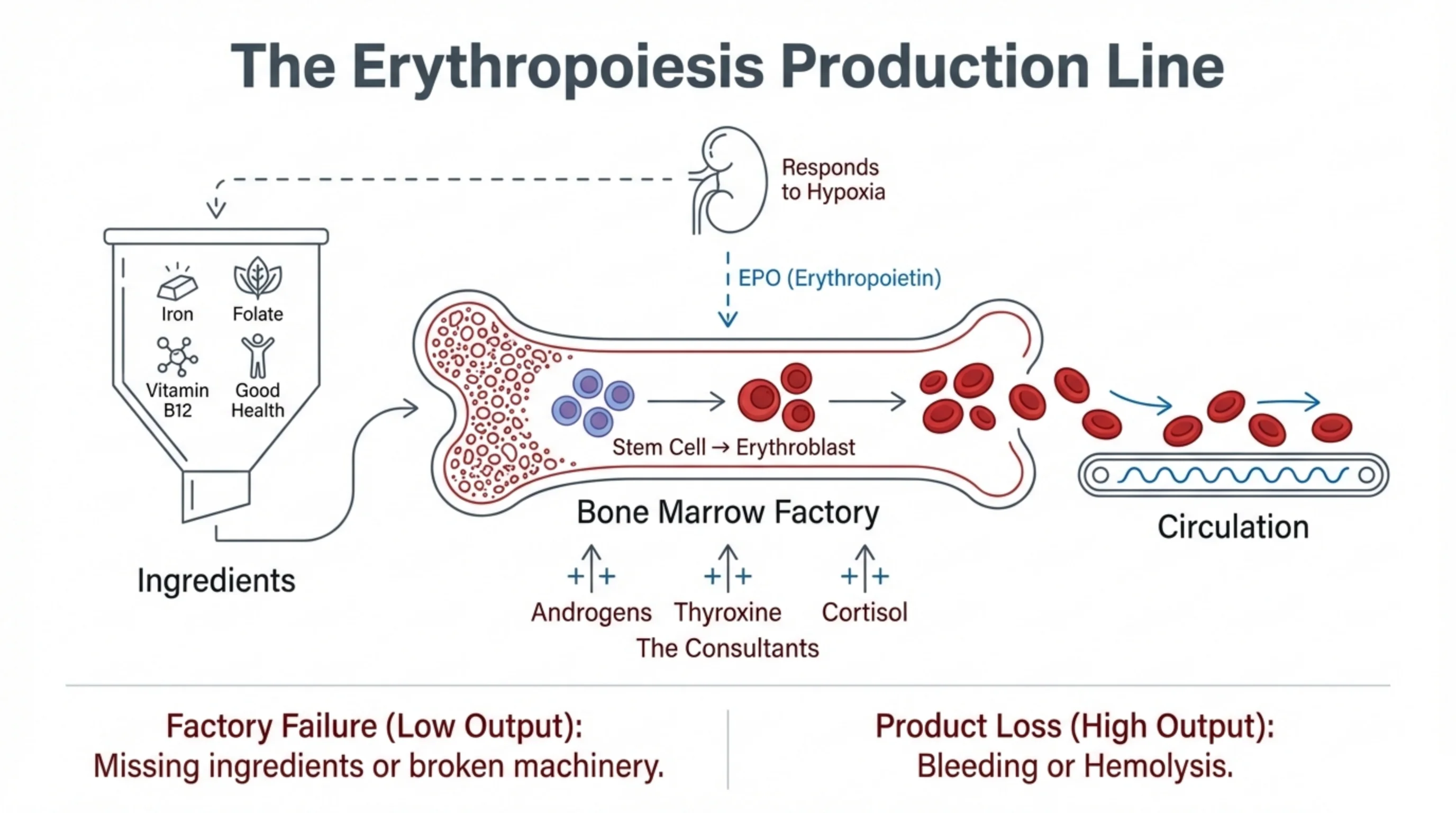
The easiest way to understand anemia is to think of red cell production as a factory. A factory needs raw materials, machinery and a manager who decides how fast to run.
The raw materials are iron (for heme), folate and vitamin B12 (for the DNA that dividing cells need), and general good health, because a body under stress diverts its resources elsewhere. The machinery is the bone marrow, where a stem cell matures step by step into an erythroblast and finally into a red cell that is released into the circulation. The manager is the kidney. When the kidney senses low oxygen (hypoxia), it releases erythropoietin, or EPO, which tells the marrow to speed up. A few hormones act as consultants and nudge production upward: androgens, thyroxine and cortisol.
With this picture, every anemia falls into one of two groups. Either the factory is not producing enough because a raw material is missing (iron, B12, folate) or the machinery is broken (marrow failure, marrow replaced by tumor, no EPO because the kidney has failed); or the factory is fine but the product is being lost faster than it can be replaced, through bleeding or through destruction of red cells (hemolysis). The next slide shows how to tell those two groups apart with a single test.
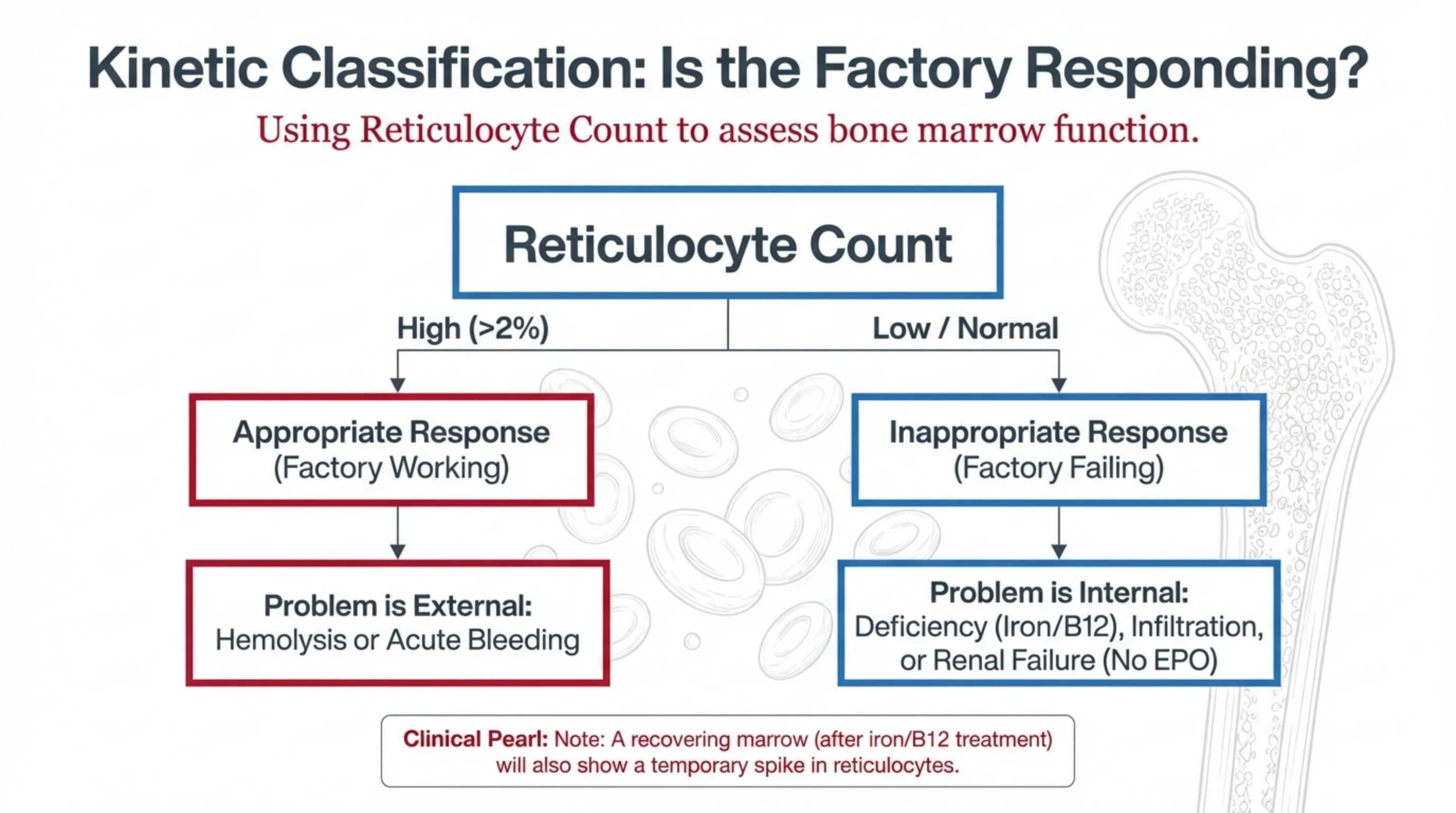
The reticulocyte count tells you whether the marrow is working
A reticulocyte is a red cell that left the marrow within the last day or so. It still carries a little RNA, which is why it can be counted separately. Because young cells are only released when the marrow is producing, the reticulocyte count is a direct read-out of factory output. This is the "kinetic" classification: it asks how the marrow is behaving over time.
If the reticulocyte count is high (above about 2%), the marrow has noticed the anemia and is responding appropriately. The factory is working; the problem lies outside it. Cells are being made and then lost, either by bleeding or by hemolysis.
If the reticulocyte count is low or normal in an anemic patient, that is an inappropriate response. A healthy marrow should be pouring out young cells; if it is not, the factory itself is the problem. Look for a missing ingredient (iron, B12, folate), a marrow that has been infiltrated or has failed, or a kidney that is no longer producing EPO.
One trap. A patient who has just started iron or B12 replacement will show a burst of reticulocytes as the marrow restarts. That is a sign of recovery, not of hemolysis, so always interpret the count in the light of recent treatment.
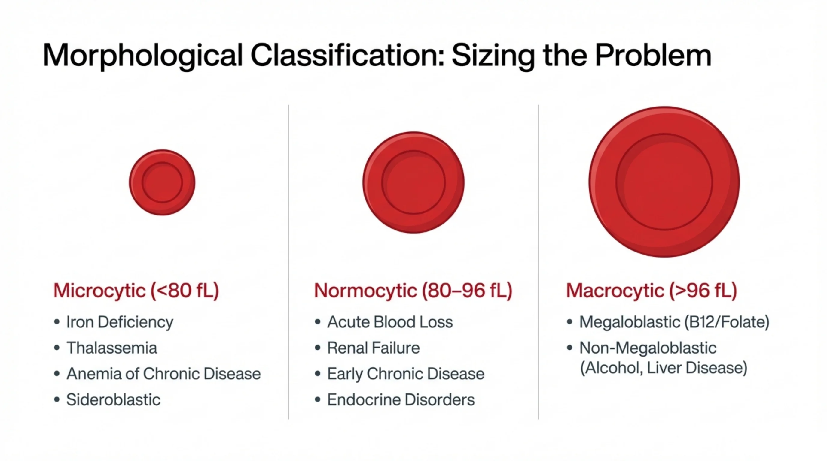
Cell size is the second axis
The mean corpuscular volume, or MCV, is the average size of the red cells, measured in femtolitres by the same machine that counts them. It costs nothing extra and it splits the differential diagnosis into three groups, because different diseases shrink or enlarge the cell for different reasons.
Microcytic cells (MCV below 80 fL) are small because they contain too little hemoglobin. Anything that limits hemoglobin synthesis produces them: iron deficiency, thalassemia, anemia of chronic disease, and the rare sideroblastic anemias. Slide 7 explains why.
Normocytic cells (80 to 96 fL) are normal in size but too few in number. The marrow is either losing cells it has made correctly (acute blood loss) or is being told to make fewer (renal failure with no EPO, early chronic disease, endocrine disorders such as hypothyroidism).
Macrocytic cells (above 96 fL) are too large. There are two quite different reasons for this, a DNA problem or a membrane problem, and slide 10 separates them.
Combine the MCV with the reticulocyte count and you already have a short list. A microcytic anemia with a low reticulocyte count in a young woman is iron deficiency until proven otherwise.
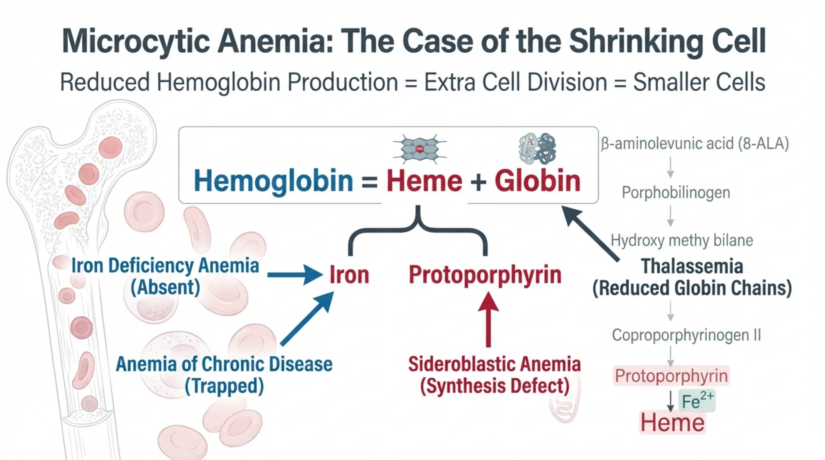
Why some anemias make small cells
A developing red cell in the marrow keeps dividing until it has accumulated a certain concentration of hemoglobin. If hemoglobin is being made slowly, the cell needs extra divisions to reach that concentration, and each division makes it smaller. So a small red cell is the signature of a hemoglobin supply problem.
Hemoglobin is built from two parts: heme and globin. Heme is itself made of an iron atom fitted into a ring called protoporphyrin, which the cell assembles through a chain of intermediates (δ-aminolevulinic acid, porphobilinogen and so on) ending in protoporphyrin, into which iron is inserted. Globin is the protein scaffold, made of α and β chains, that holds the heme.
Each microcytic anemia breaks one link in this chain:
- Iron deficiency: the iron is simply absent.
- Anemia of chronic disease: iron is present in the body but trapped in storage cells and unavailable to the marrow (the hepcidin story on the next slide).
- Sideroblastic anemia: iron is available but the cell cannot build the protoporphyrin ring to put it in, so iron piles up in the mitochondria.
- Thalassemia: heme is fine but the cell cannot make enough globin chains.
Knowing which link is broken tells you which test will confirm it: iron studies for the first two, and hemoglobin electrophoresis for thalassemia.
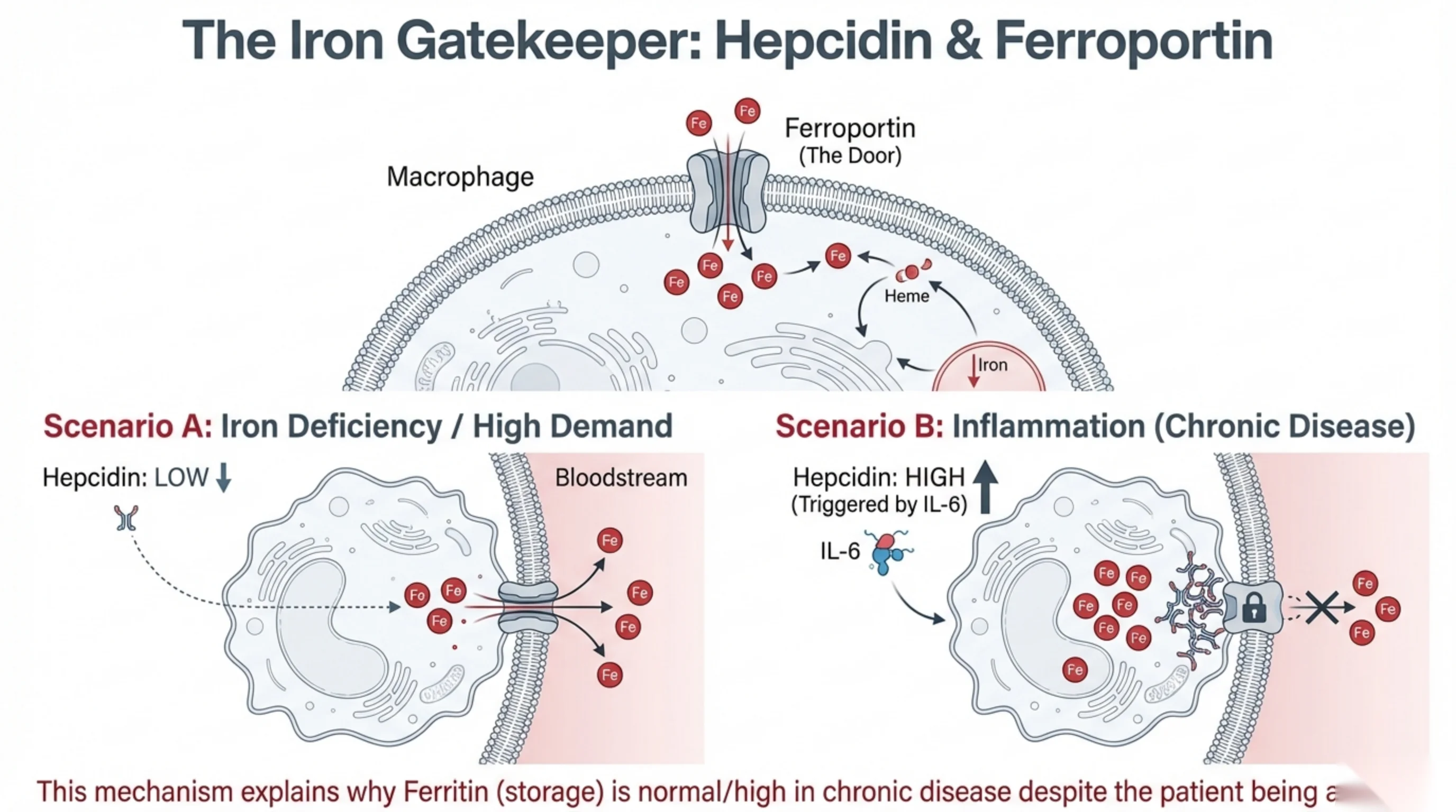
Hepcidin decides whether stored iron can be used
Most of the iron the marrow uses every day is not new iron from food. It is recycled iron, recovered by macrophages that break down old red cells. To be useful, that iron has to leave the macrophage and enter the blood, and there is only one exit: a protein channel in the cell membrane called ferroportin. Think of it as a door.
The liver produces a hormone, hepcidin, whose only job is to lock that door. When hepcidin is high, ferroportin is pulled inside the cell and destroyed, iron stays trapped in the macrophage, and the marrow is starved.
Scenario A, iron deficiency. The body needs iron, so hepcidin falls, the door stays open, and every recoverable atom of iron is pushed into the bloodstream. Stores empty out, and ferritin, which reflects those stores, falls.
Scenario B, inflammation. Infection or chronic disease raises the cytokine IL-6, and IL-6 tells the liver to make hepcidin. The door locks. Iron accumulates inside the macrophages, so ferritin is normal or even high, yet the marrow cannot get at it and the patient becomes anemic. This is the mechanism behind anemia of chronic disease, and it is why a normal ferritin does not exclude an iron problem in an inflamed patient. Evolution probably built this switch to deny iron to invading bacteria; the price is anemia in anyone with a long-running inflammatory illness.
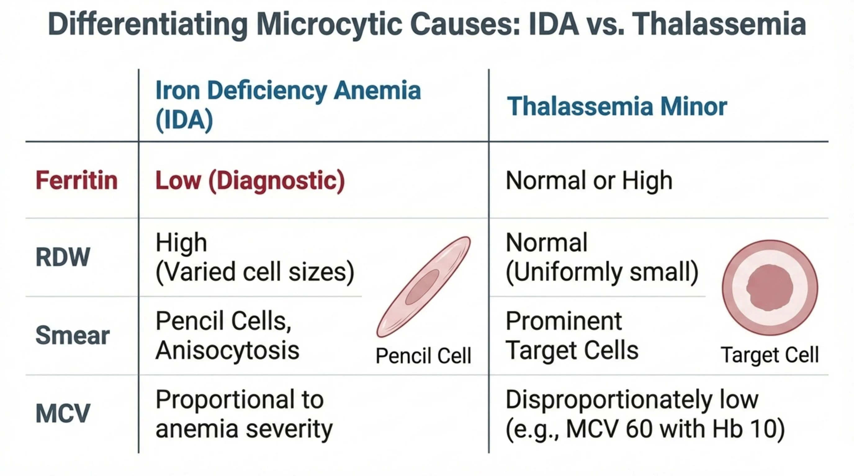
Two small-cell anemias that look alike on the CBC
Iron deficiency anemia and thalassemia minor are the two microcytic anemias you will meet most often, and both are common in Saudi Arabia. Confusing them matters: one is treated with iron and one must not be, because a thalassemic patient absorbs iron avidly and can become overloaded. Four simple clues tell them apart.
- Ferritin. Ferritin reflects iron stores. In iron deficiency it is low, and a low ferritin is essentially diagnostic. In thalassemia minor iron stores are normal or high.
- RDW (red cell distribution width) measures how varied the cells are in size. Iron deficiency develops gradually, so old normal cells mix with new small ones and the RDW is high. In thalassemia every cell has been small since birth, so the population is uniform and the RDW is normal.
- The smear. Iron deficiency produces elongated "pencil cells" and cells of many sizes (anisocytosis). Thalassemia produces prominent target cells, which look like a bull's-eye because the thin, hemoglobin-poor cell folds in on itself.
- MCV in proportion to the anemia. In iron deficiency the cells shrink in step with the hemoglobin. In thalassemia the cells are far smaller than the mild anemia would predict: an MCV of 60 with a hemoglobin of 10 is typical of thalassemia and would be unusual for iron deficiency of that degree.
When ferritin is normal and the RDW is normal in a microcytic patient, order hemoglobin electrophoresis before you order iron.
See it online.
- Pencil cells in iron deficiency ASH Image Bank
- Target cells in beta-thalassemia ASH Image Bank
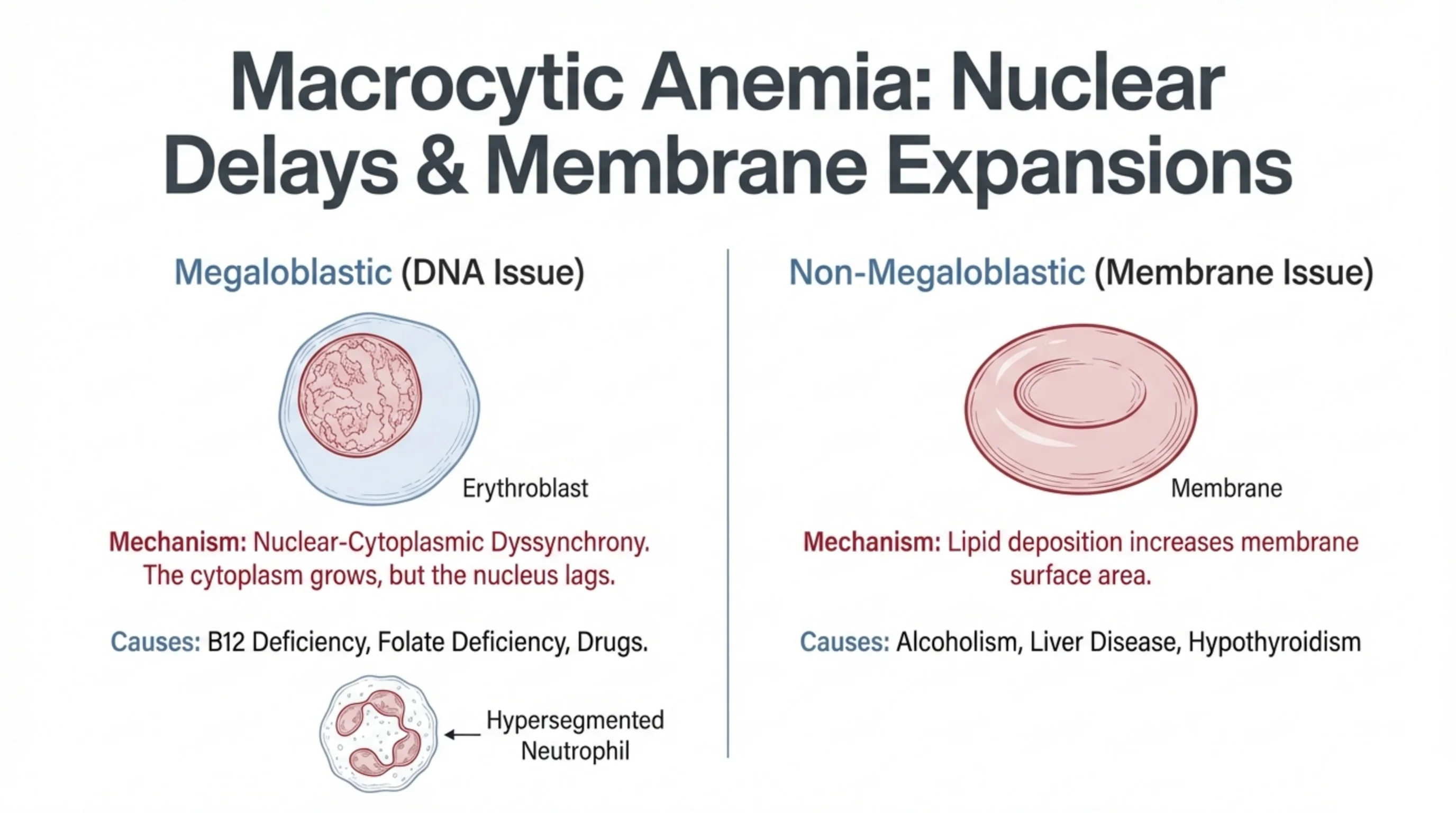
Two very different reasons for a big red cell
A macrocytic anemia can arise from a problem in the nucleus or from a problem in the membrane, and the distinction guides the whole workup.
Megaloblastic anemia is a DNA problem. Vitamin B12 and folate are needed to make the building blocks of DNA. Without them, a developing red cell cannot copy its DNA fast enough to divide, but its cytoplasm keeps growing and filling with hemoglobin. The nucleus lags behind the cytoplasm, a state called nuclear-cytoplasmic dyssynchrony, and the cell is released large, oval and often fragile. The same defect affects every dividing cell in the marrow, which is why the neutrophils are affected too: a hypersegmented neutrophil, with five or more nuclear lobes, is the tell-tale finding on the smear. Causes are B12 deficiency, folate deficiency, and drugs that interfere with DNA synthesis.
Non-megaloblastic macrocytosis is a membrane problem. Here DNA synthesis is normal, but extra lipid is laid down in the red cell membrane, so the cell has more surface and is larger and rounder. The classic causes are alcohol, liver disease and hypothyroidism. There are no hypersegmented neutrophils, and B12 and folate levels are normal.
So when the MCV is high, look at the neutrophils on the smear, then check B12 and folate, and take an alcohol and thyroid history.
Read next. Case 50 · Macrocytic and Haemolytic Anaemia in the casebook is this slide in one patient: oval macrocytes and hypersegmented neutrophils on the film, the B12 workup, and the neurological signs that can come before the anemia does.
See it online.
- A hypersegmented neutrophil in megaloblastic anemia ASH Image Bank
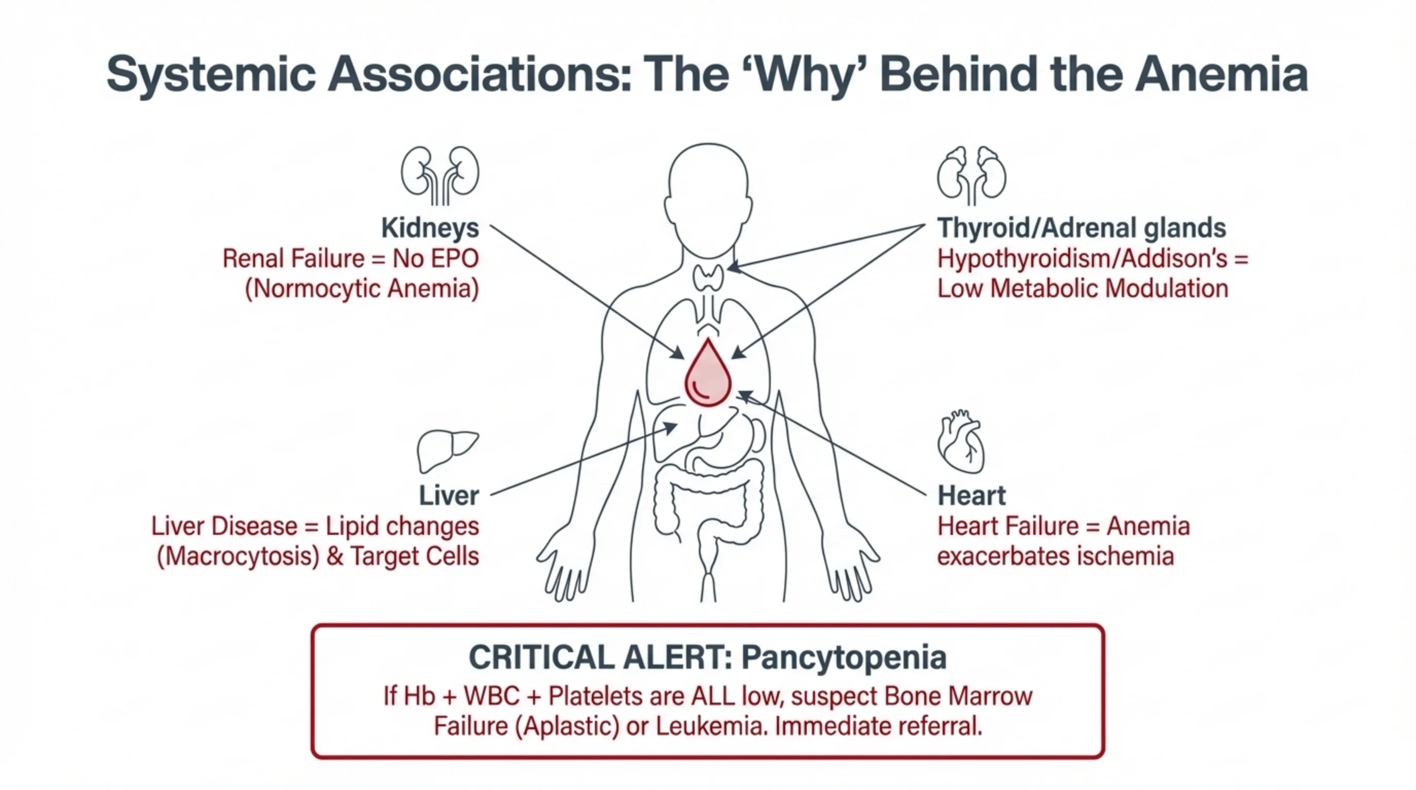
Anemia is often a message from another organ
Many anemias are not diseases of the blood at all. They are the blood's response to a failing organ elsewhere, and the pattern of anemia can point you to that organ.
- Kidney. The kidney makes EPO, the signal that drives red cell production. In renal failure the signal disappears and the marrow simply produces less: a normocytic anemia with a low reticulocyte count. Nothing is wrong with the cells; there are just too few of them.
- Thyroid and adrenal glands. Thyroxine and cortisol are among the hormones that keep the marrow ticking over. In hypothyroidism and Addison's disease the metabolic drive falls and a mild anemia follows.
- Liver. Liver disease alters the lipids in the red cell membrane, producing macrocytes and target cells, and it is often accompanied by alcohol and folate deficiency.
- Heart. The heart does not usually cause anemia, but anemia hurts the heart: less oxygen-carrying capacity means more ischemia in a patient with coronary disease or heart failure, which is why these patients tolerate anemia poorly.
One finding deserves a red flag of its own. If the hemoglobin, the white cell count and the platelet count are all low (pancytopenia), the problem is in the marrow itself, either failure of the marrow (aplastic anemia) or replacement of the marrow by leukemia. That patient needs urgent referral, not a trial of iron.
Read next. Case 29 · Chronic Kidney Disease in the casebook takes the kidney line of this slide onto the ward. Two rules from it are worth carrying: do not call an anemia renal until iron deficiency has been excluded and corrected, and when you do give an erythropoiesis-stimulating agent, aim for a hemoglobin below normal, not a normal one.
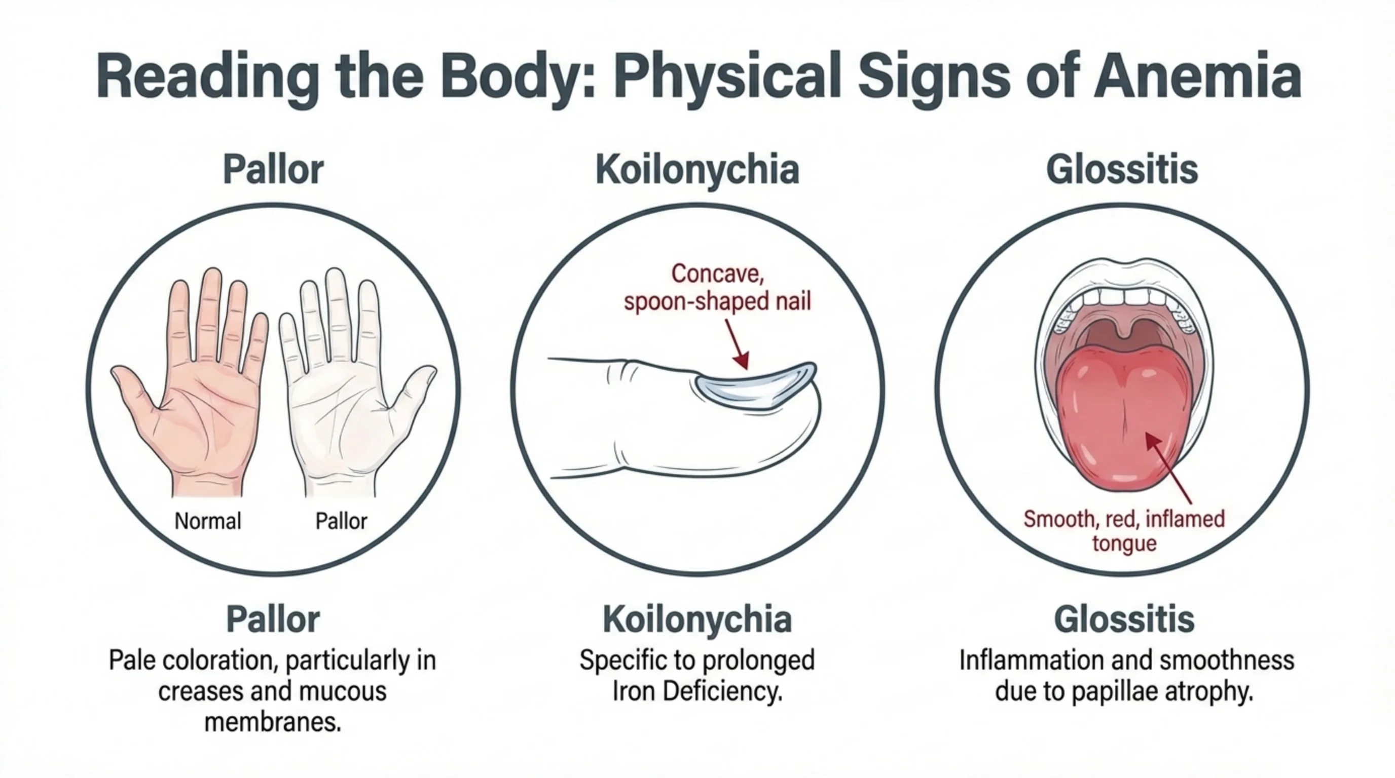
Three signs you can find with your eyes
Before any laboratory result arrives, the body often shows you the anemia and sometimes its cause.
Pallor is the general sign. Skin colour is unreliable because it varies so much between people, so look where pigment does not interfere: the conjunctiva inside the lower eyelid, the mucous membranes of the mouth, the nail beds, and the creases of the palm. A palmar crease that is as pale as the surrounding skin suggests a markedly low hemoglobin.
Koilonychia means spoon-shaped nails. The nail plate becomes thin and concave, so that a drop of water would sit in it. It takes months to develop and is a sign of prolonged iron deficiency in particular, because the rapidly growing nail matrix is one of the first tissues to suffer when iron runs short.
Glossitis is a smooth, red, sometimes sore tongue. The tiny papillae on the surface of the tongue are constantly renewed and, like the nail matrix, they fail early when the body lacks iron, B12 or folate. A smooth tongue therefore points to a nutritional anemia.
These signs are not sensitive, so their absence proves nothing, but when they are present they are useful and free. Look at the hands, the eyes and the tongue of every patient you suspect of anemia.
Read next. Open Case 49 · Microcytic Anaemia and the Iron Deficiency Workup in the casebook: the woman in it has pale conjunctivae and koilonychia, and the case carries those findings through to the investigations you would order and the cause you must not miss.
See it online.
- Koilonychia in iron deficiency anemia Wikimedia Commons · CC BY 2.0 · Corey Heitz
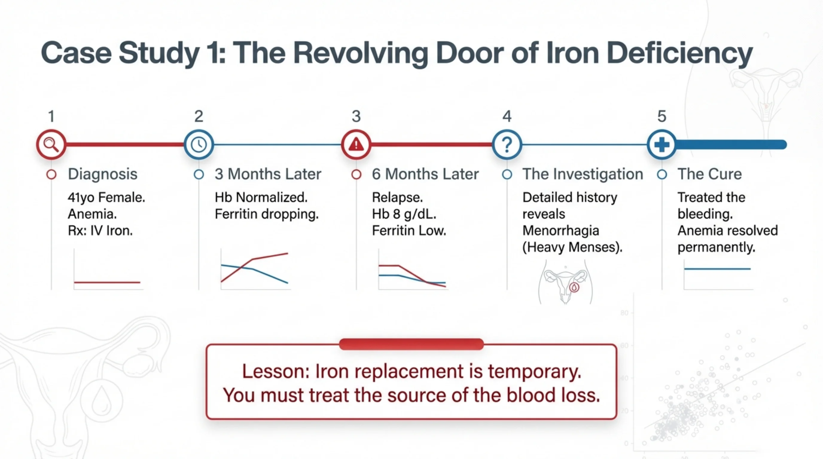
Case 1: iron replaces the iron, but not the cause
This case is here to teach a single habit: when you diagnose iron deficiency, you have not finished. You have found what is wrong; you still need to find why.
A 41-year-old woman was found to be anemic and was given intravenous iron. Three months later her hemoglobin was normal, and everyone was pleased, but the ferritin was already drifting downward, a quiet warning that iron was leaving the body faster than it should. By six months she had relapsed, with a hemoglobin of 8 g/dL and an empty iron store again.
Only then did someone take a careful menstrual history and discover heavy periods. Every month she was losing more iron than her diet could replace. Once the menorrhagia was treated, the iron stayed where it was put and the anemia never came back.
The lesson is general. Iron replacement, oral or intravenous, only refills a leaking tank. In a woman of reproductive age the leak is usually menstrual; in a man or a post-menopausal woman it is assumed to be the gastrointestinal tract until endoscopy proves otherwise. Ask about the bleeding, and if the patient cannot tell you where it is, go and look.
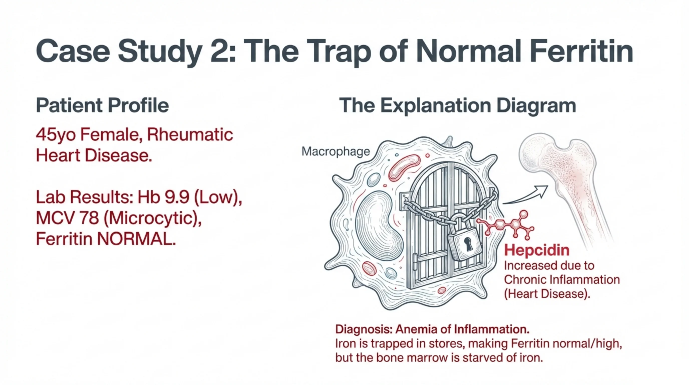
Case 2: the ferritin is normal, so it cannot be iron deficiency?
A 45-year-old woman with rheumatic heart disease has a hemoglobin of 9.9 g/dL and an MCV of 78 fL. The cells are small, so hemoglobin synthesis is limited, and the first thought is iron deficiency. But her ferritin is normal. Does that rule out an iron problem?
No, and this is the hepcidin lesson from slide 8 in action. Rheumatic heart disease is a chronic inflammatory condition. Inflammation raises IL-6, IL-6 raises hepcidin, and hepcidin locks the ferroportin door on the macrophages. Iron is still in the body, indeed it piles up in the storage cells, which is exactly why the ferritin reads normal. But the marrow cannot reach it. The factory has raw material sitting in a locked warehouse.
The diagnosis is anemia of inflammation (anemia of chronic disease). The clues that separate it from true iron deficiency are the normal or high ferritin, the presence of an inflammatory disease, and often a normal RDW. The treatment is not iron, which would simply add to the locked store; it is control of the underlying inflammation. Once the hepcidin falls, the door opens and the marrow recovers on its own.
Remember also that ferritin rises with inflammation for its own reasons, as an acute-phase protein, so in an inflamed patient a "normal" ferritin may hide a genuinely low iron store. Interpret ferritin alongside the clinical picture, never alone.
The four iron tests, and what each one is for
Everything so far has rested on ferritin. Ferritin is the best single test, but it is one of four, and the other three are what rescue you when ferritin misleads.
Ferritin is the iron in store, and it is the most specific of the four: a low ferritin confirms iron deficiency on its own. Serum iron is the iron in transit, riding on transferrin at the moment the sample was taken. TIBC, the total iron-binding capacity, measures the transferrin itself — how much carrier the body has put into the blood. When iron is short the body makes more carrier, so the TIBC rises. Transferrin saturation is the serum iron divided by the TIBC: how full the carrier is. Less iron spread over more carrier means the saturation falls.
So iron deficiency reads low, low, high, low. None of the four is interpreted alone; they work as a set, and they answer two different questions. Ferritin asks how much iron is in store. The saturation asks how much is reaching the marrow now. Those two answers can disagree, and the next slide is about what it means when they do.
The trap is the one from slide 14, stated as a rule. Ferritin is an acute-phase protein, so infection, inflammation and liver disease raise it whatever the stores are doing, and a normal ferritin in a sick patient does not exclude iron deficiency — check the saturation. One practical point: take the iron studies before you transfuse or start iron, because treatment changes all four numbers.
The handbook covers the panel in Interpreting the Core Hematology Lab Tests and the disease itself in Iron Deficiency Anemia and Iron Overload.
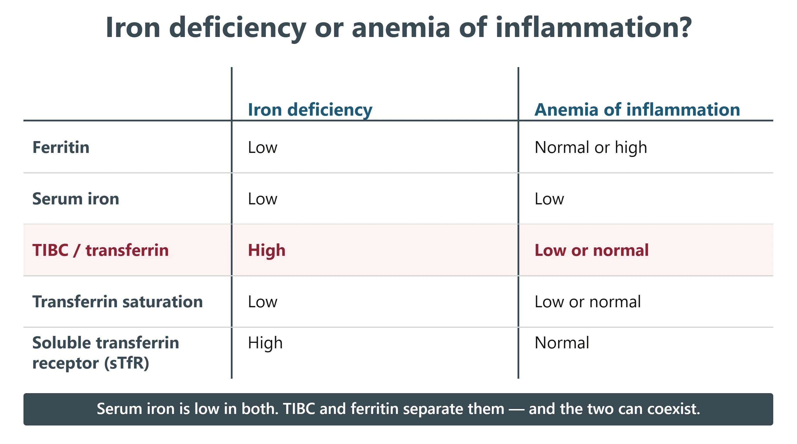
Telling iron deficiency from anemia of inflammation
Slide 14 gave you the patient and the mechanism. This is the laboratory version of the same thing, and it is worth learning as one pattern rather than five separate facts. Slide 9 separated iron deficiency from thalassemia minor; this separates it from the other mimic.
Start with what the two share. Serum iron is low in both, for opposite reasons. In iron deficiency it is low because there is no iron. In anemia of inflammation it is low because hepcidin has shut the ferroportin door, as on slide 8, and the iron cannot get out of the macrophage into the plasma. A low serum iron therefore does not tell you which of the two you are looking at, and neither does a low transferrin saturation, which can fall in both.
What separates them is the carrier and the store, and they move in opposite directions. In iron deficiency the body makes more transferrin, so the TIBC is high, and the store is empty, so the ferritin is low. In inflammation the TIBC is low or normal and the ferritin is normal or high, because the iron is there — it is simply locked away. That pair, a low TIBC with a preserved ferritin in a patient who has an inflammatory disease, is what makes the diagnosis, and it is why the treatment is control of the underlying disease and not iron.
Soluble transferrin receptor is the test for the difficult case. Marrow that is genuinely short of iron puts out more receptor, so the sTfR is high in iron deficiency and normal in inflammation.
And the two do coexist. A patient with rheumatoid arthritis can also be bleeding from an ulcer, and the inflammatory rise in ferritin will hide it. When the picture is mixed, a low transferrin saturation alongside a normal-range ferritin, or a high sTfR, is what unmasks the real deficiency. Test for it rather than assuming that either diagnosis has explained the whole anemia.
Further reading in the handbook: Iron Deficiency Anemia and Iron Overload and Anemia of Inflammation (Chronic Disease). The same reasoning applied to the large cells is in Megaloblastic Anemias: B12 and Folate, and the method behind all of it is in A Structured Approach to Anemia and Reading the Complete Blood Count (CBC). In the casebook, Case 49 · Microcytic Anaemia and the Iron Deficiency Workup sets out the trap on this slide: ferritin is an acute phase protein, so in a sick patient a normal ferritin does not exclude iron deficiency.
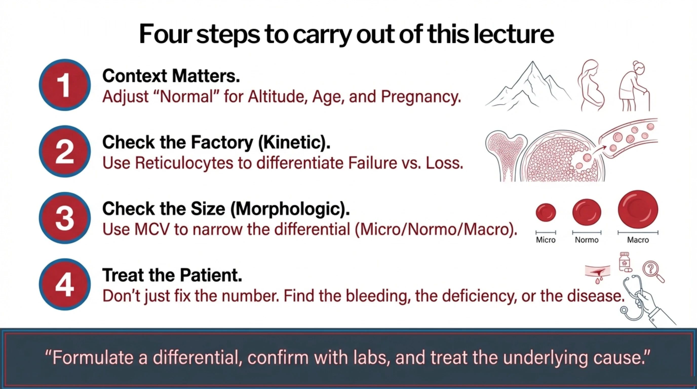
Putting the method together
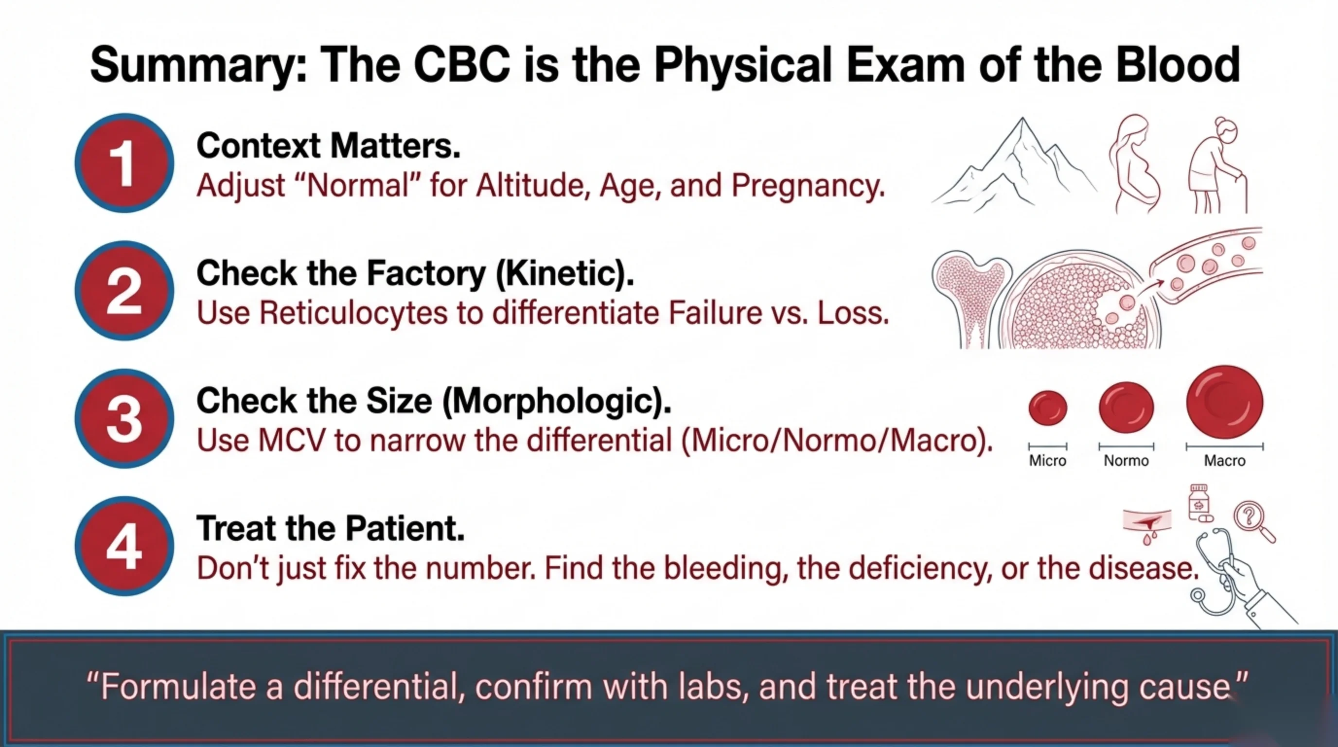
Everything in this lecture can be compressed into four steps, and they are worth memorising in order.
- Context first. Decide what "normal" means for this patient: their sex, their age, whether they are pregnant, and the altitude at which they live. A hemoglobin that is normal in Jeddah may be anemia in Abha.
- Check the factory. The reticulocyte count tells you whether the marrow is responding. High means the cells are being lost after production (bleeding or hemolysis); low means production itself has failed.
- Check the size. The MCV sorts the causes into microcytic, normocytic and macrocytic, and each group has its own short list and its own confirmatory tests.
- Treat the patient, not the number. Iron, B12 or a transfusion corrects a value; finding the bleeding, the deficiency or the underlying disease corrects the patient.
The CBC is called the physical exam of the blood for good reason. Read with care, it tells you where to look before you order a single further test. Formulate a differential, confirm it with the laboratory, and then treat the cause.
Acquired Hemolytic Anemia
Where Part 2 fits
These slides are an outline only. Read the accompanying material with them: the notes under each slide on this page, and the Hematology Handbook. Then work through the Internal Medicine Casebook and the Hematology Question Bank.
Part 1 gave the method for approaching any anemia. One of its branches was the anemia in which the marrow is working hard, the reticulocyte count is high, and the hemoglobin still falls because cells are being destroyed. That is hemolytic anemia, and it is the subject of this lecture and the next.
Hemolysis has two broad origins. Either the red cell was built with a flaw and was always going to fail early, which is inherited hemolysis and the subject of Part 3, or the cell was built normally and something in its environment is destroying it, which is acquired hemolysis and the subject of this lecture.
Two acquired mechanisms account for most of what you will see. In the first, the patient's own immune system coats the red cells with antibody and the coated cells are removed, mostly by the spleen. That is autoimmune hemolytic anemia, AIHA. In the second, red cells are torn apart mechanically as they pass through small vessels blocked by platelet and fibrin thrombi. That is microangiopathic hemolytic anemia, MAHA.
The lecture runs in the order the problem is met clinically. First the cell, and why it is so easily destroyed. Then where destruction happens, and the laboratory panel that establishes that it is happening at all. Then the two mechanisms in turn, each introduced by a patient and each with its own cell shape on the film. One test, the direct antiglobulin test, separates them. The last two slides put the sequence back together.
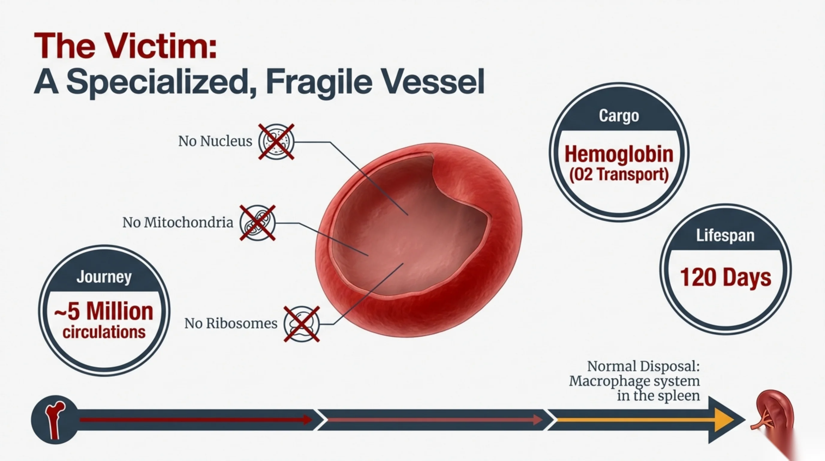
Why the objectives are in this order
The five objectives follow the order of the work at the bedside, and each one can be examined on its own.
The first is the gate. Until the laboratory pattern shows that cells are being destroyed, there is no point asking what is destroying them. The second locates the destruction, which changes the signs to look for and the results that move. The third and fourth cover the immune mechanism: one test to ask whether antibody is on the cell, then the warm and cold forms, which differ in site, in association and in treatment.
The fifth objective is different in kind. The first four are understanding, and understanding can be rebuilt from the mechanism if it is forgotten. The fifth is a decision that has to be made on the day the film is reported, because untreated thrombotic thrombocytopenic purpura is fatal within days.
The red cell cannot replace what it loses
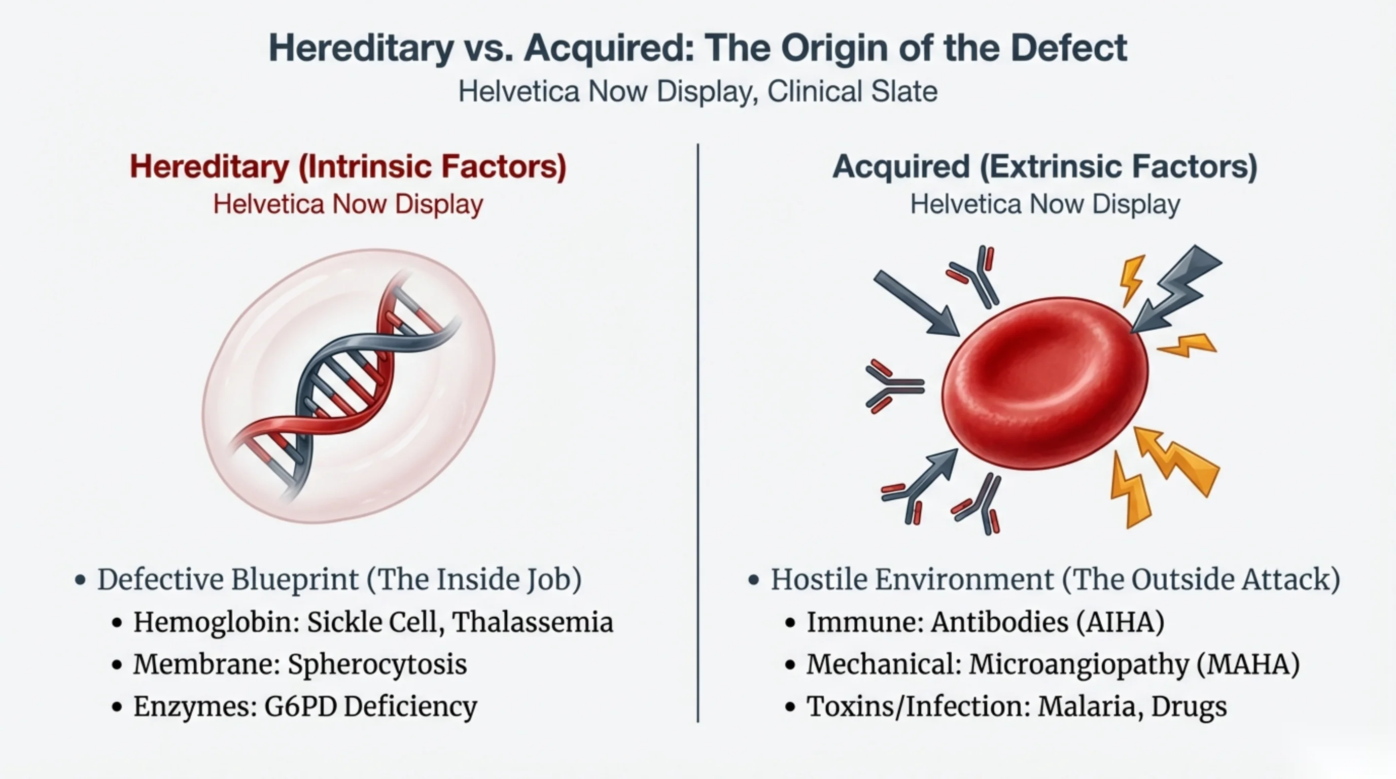
The mature red cell has no nucleus, so it cannot transcribe a gene or make a new protein. It has no ribosomes, so the enzymes it carried out of the marrow are the only ones it will ever have. It has no mitochondria, so it depends on glycolysis for energy. What it has instead is a very large amount of hemoglobin and a membrane flexible enough to fold through capillaries narrower than the cell itself.
That arrangement lasts about 120 days. The cell gradually loses flexibility, and in the end it can no longer pass the narrow slits of the spleen. Macrophages there remove it and recycle its iron. This is the normal route, and it produces no symptoms and no abnormal results.
The consequence of having no repair machinery is that every injury is permanent. A cell that loses membrane does not rebuild it. A cell whose hemoglobin is damaged cannot replace it. Injury therefore accumulates across the cell's remaining passages through the circulation, and both mechanisms in this lecture end in destruction rather than recovery for that reason.
Hemolysis is the normal process happening early and fast. Anything that makes a red cell stiff, damaged, or recognisable as abnormal will send it to the spleen ahead of schedule, or rupture it in the bloodstream. The marrow can raise its output several-fold, so mild hemolysis may produce no anemia at all. Anemia appears only when destruction outruns a marrow that is already working hard.
The defect is either in the cell or in its environment
The first question in any hemolysis is whether the defect is inside the cell or outside it. The answer changes the workup, the treatment, and what you tell the family.
Inherited hemolysis means the cell was built wrong. The membrane skeleton may be weak, as in hereditary spherocytosis. An enzyme may be deficient, as in G6PD deficiency. The hemoglobin may be abnormal in amount, as in thalassemia, or in structure, as in sickle cell disease. These patients usually have had the problem since childhood, often have a family history, and the hemolysis is lifelong. Part 3 covers them. All three groups are common in Saudi Arabia, so an inherited cause is never a remote possibility in this population, whatever the patient's age.
Acquired hemolysis means the cell was normal and something outside it is destroying it. The common causes are antibody made by the patient's own immune system, physical shearing in small vessels, and infections and toxins that damage the cell directly, of which malaria is the standard example. These patients are often previously well adults with a new illness, and the hemolysis stops if the cause is removed.
One test of the distinction is worth keeping. If normal donor cells were transfused into this patient, would they be destroyed as well? In acquired hemolysis they would, because the cause is in the patient's plasma or circulation. In inherited hemolysis they would not, because the fault was in the patient's own cells.
The site of destruction determines the findings
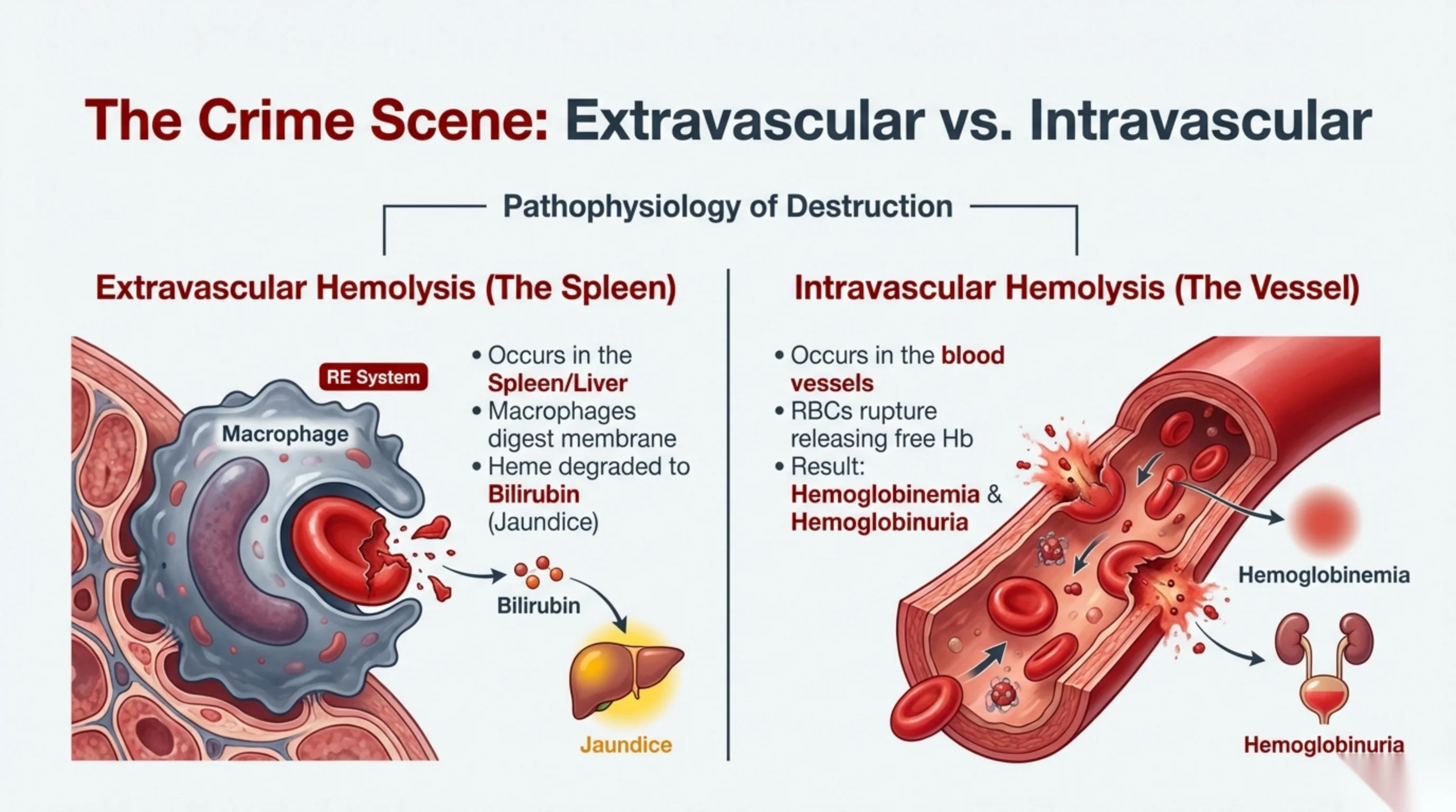
Red cells are destroyed in one of two places, and each site produces a different set of clinical and laboratory findings.
Extravascular destruction happens outside the vessels, inside the macrophages of the spleen and liver. It is the normal route of disposal, accelerated. The macrophage digests the cell, returns the iron for reuse, and breaks the heme down to bilirubin. That bilirubin is unconjugated, because the liver has not yet processed it, and when the load exceeds what the liver can conjugate the level rises and the patient becomes jaundiced. Over years the extra pigment also forms gallstones, so pigment stones in a young patient are a reason to ask about hemolysis. The spleen is often enlarged from the work it is doing. Haptoglobin is normal or only modestly reduced, because little free hemoglobin ever reaches the plasma. This is the commoner route, and it is how antibody-coated cells are usually cleared.
Intravascular destruction happens inside the vessel. The cell ruptures and releases hemoglobin directly into the plasma. Free hemoglobin is toxic and is normally bound by haptoglobin and carried to the liver, but in brisk hemolysis the haptoglobin is consumed and is often undetectable. Hemoglobin then circulates unbound, which is hemoglobinemia, is filtered by the kidney, and appears in the urine as hemoglobinuria, turning it dark brown. With continuing hemolysis iron is deposited in the tubular cells and later appears in the urine as hemosiderin. Lactate dehydrogenase sits in the cell cytoplasm and is released in bulk, so it rises markedly. This route is seen with mechanical shearing, with complement-fixing antibodies, and with some infections and toxins.
Most patients show a mixture of the two. The useful question is which one predominates, because that is what the laboratory panel on the next slide will show.
Both routes can be found at the bedside. Look for icterus in the sclera, in daylight. Start the spleen in the right iliac fossa, percuss before you palpate, and roll the patient onto the right side before calling a spleen impalpable.
Read next. Open two cases in the casebook. Case 50 · Macrocytic and Haemolytic Anaemia sets the two routes side by side, with the findings that belong to each. Case 51 · Acquired Haemolytic Anaemia and the Direct Antiglobulin Test is the extravascular route worked through in one patient: spherocytes, a raised unconjugated bilirubin, an undetectable haptoglobin and a palpable spleen.
Four results together establish hemolysis
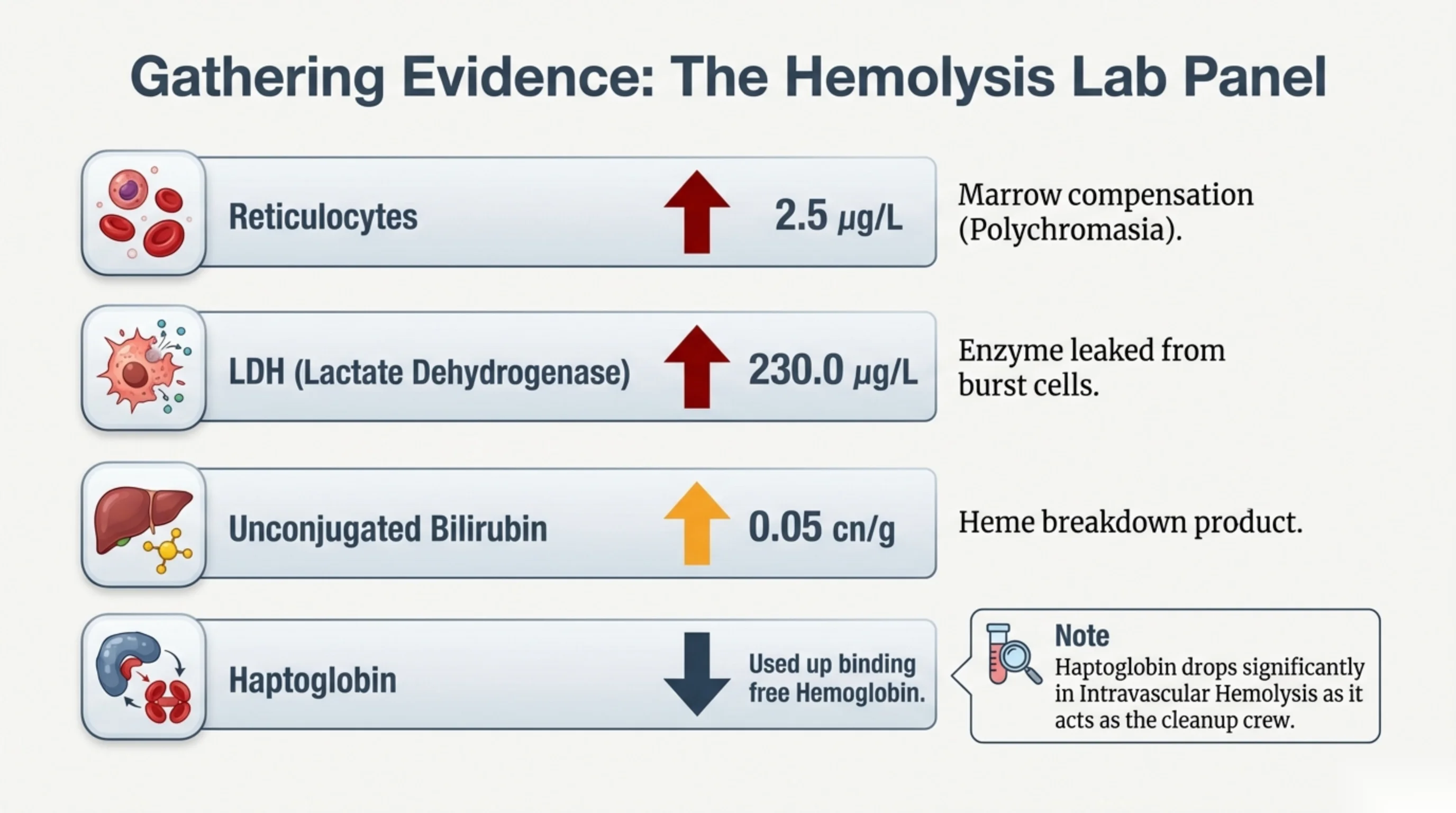
Before looking for the cause of hemolysis, establish that hemolysis is occurring. Four results do this when they are read together, and each one has a straightforward explanation.
- The reticulocyte count rises above a usual 0.5–2.5 % (25–100 × 109/L). The marrow responds to the anemia by releasing young cells early. On the film they appear slightly blue-grey because of residual RNA, which is polychromasia. A raised count means the marrow is replacing cells that are being lost, not failing to make them.
- Lactate dehydrogenase rises above a usual 140–280 U/L. It is a cytoplasmic enzyme of the red cell and leaks into the plasma when cells lyse. It is not specific, because many tissues contain it, but a raised level in an anemic patient supports cell destruction.
- Unconjugated bilirubin rises above a usual < 1.0 mg/dL (< 17 µmol/L). Heme from destroyed cells is converted to bilirubin faster than the liver can conjugate it, so the unconjugated fraction accumulates. This is the bilirubin that produces the jaundice of hemolysis. A raised conjugated fraction points instead to liver or biliary disease.
- Haptoglobin falls below a usual 30–200 mg/dL (0.3–2.0 g/L). Haptoglobin binds free hemoglobin and carries it to the liver, and each molecule is consumed in doing so. In hemolysis it is used faster than the liver replaces it, so the level falls and may become undetectable. Free hemoglobin reaches the plasma mainly in intravascular hemolysis, so the fall is steepest there.
Two cautions about the numbers. The ranges above are typical adult values, and they differ between laboratories and with assay method, so confirm what your own laboratory reports before calling a result abnormal. And haptoglobin is an acute-phase protein, so in a patient who is also infected or inflamed it is being produced faster than usual; a genuine fall can be pulled back into the reference range and the hemolysis missed.
No single result is diagnostic. A raised reticulocyte count with a raised lactate dehydrogenase and a low haptoglobin, together, makes hemolysis close to certain, and only then is it worth asking what is causing it.
The handbook works through the same four in Hemolytic Anemias: Overview and Proving Hemolysis, which calls them the hemolysis quartet and adds one refinement worth carrying: haptoglobin falls earliest and furthest when the destruction is intravascular, so a haptoglobin that is undetectable rather than merely low is itself a pointer to the route. Interpreting the Core Hematology Lab Tests sets this panel beside the others — the iron studies, the electrophoresis, the coagulation screen — with each one framed as the single question it answers.
See it online.
- Polychromasia: the young red cells of a working marrow ASH Image Bank
The reticulocyte count, seen on the film
This is a real blood film. Most of the red cells are the usual pink-brown. A few, scattered through the field, are a dull blue-grey. Those are the polychromatic cells.
They are young red cells. When the marrow is pushed to replace cells that are being lost, it releases them early, while they still carry some of the RNA they were built with. RNA takes up the blue part of the stain, so these cells look grey-blue rather than pink. They are often a little larger than the older cells around them.
Polychromasia is the reticulocyte count seen through the microscope. The count on the previous slide is a number from the analyser; this is the same finding on the glass. In an anemic patient, plenty of blue-grey cells means the marrow is working hard and the anemia comes from cells being lost, not from cells not being made. That is the first gate of the lecture.
One practical point follows. Because these young cells are larger, a brisk reticulocyte response can raise the MCV. A macrocytosis in a hemolysing patient is the reticulocytes, not a vitamin B12 deficiency.
Image on the slide. “Polychromasia 1” by Osaretin, Wikimedia Commons, CC BY-SA 4.0. Cropped for the slide.
Following bilirubin from the red cell to the urine
Every red cell that dies leaves its hemoglobin behind, and the heme in it has to be cleared. The diagram follows that heme in six steps. Each result in the laboratory panel and on the urine dipstick comes from one of them.
- The macrophage. In the spleen and liver, macrophages take in the old or damaged cell. The enzyme heme oxygenase opens the heme ring. The iron comes out and is kept for reuse, and what is left is green biliverdin. A second enzyme, biliverdin reductase, turns biliverdin into bilirubin. This is unconjugated bilirubin.
- The blood. Unconjugated bilirubin does not dissolve in water, so it travels bound to albumin. The pair is too large to pass the glomerulus. Unconjugated bilirubin is therefore not filtered, and it never appears in urine.
- The liver. The liver cell takes the bilirubin up, and the enzyme UGT1A1 joins it to glucuronic acid. This conjugated bilirubin dissolves in water, and the liver secretes it into bile.
- The gut. In the large intestine, bacteria turn bilirubin into urobilinogen.
- The stool. Most of the urobilinogen becomes stercobilin, which gives stool its brown colour, and leaves the body.
- Back to the liver, and a little to the kidney. Some urobilinogen is absorbed from the gut into the portal vein. The liver takes most of it up and sends it back out in bile. That loop, from gut to liver and back to gut, is the enterohepatic circulation. A small amount gets past the liver into the general circulation, and the kidney excretes it as urinary urobilinogen.
One physiology text gives rough shares for the last three steps: about 80 % leaves in the stool, about 18 % is reabsorbed, and about 2 % reaches the urine. Treat these as teaching figures that show the order of size, not as values to apply to a patient.
What hemolysis does to the loop. Hemolysis pushes far more heme into step 1, and each step downstream shows it.
- The liver cannot conjugate bilirubin as fast as it now arrives, so unconjugated bilirubin builds up in the blood. That is the jaundice of hemolysis, and it is why the panel on slide 6 asks for the unconjugated fraction.
- Unconjugated bilirubin rides on albumin and is not filtered, so there is no bilirubin in the urine. The patient can be visibly yellow with a dipstick that is negative for bilirubin. The old name is acholuric jaundice: jaundice without bile in the urine.
- More conjugated bilirubin reaches the gut, so more urobilinogen is made and reabsorbed, and more reaches the urine. Urinary urobilinogen rises. It also rises in liver disease, so it supports hemolysis without proving it.
- Over years, the extra bilirubin in bile can form pigment gallstones. Gallstones in a young patient are a reason to ask about hemolysis.
Set this against a blocked bile duct, where the loop breaks at step 3. Conjugated bilirubin backs up into the blood. It dissolves in water, so it is filtered and darkens the urine. Little reaches the gut, so urinary urobilinogen falls. The two patterns separate at the dipstick.
Dark urine has three separate causes
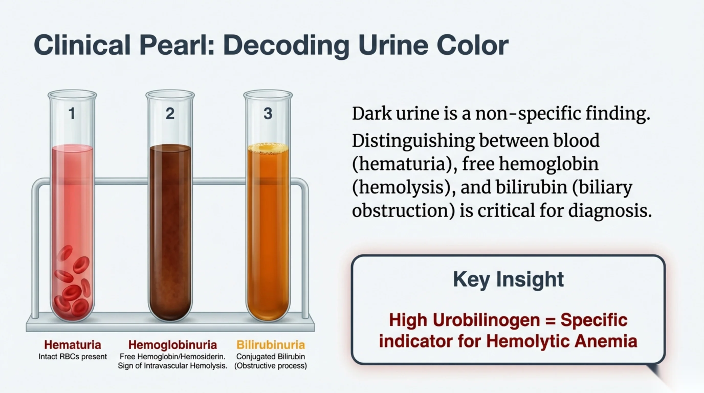
Patients and doctors both report dark urine as though it were a single finding. Three different substances darken urine, and separating them points to three different diseases.
- Hematuria is intact red cells in the urine. The urine is red or pink and microscopy shows whole red cells. The source is the urinary tract: a stone, an infection, a tumour, or glomerular disease. It is unrelated to hemolysis.
- Hemoglobinuria is free hemoglobin with no intact cells. The urine is dark brown. The dipstick reads positive for blood, because it detects heme, but microscopy shows no red cells. This pattern indicates intravascular hemolysis: the cells lysed in the vessel, the hemoglobin exceeded what haptoglobin could bind, and the kidney filtered it. Continuing intravascular hemolysis also deposits iron in the renal tubular cells, which are later shed as hemosiderin in the urine.
- Bilirubinuria is conjugated bilirubin in the urine. It looks orange or yellow-brown and froths on shaking. Only conjugated bilirubin is water-soluble enough to be excreted in urine, and it rises when bile cannot drain, so this points to biliary obstruction or liver disease rather than hemolysis.
The combination of a dipstick positive for blood with no red cells on microscopy is the one to carry away, because it separates hemoglobinuria from hematuria using two tests that are already at the bedside.
Two findings help less than students expect. Unconjugated bilirubin, which is the fraction that rises in hemolysis, is bound to albumin and is not filtered, so it never appears in urine; a patient can be deeply jaundiced from hemolysis with no bilirubin on the dipstick. Urinary urobilinogen does rise in hemolysis, because more bilirubin reaches the gut and more is reabsorbed, but it also rises in liver disease and falls in biliary obstruction. It supports the diagnosis; it does not make it.
Autoimmune hemolysis in one patient
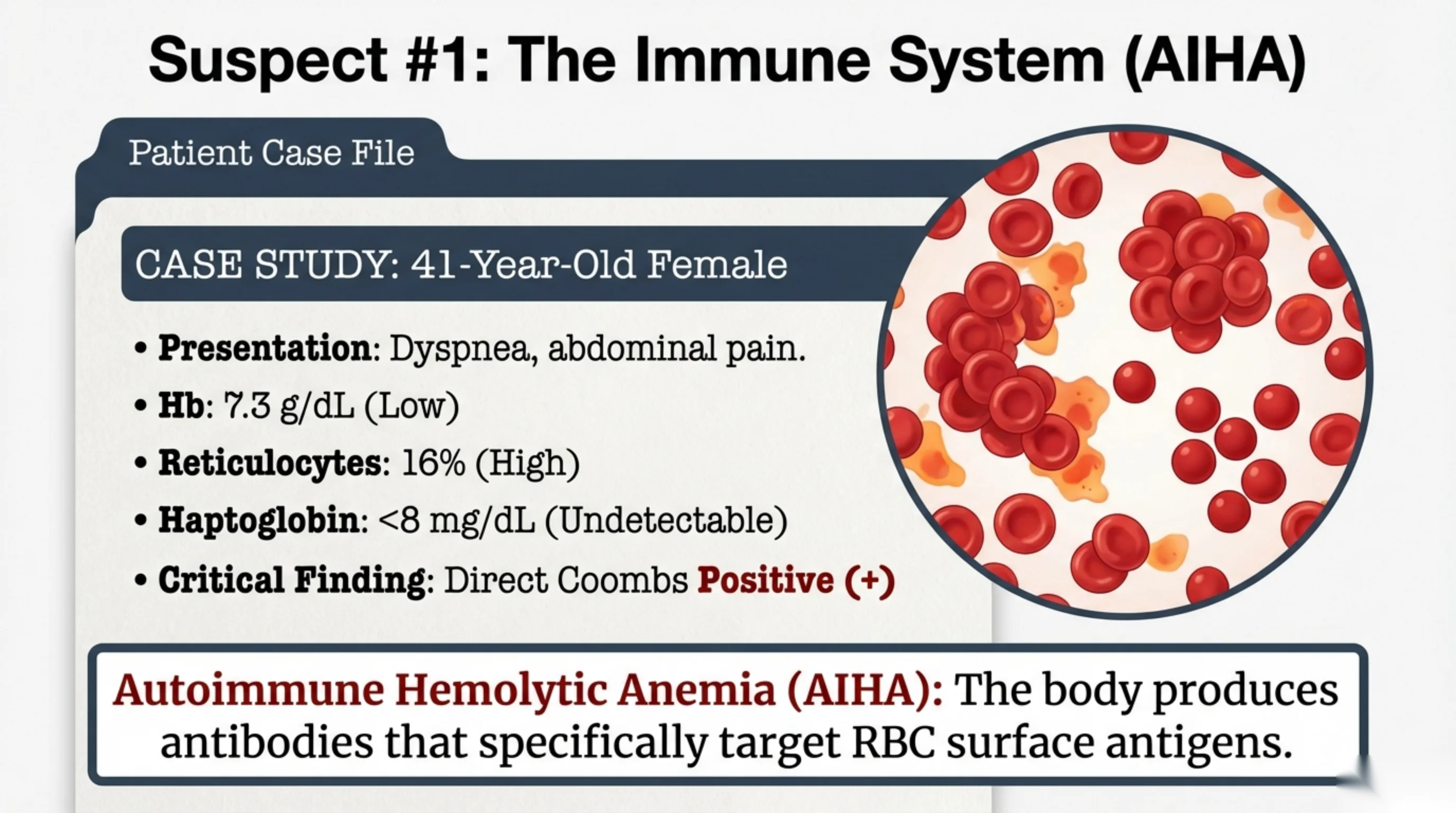
A 41-year-old woman presents with breathlessness and abdominal pain. Her hemoglobin is 7.3 g/dL, which is severe anemia. Her reticulocyte count is 16 %, so the marrow is responding strongly and the cells are being lost rather than under-produced. Her haptoglobin is below 8 mg/dL, effectively undetectable, so free hemoglobin has consumed it. The panel on slide 6 is satisfied and hemolysis is established.
The direct antiglobulin test is positive, and that result assigns the mechanism. It means her red cells are coated with antibody. The diagnosis is autoimmune hemolytic anemia: her immune system is producing antibody against antigens on the surface of her own red cells. Sometimes a cause is found, such as a lymphoma, systemic lupus erythematosus, or a drug; often none is. Once coated, the cells are recognised by splenic macrophages and removed, or, with some antibodies, are lysed by complement inside the vessel.
The film supports the diagnosis. An antibody has two binding arms, so one molecule can bridge two red cells and the cells stick together in clumps. That is agglutination, and it is what the microscope field on this slide shows. Agglutinated red cells on a film should raise antibody-mediated hemolysis immediately.
The order of work in this case is the order of the whole lecture. Establish hemolysis with the panel, then ask one question: is there antibody on the cell? The next two slides cover the kinds of antibody and the test that detects them.
See it online.
- Red cells clumped together in autoimmune hemolytic anemia ASH Image Bank
The binding temperature predicts everything else
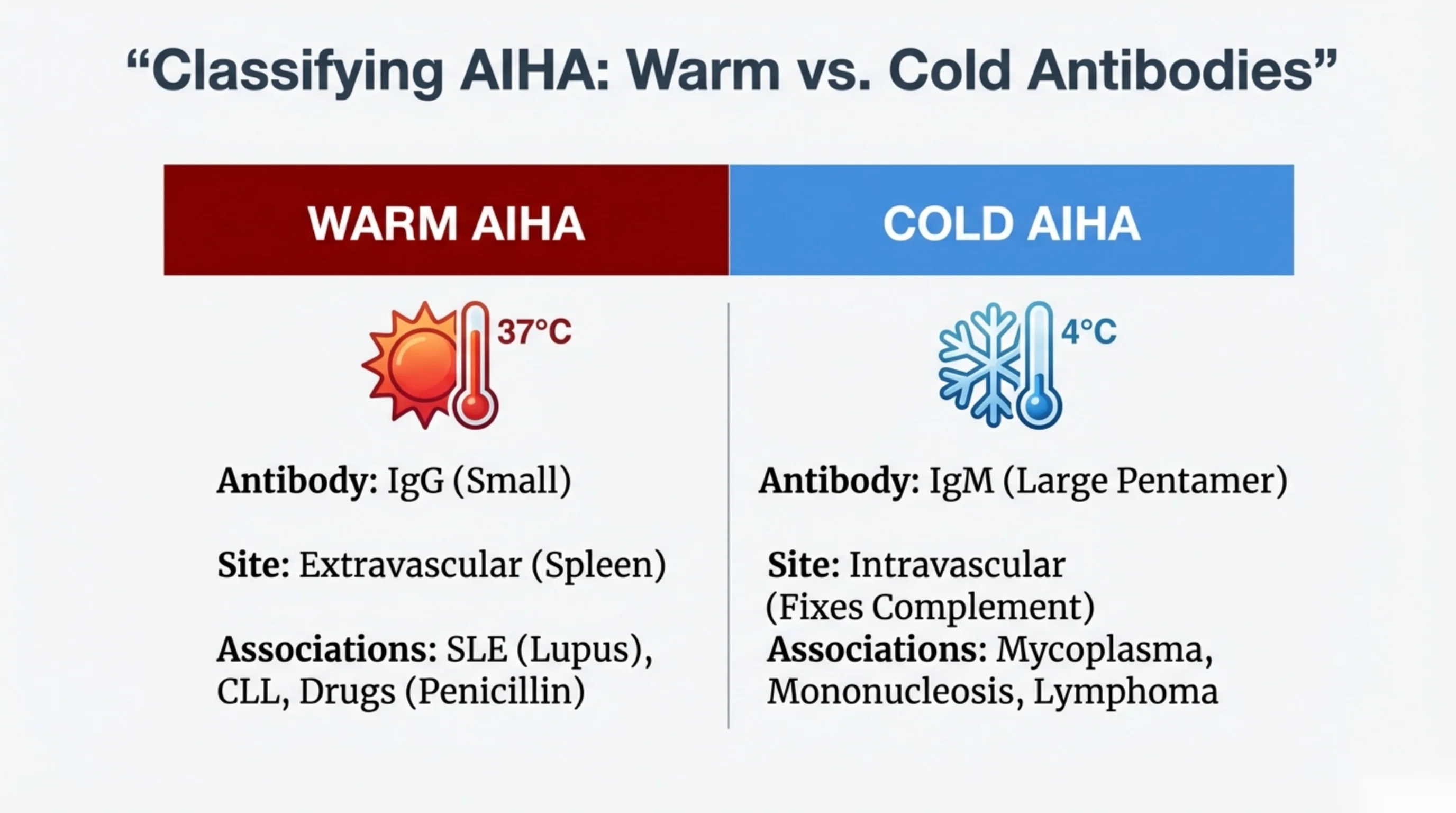
Autoimmune hemolytic anemia takes two forms, named for the temperature at which the antibody binds best. The distinction is not a classification exercise. The binding temperature determines the antibody class, where the cells are destroyed, what the antiglobulin test shows, and which treatments work.
Warm AIHA. The antibody is IgG, which binds best at body temperature, 37 °C. IgG-coated cells are not usually lysed in the circulation. Splenic macrophages carry receptors for the tail of IgG, recognise the coated cells and remove them, so the destruction is extravascular, with jaundice and an enlarged spleen. The antiglobulin test reads IgG on the cell, with or without complement C3d alongside it. Warm disease is much the commoner of the two. It occurs on its own, or with systemic lupus erythematosus, chronic lymphocytic leukemia, or a drug. Corticosteroids are the usual first treatment, together with treatment of whatever underlies it.
Cold agglutinin disease. The antibody is IgM, a large pentamer that binds best at 0–4 °C. In the body that means the fingers, toes, ears and nose, where blood is coolest. IgM activates complement efficiently, and complement lyses the cell where it sits, so the destruction is intravascular, with hemoglobinuria. The antiglobulin test finds complement C3d alone, because the IgM has usually dissociated by the time the sample is warmed in the laboratory. Patients notice symptoms in cold weather and may have bluish extremities. The disease follows infection with Mycoplasma pneumoniae or infectious mononucleosis, or accompanies a lymphoma.
The same temperature dependence explains the treatment. Steroids work poorly here. Keeping the patient warm is treatment rather than comfort, because an antibody that does not bind cannot fix complement. Rituximab, which removes the B cells producing the IgM, is the drug that works.
One practical point for the laboratory: IgM cold agglutinins can clump cells in the sample tube, giving a falsely high MCV and a falsely low red cell count until the sample is warmed and re-run.
How the antiglobulin test detects antibody
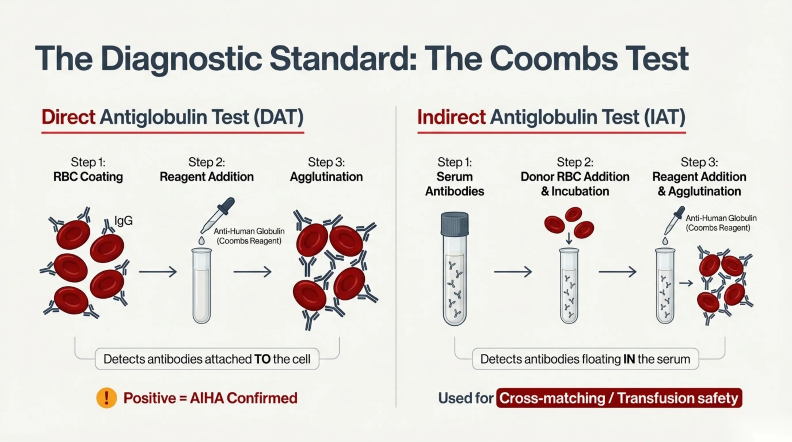
The antiglobulin test, usually called the Coombs test after the man who devised it, depends on one reagent: anti-human globulin, an antibody raised in animals that binds human antibody. Red cells coated with human antibody do not clump on their own, because the antibody molecules are too small to bridge the distance between two cells. Anti-human globulin links the coated cells together and they agglutinate visibly. Agglutination therefore means antibody was present on the cells.
The direct test asks whether antibody is already bound to this patient's red cells. The patient's washed red cells are mixed with the reagent and examined for clumping. If the cells were coated with IgG, or with complement, in the patient's circulation, they clump. A positive direct test in a patient with proven hemolysis confirms autoimmune hemolytic anemia. The test can be refined to report whether IgG, complement, or both are present, and that pattern is what separates warm disease from cold agglutinin disease on the previous slide.
The indirect test asks a different question: whether there is antibody free in the patient's serum that could attack red cells. The patient's serum is mixed with donor red cells and incubated so that any antibody can bind, then the reagent is added and the mixture examined for clumping. Its main use is not diagnosis but transfusion safety: it is the basis of the antibody screen and the cross-match, which confirm that the blood about to be given will not be destroyed by antibody the patient already carries.
Direct means the antibody is on the cell. Indirect means it is in the serum. That is the single distinction to remember, for the examination and for every transfusion request.
A negative direct test needs care. It does not say the patient is not hemolysing. It says the hemolysis is not antibody-mediated, which is a narrower statement. The second half of this lecture is about patients who are destroying red cells rapidly with a negative test.
Further reading. Acquired Hemolytic Anemias: Autoimmune and Microangiopathic carries the detail this slide leaves out: the direct test does not simply read positive or negative, and the pattern is the point — IgG with or without complement in warm disease, complement alone in cold agglutinin disease. The indirect test's real home is Transfusion Medicine, where it is the antibody screen and the crossmatch, and where you find why the worst transfusion accident of all is a clerical error at the bedside rather than a laboratory failure. Then read Case 51 · Acquired Haemolytic Anaemia and the Direct Antiglobulin Test in the casebook, which is this slide worked through as a patient: it reads the test as four patterns rather than as positive or negative, and it answers the question the vignette raises when the laboratory telephones to say that nothing is crossmatch-compatible.
What agglutination looks like on a film
This film is from a patient with cold agglutinin disease. The red cells are not lying separately. They are stuck together in large, irregular clumps, with only a few single cells between them. Ignore the green tint of the background and look at how the cells are arranged.
The previous slide explained why the antiglobulin test needs a reagent. IgG on a red cell usually cannot bridge the gap to the next cell, so the reagent is added to link the coated cells together. IgM is different. It is a large pentamer, big enough to reach from one cell to another on its own, so IgM clumps red cells without help.
The clumping depends on temperature. The antibody binds best in the cold, so clumps form as blood cools: in the fingers and ears, and in the sample tube on the bench. Warming the sample to 37 °C breaks them up.
The clumps also mislead the analyser. It counts a clump as one large cell, so the MCV reads falsely high and the red cell count falsely low, and the hematocrit calculated from them cannot be trusted. An improbable MCV with a low red cell count in an anemic patient is a reason to look at the film and to ask for the sample to be warmed and run again.
Agglutinates on a film should raise antibody-mediated hemolysis at once, and cold agglutinin disease above all. The direct antiglobulin test then finds complement on the cells, as the table on slide 11 set out.
Image on the slide. “Red cell agglutination” by Prof. Erhabor Osaro, Wikimedia Commons, CC BY-SA 4.0.
How complement bursts a red cell
Complement is a set of plasma proteins that act in order, each one switching on the next. The strip follows the classical pathway, the one that antibody starts, from antibody on the cell to a hole in its membrane.
- IgM binds and C1 attaches. The first protein, C1, attaches to antibody bound on the cell. One IgM molecule is enough to do this. IgG needs two or more molecules sitting close together, so IgM starts complement far more easily. That is why cold agglutinin disease is a complement disease.
- C3b coats the cell. C1 activates C4 and C2, which join to form the C3 convertase, written C4b2a (newer sources write C4b2b). This cuts C3 and leaves C3b stuck to the membrane. C3b marks the cell for macrophages. On the cells that survive, C3b is cut down to a smaller piece, C3d, which stays on the membrane. C3d is what the complement part of the direct antiglobulin test detects.
- C5 is cut. When C3b joins the convertase, it becomes a C5 convertase. This splits C5 into C5a, which floats off and acts as an inflammatory signal, and C5b, which stays on the cell.
- The pore. C5b binds C6, C7, C8 and C9 in turn. Together they form the membrane attack complex, C5b-9: a ring-shaped pore through the membrane. Water and salt pour in, the cell swells, and it bursts inside the vessel. This is intravascular hemolysis, with the free hemoglobin, the hemoglobinuria and the undetectable haptoglobin of slide 5.
Three things in this lecture sit on that strip.
- Cold agglutinin disease. IgM binds in the cool periphery and starts the cascade. In most patients with stable disease, much of it stops at C3b, and the coated cells are removed by macrophages, mainly in the liver. That is extravascular hemolysis. In some patients the cascade runs on to the pore, mostly in severe disease and during flares, which often follow a feverish illness. Cells then burst in the vessel and hemoglobinuria appears. This is why the handbook and the casebook describe the destruction in cold agglutinin disease as both intravascular and hepatic.
- Paroxysmal nocturnal hemoglobinuria (PNH). This is an acquired hemolysis too, but the fault is in the cells, not in an antibody. A mutation in the PIGA gene arises in one blood stem cell. The cells descended from it cannot make the GPI anchor that holds certain proteins on the cell surface. Two of those proteins are the cell’s brakes on complement: CD55, which breaks down the convertases, and CD59, which stops C9 forming the pore. Without them, complement runs through to the pore and the cells burst in the vessel. There is no antibody, so the direct antiglobulin test is negative. The diagnosis is made by flow cytometry, which shows the missing proteins. Think of it in unexplained intravascular hemolysis with thrombosis or low blood counts.
- Eculizumab. A monoclonal antibody that binds C5 and stops it being cut. No C5b means no pore, so lysis in the vessel stops. It is licensed for PNH and for atypical hemolytic uremic syndrome. It carries a boxed warning for life-threatening meningococcal infection, and patients are vaccinated against meningococcus at least two weeks before the first dose unless the delay is the greater risk. Because it acts at C5, it leaves C3b coating untouched. In PNH, some patients on eculizumab are reported to go on losing C3b-coated cells to macrophages instead, with a direct antiglobulin test positive for C3.
Further reading. Acquired Hemolytic Anemias: Autoimmune and Microangiopathic places PNH among the other acquired causes, and Thrombotic Microangiopathies: TTP and HUS covers atypical HUS, the other disease in which eculizumab is used.
What a spherocyte is and what it indicates
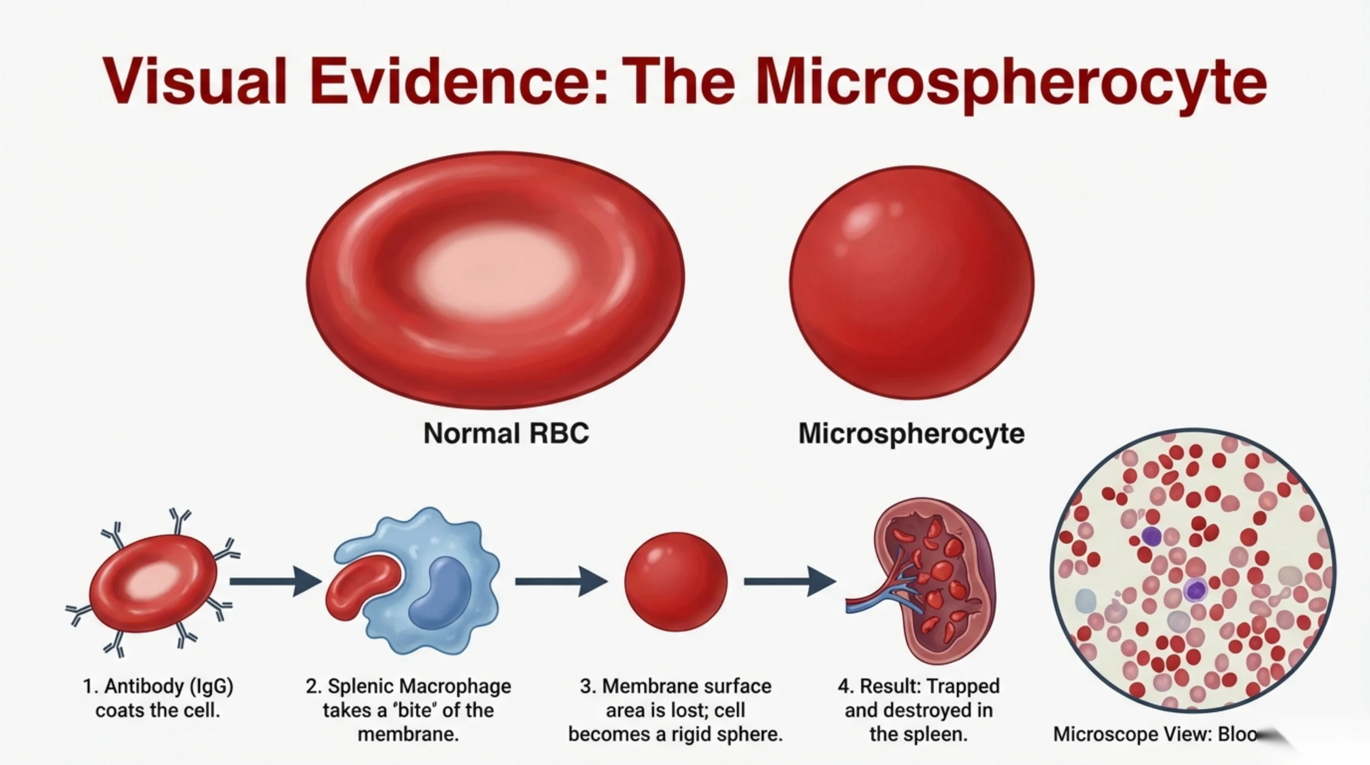
One feature separates a spherocyte from a normal red cell on the film. A normal cell is a biconcave disc, thin through the middle, so it has a pale centre occupying about a third of its diameter. A spherocyte has no central pallor. It is small, round and uniformly dark. Look for cells that are too dark rather than cells that are too small; the colour is the easier finding at low power.
The appearance follows from the mechanics. The cell has lost membrane surface area while keeping its hemoglobin, so it has been forced into a rounder shape and has no spare membrane left. A normal red cell carries considerably more membrane than it needs to enclose its contents, and that excess is what allows it to fold and deform. Remove the excess and the cell becomes rigid.
A rigid cell cannot pass the narrow slits between the splenic cords and the sinusoids. It is retained there and destroyed, which is why spherocytosis of any cause produces extravascular hemolysis with an enlarged spleen.
The caution on this slide matters more than the morphology. Spherocytes show that membrane has been lost; they do not show why. Two common diseases lose membrane: warm autoimmune hemolytic anemia, in which an antibody is responsible, and hereditary spherocytosis, in which the membrane skeleton is congenitally weak. The cells look the same in both. The direct antiglobulin test separates them, and that is the reason to order it in a patient with spherocytes on the film.
See it online.
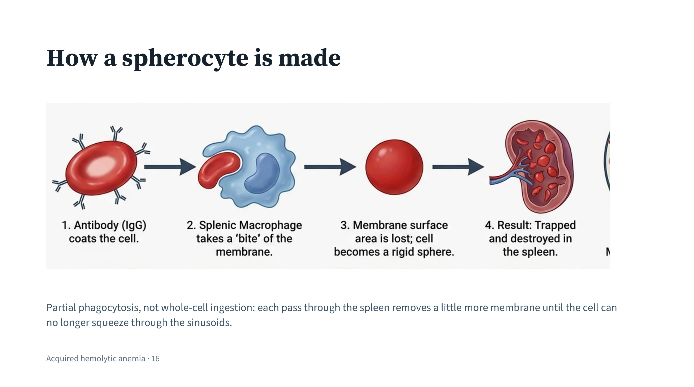
Partial phagocytosis removes membrane in stages
In warm AIHA the spleen does not usually ingest the coated cell whole. As the cell passes a macrophage, the macrophage binds the IgG on its surface and removes a portion of the membrane with it. The cell survives and continues in the circulation, but it has lost surface area while keeping the same volume of hemoglobin. This is partial phagocytosis, and it is the step that explains the shape on the previous slide.
The rest is geometry. For a given volume, the shape with the smallest surface area is a sphere, so as membrane is removed the cell is forced to round up. It becomes a microspherocyte: smaller than normal, densely filled with hemoglobin, and rigid because it has no spare membrane.
The damage accumulates because the cell cannot rebuild what it has lost. Each passage through the spleen leaves it rounder and stiffer than the last, until it can no longer pass the sinusoids at all. On that passage it is retained and destroyed. This is the extravascular death of warm AIHA, and it is why the hemolysis is gradual rather than abrupt.
The same sequence, beginning from a congenitally weak membrane skeleton instead of from an antibody, produces the spherocytes of hereditary spherocytosis. Part 3 follows that route.
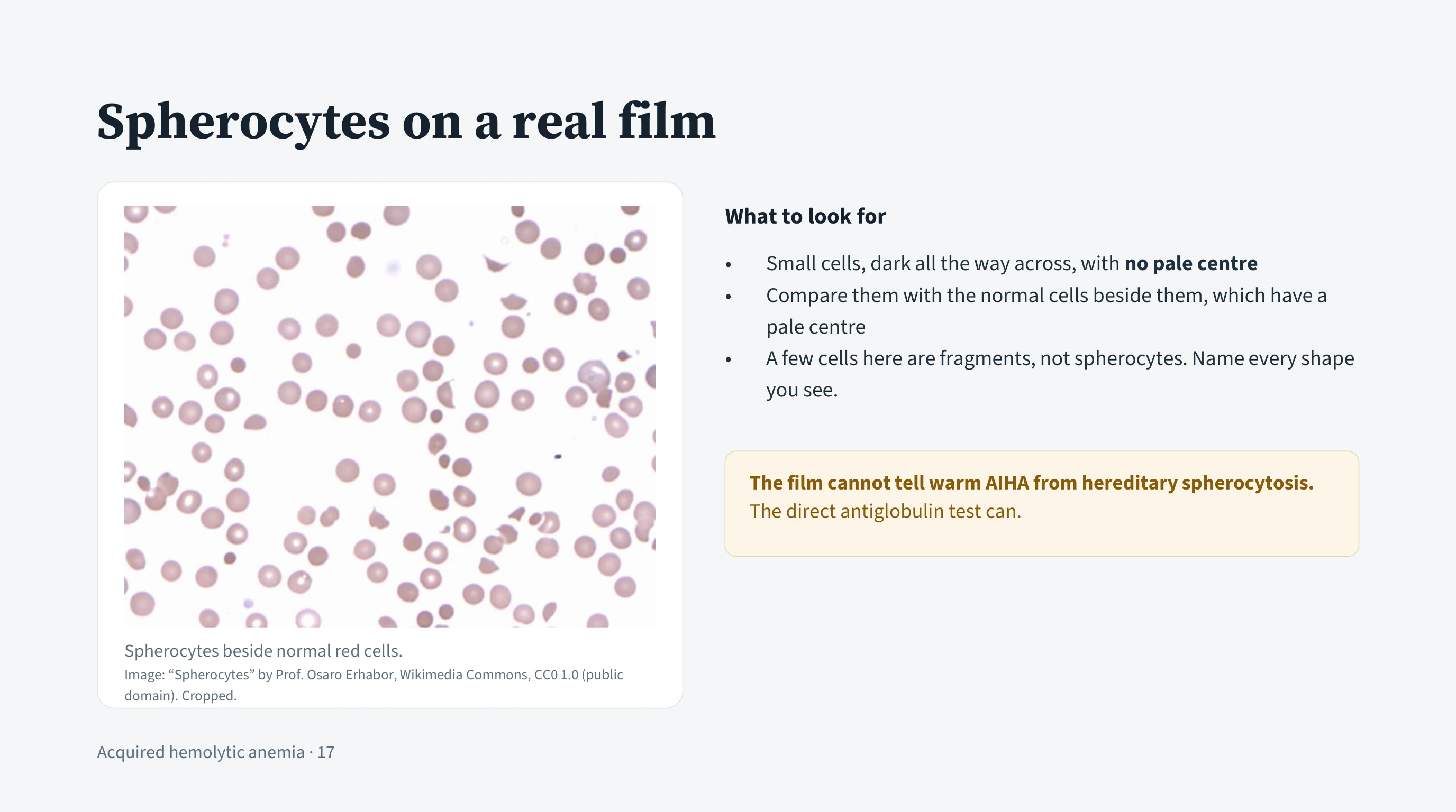
Finding spherocytes on a real film
Slide 15 drew a spherocyte and slide 16 showed how the spleen makes one. On a real film they are mixed in with everything else, and this field is practice at finding them.
Scan for cells that are dark all the way across. A normal red cell is thin in the middle, so it has a pale centre. A spherocyte has lost that pale centre and is usually a little smaller. Colour is easier to judge than size at low power, so find the dense cells first, then compare each with the normal cells beside it.
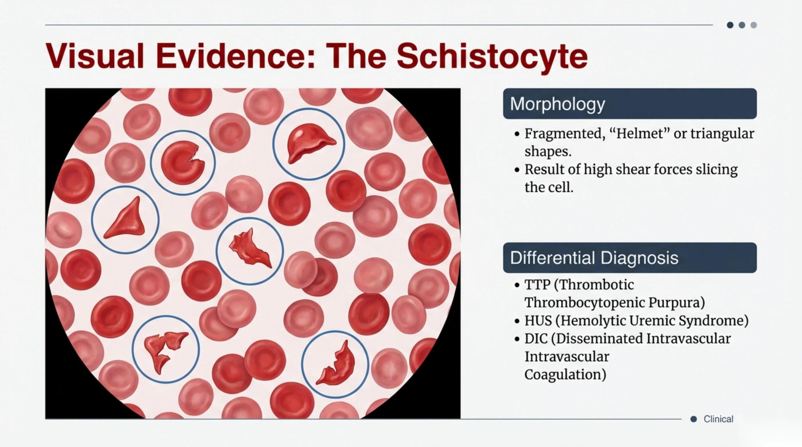
Not every small dark shape here is a spherocyte. A few cells in this field have points and straight edges: those are fragments, cut cells that belong to the mechanical half of the lecture. Describe every shape you see rather than naming the first one you recognise.
The caution from slide 15 still holds. Spherocytes show that membrane has been lost, not why. Warm AIHA and hereditary spherocytosis look the same on the film. The direct antiglobulin test separates them.
Image on the slide. “Spherocytes” by Prof. Osaro Erhabor, Wikimedia Commons, CC0 1.0 (public domain). Cropped for the slide.
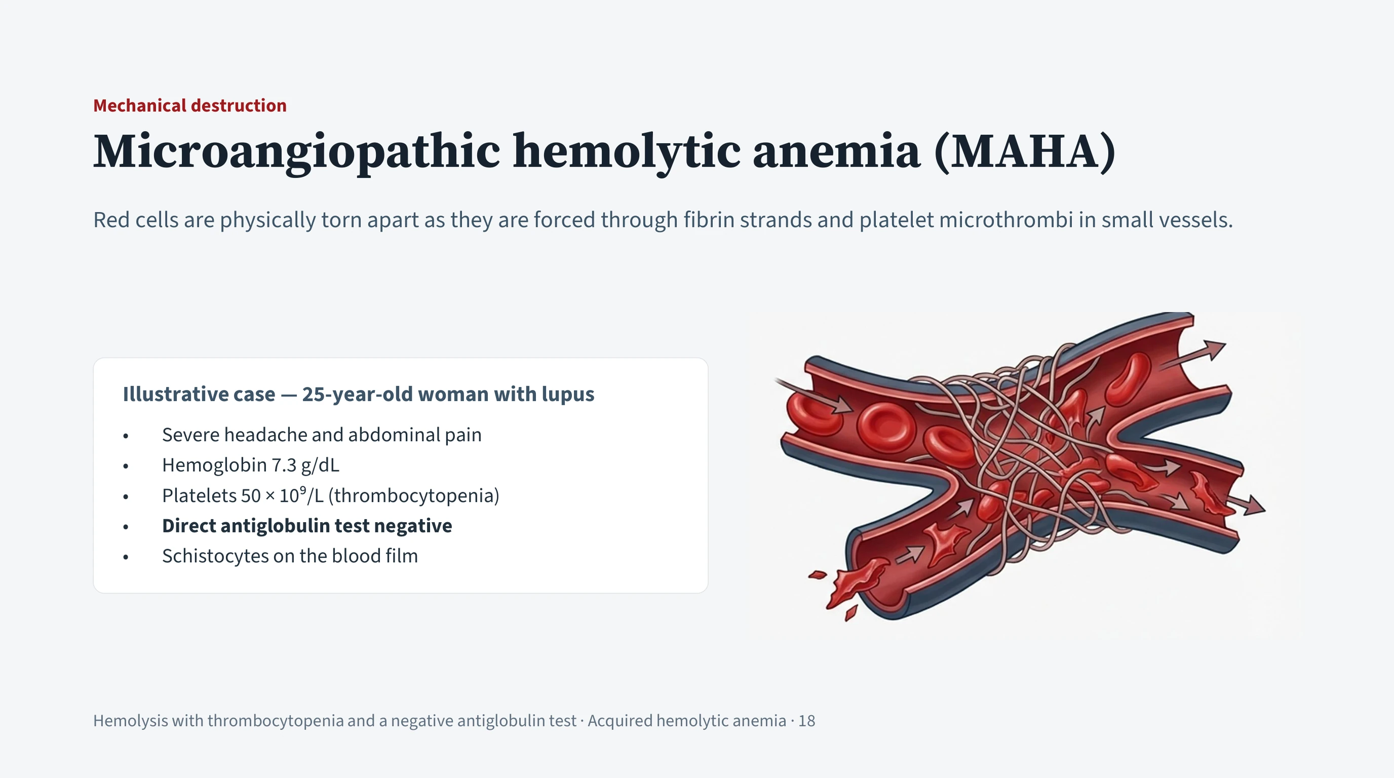
Mechanical hemolysis in one patient
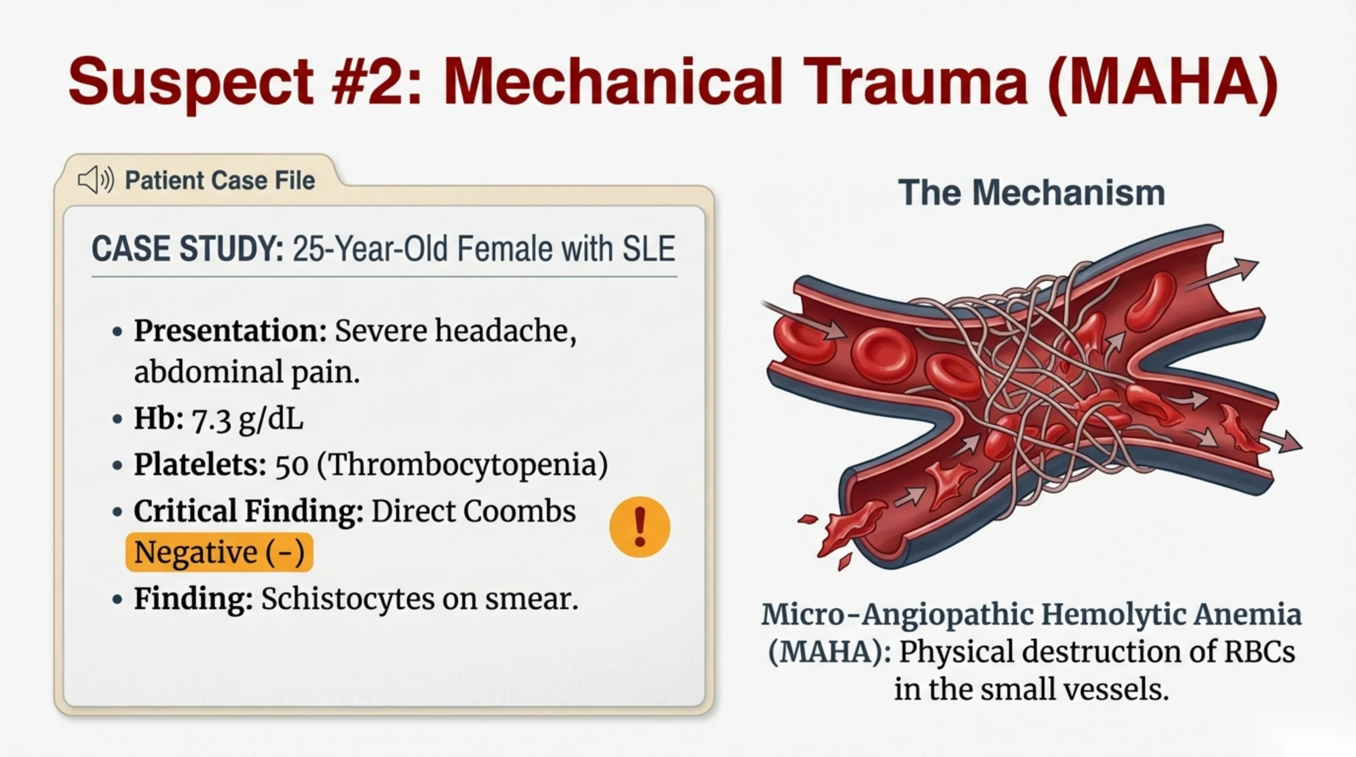
A 25-year-old woman with systemic lupus erythematosus presents with a severe headache and abdominal pain. Her hemoglobin is 7.3 g/dL, the same as the patient on slide 10. Two findings make this a different problem. Her platelet count is 50 × 109/L, well below normal, and her direct antiglobulin test is negative. There is no antibody on her red cells.
The film gives the mechanism: schistocytes, fragments of red cells. Fragmentation means the cells were broken physically, and the diagnosis is microangiopathic hemolytic anemia. Platelet and fibrin thrombi have formed in the smallest vessels, and red cells driven past them at arterial velocity are cut by the strands. The fragments are cleared from the circulation and the patient becomes anemic.
The low platelet count is part of the same process, not a separate finding. Platelets are being consumed in the microthrombi that are cutting the red cells, which is why the two cytopenias appear together.
The headache is the most concerning feature, because it suggests the thrombi are forming in the cerebral circulation.
Compare the two patients directly. The same hemoglobin, the same evidence of hemolysis, the opposite antiglobulin result. The pattern to recognise is hemolysis with a low platelet count, fragments on the film, and a negative antiglobulin test. That combination moves the problem from immunology to emergency medicine, and the next slides explain why.
See it online.
- Helmet cells in thrombotic thrombocytopenic purpura ASH Image Bank

Finding schistocytes on a real film
The patient on the previous slide had schistocytes on her film. This is what that looks like in a real field. Most of the cells are intact. The fragments are few and small, and they are easy to miss.
Look for sharp edges and straight lines. A schistocyte was cut by shear rather than nibbled by a macrophage, so it has corners: a helmet with one straight edge, a triangle, a crescent. A spherocyte, by contrast, is smooth and round.
The number matters. A few fragments occur in many conditions. Numerous fragments in a patient who is anemic and has a low platelet count mean microangiopathic hemolysis until proven otherwise.
That is why the platelet count and the film are read together. When a patient is hemolysing and the platelets are low, ask for the film to be examined for fragments. If they are there and the direct antiglobulin test is negative, treat as TTP until proven otherwise.
Image on the slide. “Schistocytes” by Prof. Osaro Erhabor, Wikimedia Commons, CC0 1.0 (public domain). Cropped for the slide.
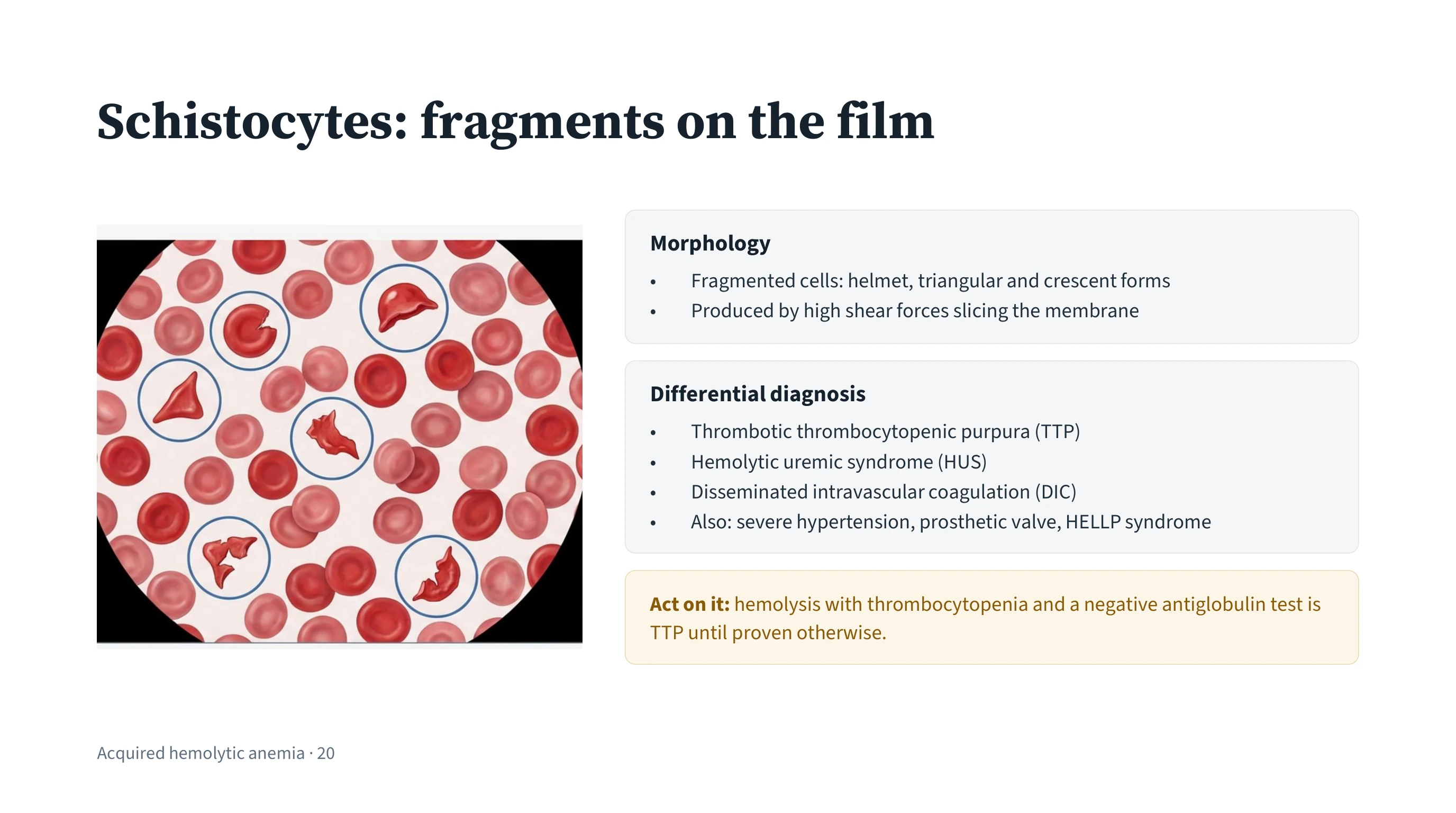
Schistocytes and the conditions that produce them
A schistocyte is a fragment of a red cell. Because it was cut by shear rather than removed piecemeal by a macrophage, it has sharp, irregular edges: a triangle, a crescent, or the helmet form with one straight edge where the cell was divided. A few schistocytes occur in many conditions. Numerous schistocytes in a patient who is anemic and thrombocytopenic mean microangiopathic hemolysis until proven otherwise.
The finding matters because of the differential it forces. Three conditions produce widespread microvascular thrombosis with fragmentation, and all three can kill within days:
- TTP, thrombotic thrombocytopenic purpura, in which platelet-rich thrombi form because von Willebrand factor is not being cleaved. The next slide is its mechanism.
- HUS, hemolytic uremic syndrome, classically following diarrhoeal infection with a toxin-producing E. coli, in which the kidneys take the greatest damage.
- DIC, disseminated intravascular coagulation, in which an underlying illness such as sepsis or an obstetric catastrophe activates the whole clotting system and consumes platelets and clotting factors together.
Four further causes shear red cells in the same way and belong on the list, because they are commoner than the three above and are managed differently: severe hypertension, a prosthetic heart valve, and, in pregnancy, HELLP syndrome. A prosthetic valve in particular should be looked for early, because the answer is an echocardiogram rather than plasma exchange.
The immediate investigations are the same whichever is suspected. Check the platelet count, the renal function and the coagulation screen, meaning the prothrombin time, the activated partial thromboplastin time and the fibrinogen, and examine the patient for fever and neurological signs. The coagulation screen is what separates them. DIC consumes clotting factors, so the times are prolonged and the fibrinogen is low. In TTP and HUS the screen is essentially normal.
TTP stays at the top of the list in every case, because it is the most rapidly fatal if it is missed and the most treatable if it is not.
Further reading. The three conditions on this list are worked through together in Thrombotic Microangiopathies: TTP and HUS, which lays them side by side on the underlying defect, the organ that suffers most, and the clotting screen — normal in TTP and HUS, deranged with a low fibrinogen in DIC. It also carries the treatments this lecture does not reach: complement blockade with eculizumab for atypical HUS, and caplacizumab added to plasma exchange in TTP. For the film itself, The Blood Film gives the habit of describing red cells in a fixed order — size, shape, colour, inclusions — and the short list of shapes that are worth a diagnosis on their own.
See it online.
- Schistocytes on the film in TTP ASH Image Bank
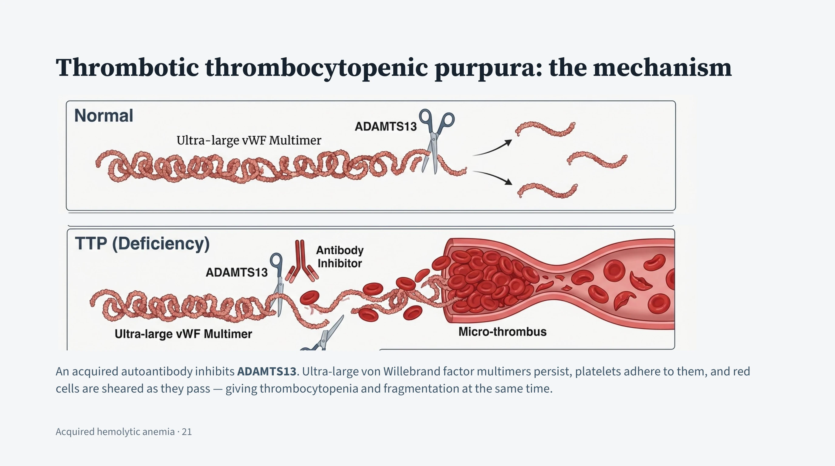
One enzyme deficiency accounts for both cytopenias
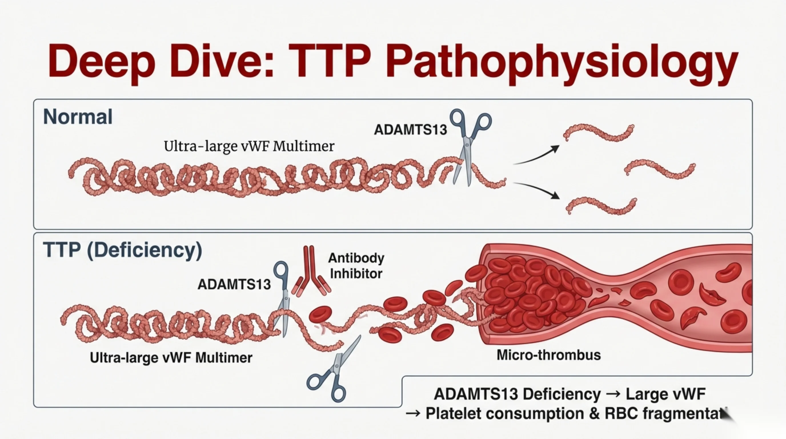
Von Willebrand factor is released by the endothelial cells lining blood vessels. Its function is to capture platelets and hold them at a site of injury. It is secreted as ultra-large multimers, long chains of many subunits, which are far too adhesive to be allowed to circulate at that length. A plasma enzyme, ADAMTS13, cleaves them down to shorter, safe lengths as they appear; the diagram on this slide draws it as a pair of scissors.
In TTP that cleavage stops. In most adult cases the patient has produced an autoantibody that binds and inhibits ADAMTS13; in rare inherited cases the enzyme is absent from birth. Either way the ultra-large multimers persist. They unfold in the rapid flow of small vessels, capture passing platelets, and build platelet-rich microthrombi throughout the body, with the brain, heart and kidneys the most vulnerable.
Every feature of the disease follows from that one defect, which is why it is worth learning as a mechanism rather than as a list. Platelets are consumed in the microthrombi, so the count falls. Red cells are fragmented as they are forced past the thrombi, so there is microangiopathic hemolysis with schistocytes. A single enzyme deficiency therefore produces both cytopenias, which is why they appear together. Organs supplied by the occluded vessels become ischemic, producing the neurological features, the renal impairment and the fever of the clinical pentad.
The mechanism also gives the treatment. If an antibody is inhibiting an enzyme, then removing the antibody and replacing the enzyme should work, and that is what plasma exchange does.
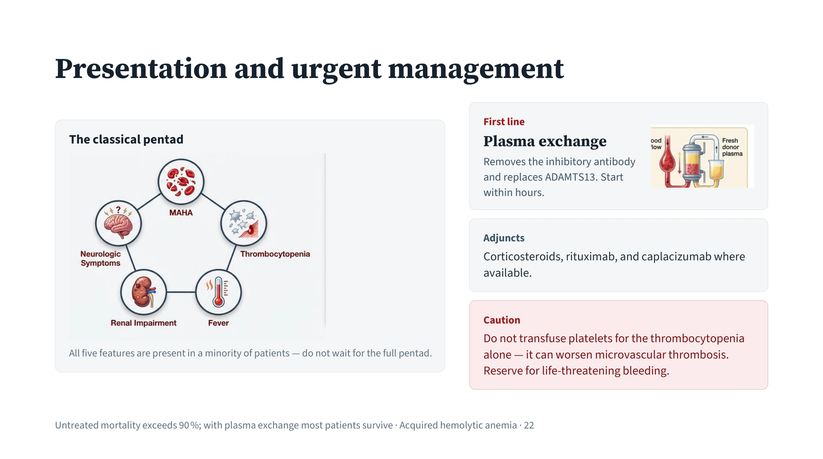
Recognising and treating TTP
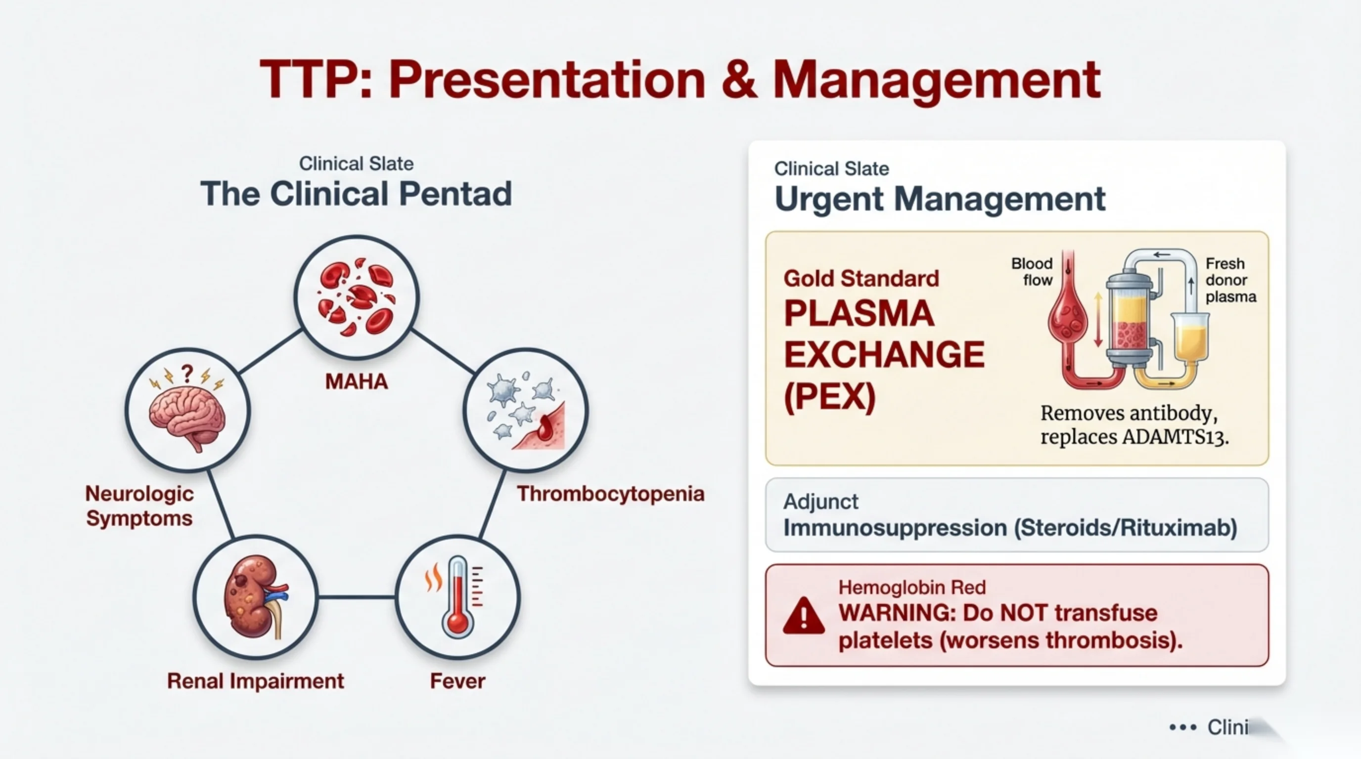
The classical description of TTP is a pentad: microangiopathic hemolytic anemia, thrombocytopenia, fever, renal impairment and neurological symptoms, which include headache, confusion, focal deficits and seizures. Know the pentad, and know equally that waiting for it is wrong. All five features are present in only a minority of patients, and when all five are present the disease is usually advanced. In practice, unexplained hemolysis with schistocytes and a low platelet count is enough to suspect TTP and to act on it.
Two figures justify treating on suspicion. Untreated mortality exceeds 90 %. With plasma exchange most patients survive. Few treatments move an outcome that far, and the cost of starting treatment in a patient who turns out not to have TTP is small against the cost of waiting.
Plasma exchange is the first-line treatment and should start within hours of the suspicion, not after the ADAMTS13 assay returns. The patient's plasma, which contains the inhibitory antibody, is removed and replaced with donor plasma, which contains functioning ADAMTS13. It removes the inhibitor and replaces the enzyme in one procedure.
Drugs are added alongside plasma exchange rather than in place of it. Corticosteroids and rituximab suppress production of the antibody, rituximab by depleting the B cells making it. Caplacizumab, where it is available, blocks the site on von Willebrand factor to which platelets bind, protecting the patient while the immunosuppression takes effect.
One rule should be learned as a reflex: do not transfuse platelets in TTP. The instinct, faced with a platelet count of 50 and falling, is to give platelets. In TTP the transfused platelets are captured by the same uncleaved von Willebrand factor multimers and add to the microthrombi, and patients have deteriorated within hours of a platelet transfusion. Reserve platelets for life-threatening bleeding. Plasma exchange comes first.
Read next. Case 51 · Acquired Haemolytic Anaemia and the Direct Antiglobulin Test in the casebook sets TTP beside HUS and DIC in one table, with the clotting screen as the fork: normal in TTP and HUS, deranged in DIC.
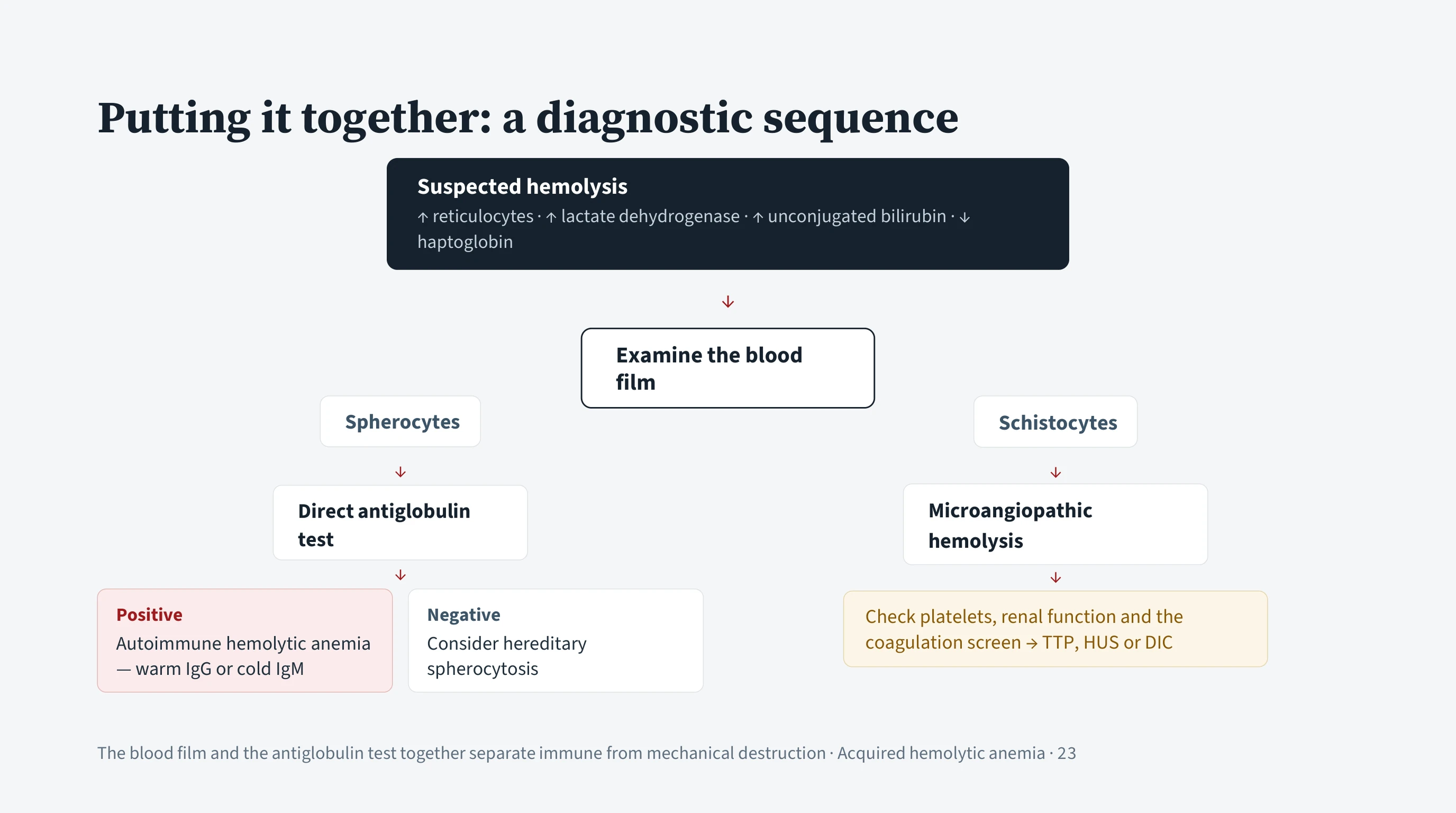
The sequence for acquired hemolysis
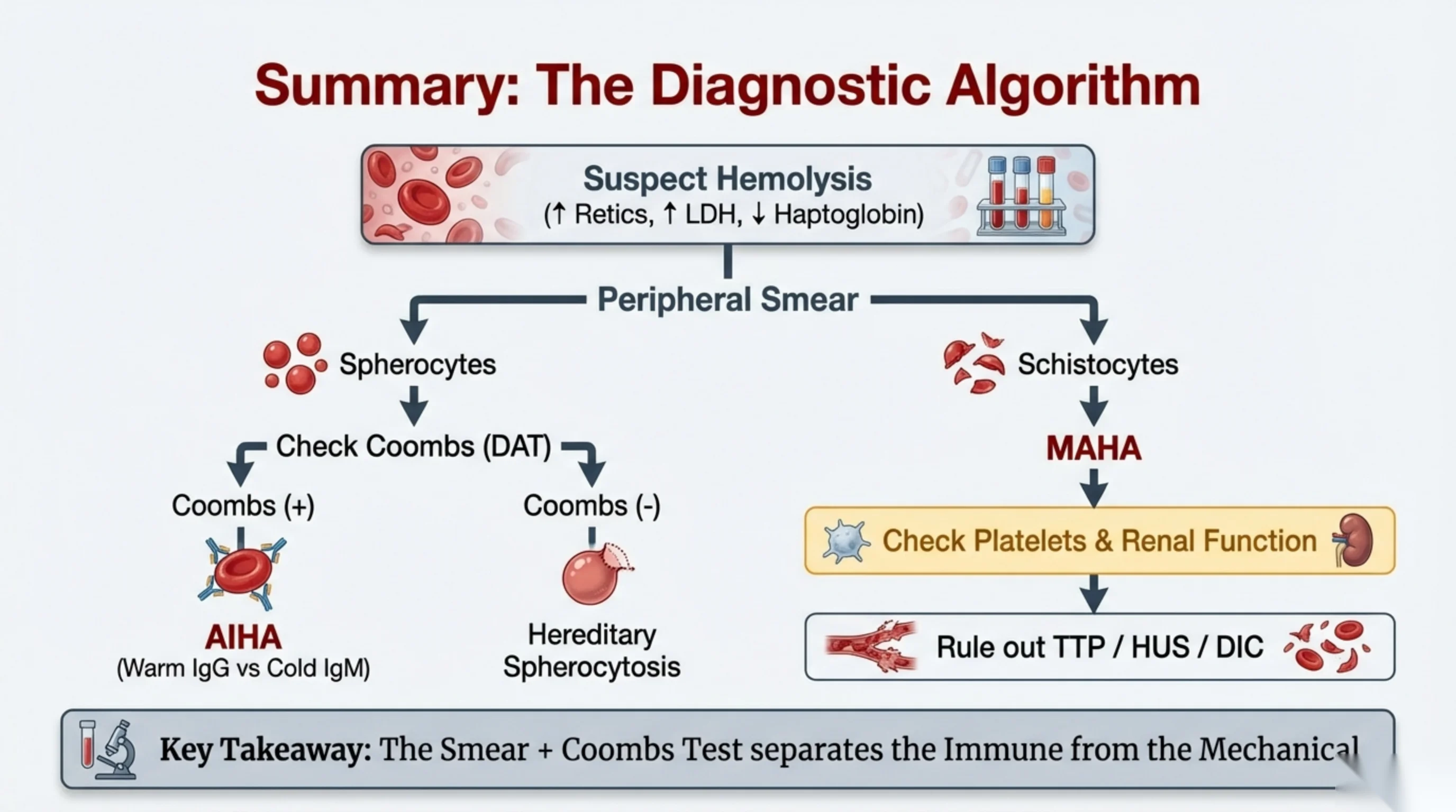
The lecture reduces to a sequence that can be run at the bedside.
Step 1: establish hemolysis. Raised reticulocytes, raised lactate dehydrogenase, raised unconjugated bilirubin, low haptoglobin. If these are absent the anemia is probably not hemolytic, and the Part 1 method is the right one to return to.
Step 2: examine the blood film. The shape of the abnormal cell gives the mechanism. This is the step most often skipped.
Spherocytes mean membrane has been lost, and there are two ways for that to happen. Order the direct antiglobulin test. A positive result means antibody-coated cells and the diagnosis is autoimmune hemolytic anemia; the antibody class and the pattern on the test then separate warm IgG from cold IgM disease. A negative result, with spherocytes and usually a family history, points to hereditary spherocytosis, which Part 3 covers.
Schistocytes mean the cells were cut, and the diagnosis is microangiopathic hemolysis. Check the platelet count, the renal function and the coagulation screen, and separate TTP, HUS and DIC, with TTP at the top of the list because plasma exchange must start early.
The two patients in this lecture run through the sequence as a check on it. The 41-year-old takes the left branch: hemolysis, spherocytes, a positive test, warm AIHA. The 25-year-old takes the right: hemolysis, schistocytes, a low platelet count, a negative test, and an emergency.
In one line: the blood film and the antiglobulin test separate immune destruction from mechanical destruction, and the rest follows from those two results.
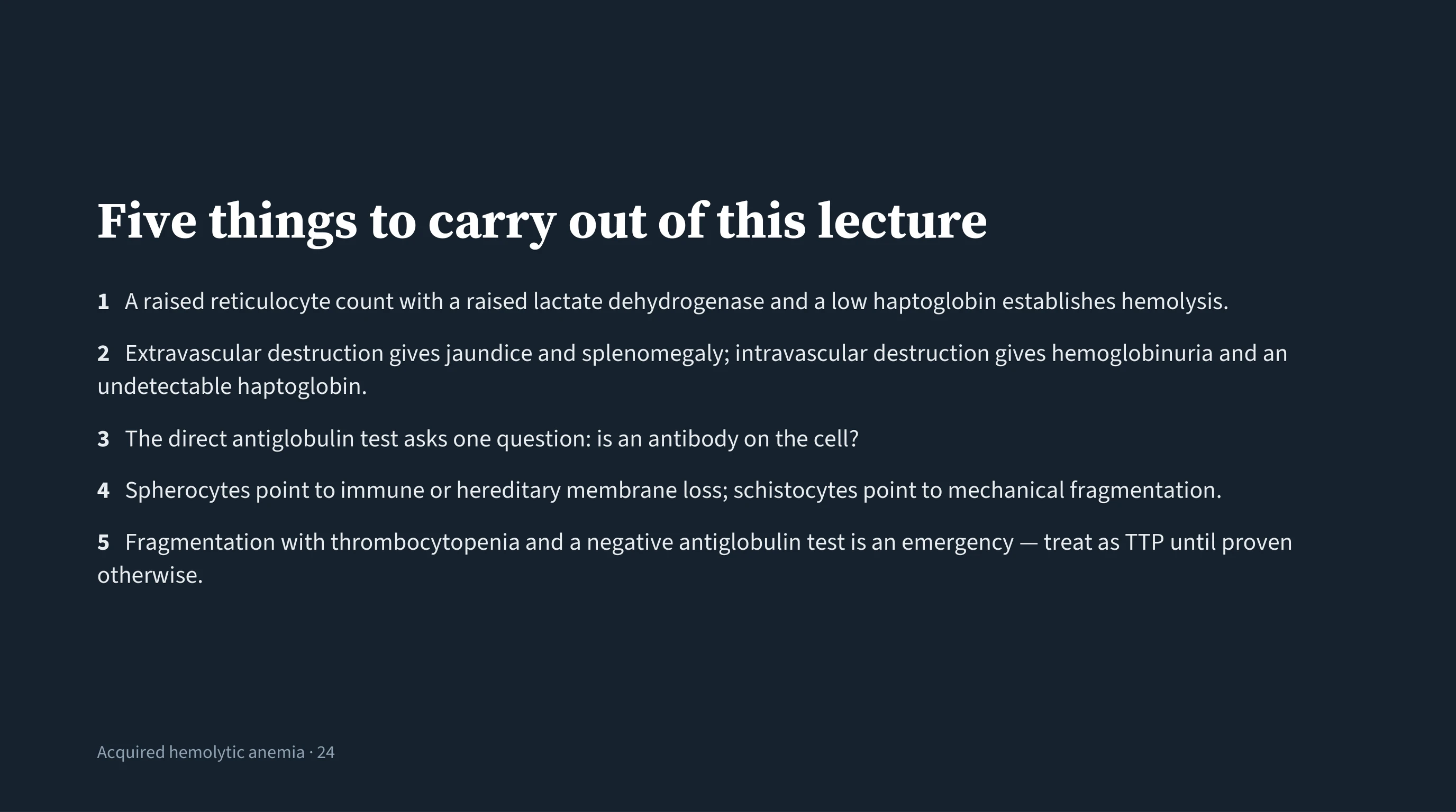
The five points worth memorising
Most of this lecture is understanding, and understanding does not need to be memorised because it can be rebuilt from the mechanism. These five are points at which something has to be recognised or decided, so they are worth carrying as they stand.
One is the gate. Three results together, not one alone, establish that cells are being destroyed. Without them the wrong problem is being investigated.
Two reads the rest of the picture. Jaundice and an enlarged spleen indicate that the spleen is doing the destroying. Dark urine and an undetectable haptoglobin indicate that the cells are lysing in the vessel.
Three keeps the antiglobulin test within its limits. It answers one question and no other, and a negative result does not mean the patient is not hemolysing.
Four is what the film contributes. Two shapes, two mechanisms: round cells with no central pallor mean membrane has been removed, by an antibody or by a hereditary fault; fragments mean the cell was cut.
Five is the only point on the list that is a decision rather than a piece of knowledge, and it is where the lecture ends. Fragments with a low platelet count and a negative antiglobulin test are not a diagnosis to be worked up over the week. Treat as TTP and start plasma exchange.
Hereditary Hemolytic Anemia
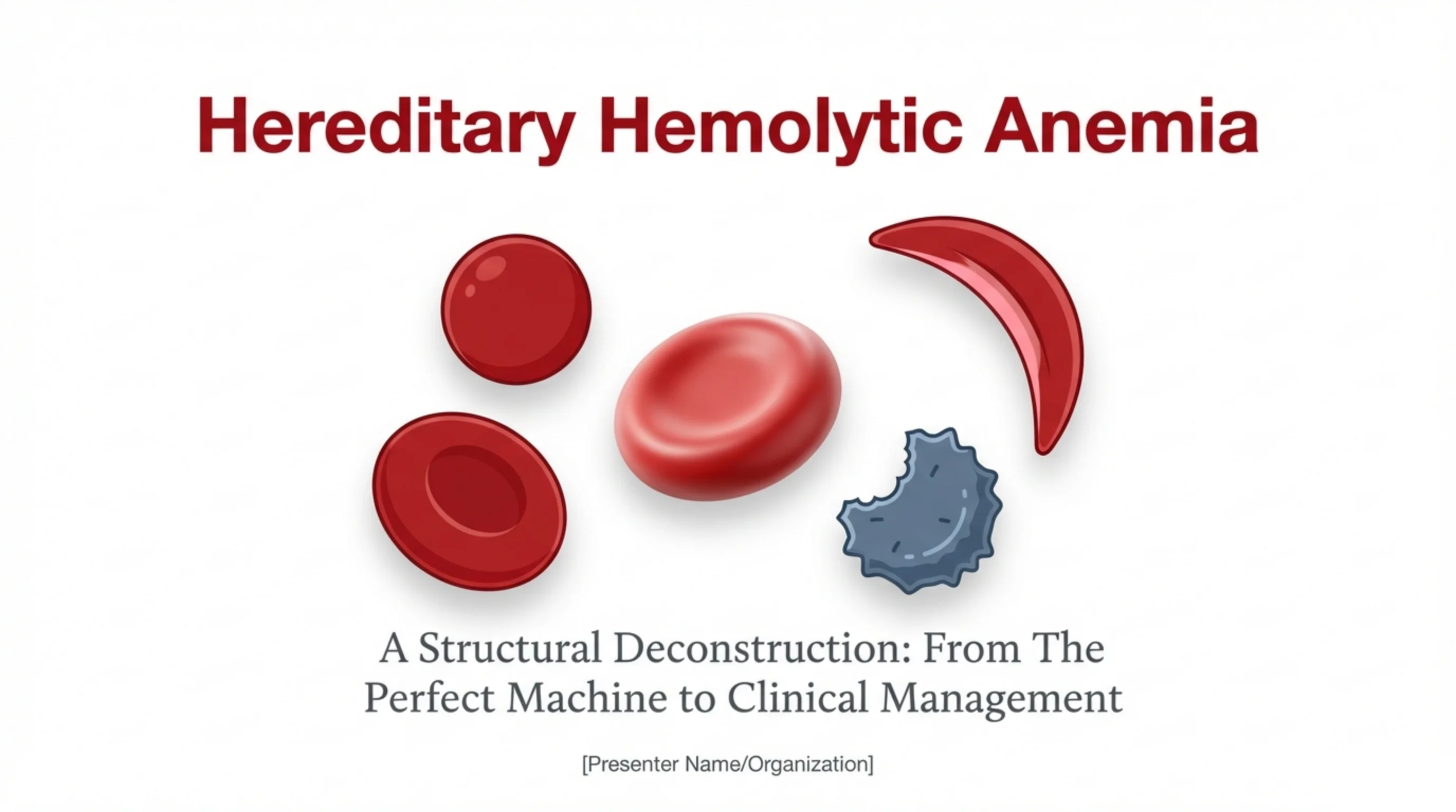
Where Part 3 fits
These slides are an outline only. Read the accompanying material with them: the notes under each slide on this page, and the Hematology Handbook. Then work through the Internal Medicine Casebook and the Hematology Question Bank.
Part 2 dealt with red cells that were built correctly and then attacked. This final lecture deals with red cells that were built with a flaw. These are the hereditary hemolytic anemias, and they are especially important in Saudi Arabia, where sickle cell disease, thalassemia and G6PD deficiency are all common.
The approach is structural. The red cell is treated as a machine with three parts, and each hereditary disease is understood as the failure of one part. Once you know which part has failed, you can predict the shape of the cell on the smear, the pattern of hemolysis, the confirmatory test and the broad lines of management. The lecture ends by folding this back into the diagnostic algorithms you already know from Parts 1 and 2.
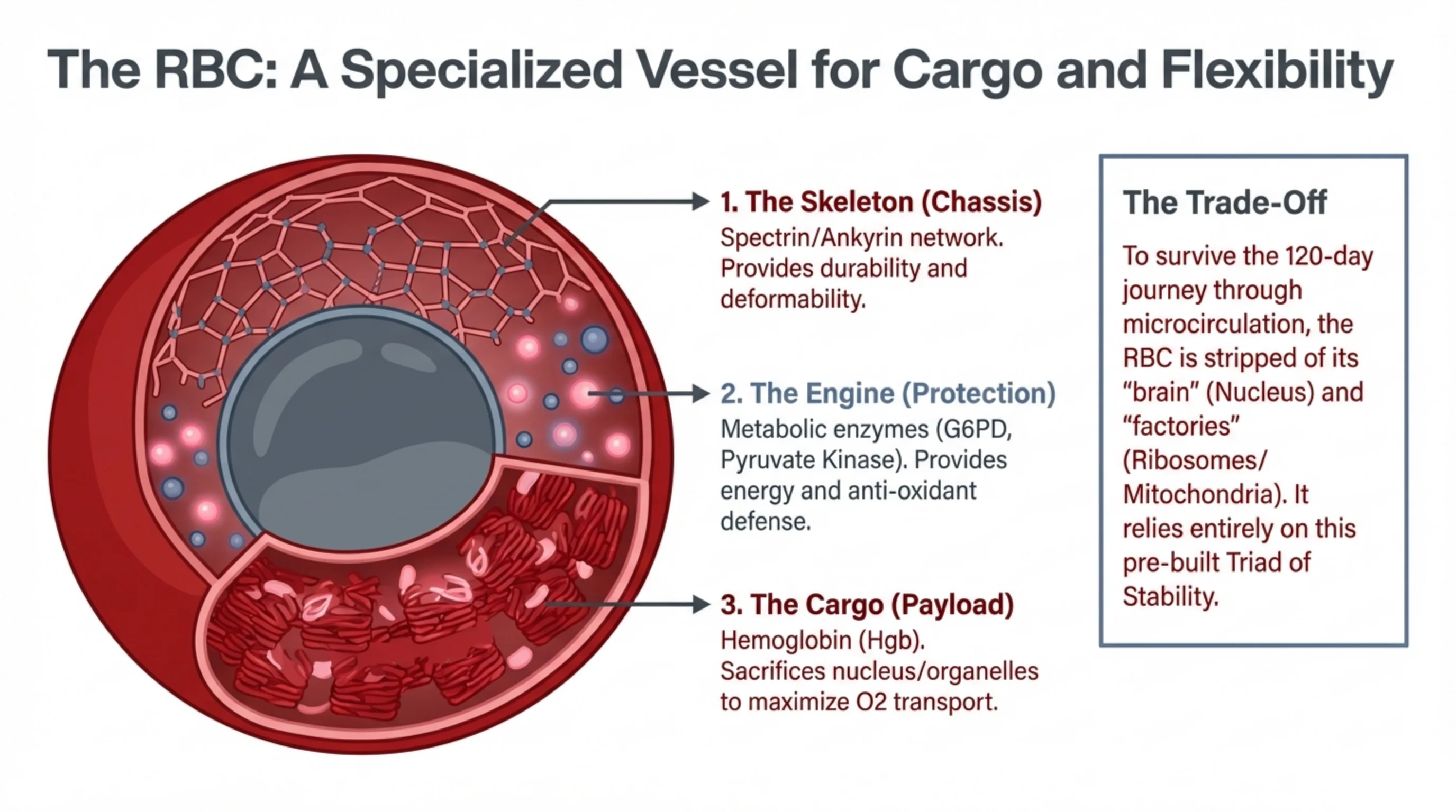
Three parts that keep a red cell alive
A red cell has to survive about 120 days of being squeezed through capillaries narrower than itself, in a bloodstream full of oxygen radicals, with no nucleus to direct repairs and no ribosomes or mitochondria to make new proteins or generate energy efficiently. It manages this because it left the marrow with three things pre-built.
The chassis: the membrane skeleton. Beneath the lipid bilayer lies a mesh of proteins, chiefly spectrin, anchored to the membrane by ankyrin and band 3. This mesh gives the cell its biconcave shape, its strength, and above all its deformability, the ability to fold and spring back millions of times.
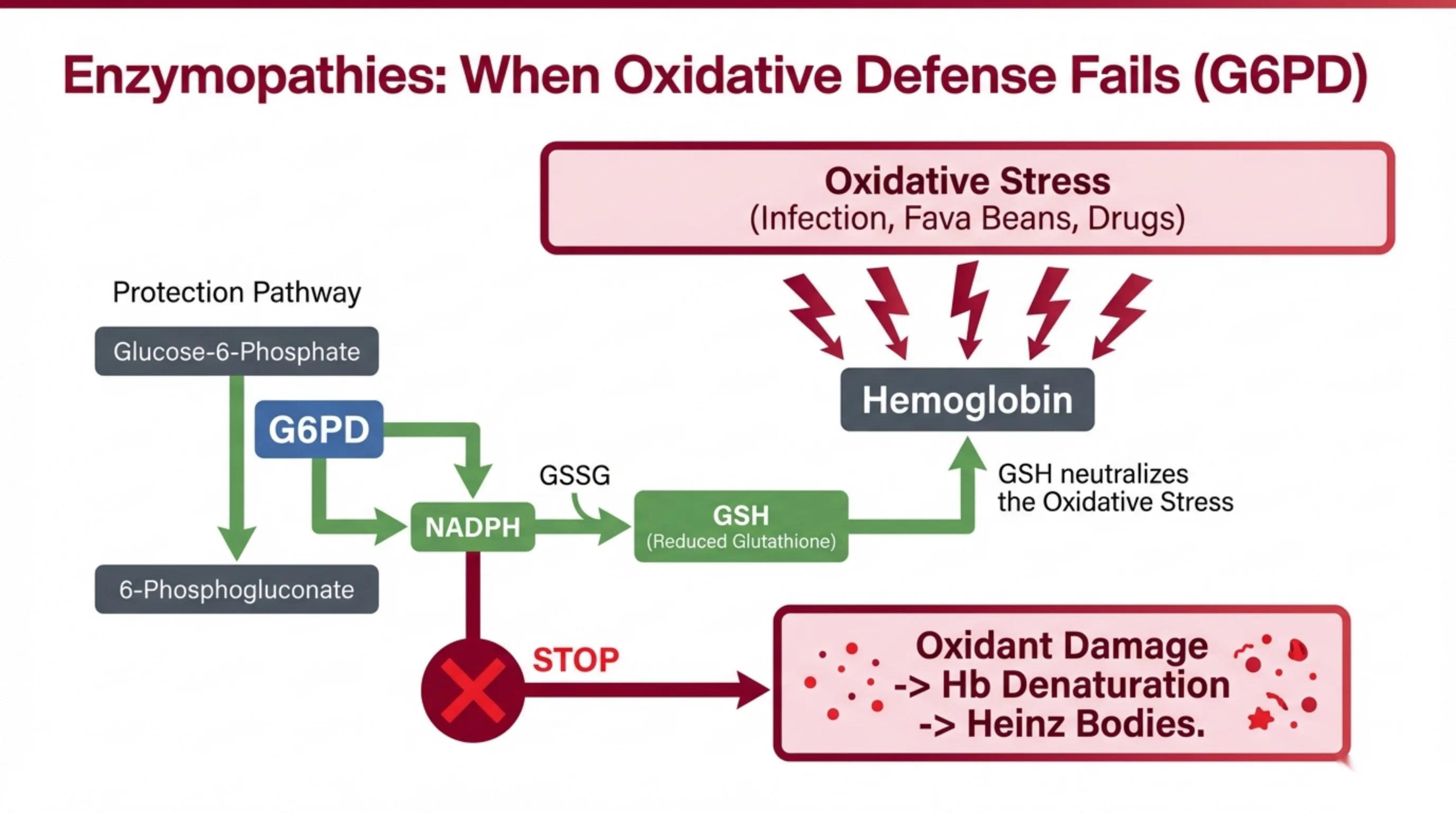
The engine: metabolic enzymes. With no mitochondria, the cell runs on glycolysis, and pyruvate kinase is a key enzyme in that pathway. A side branch, the pentose phosphate pathway, uses G6PD to generate NADPH, the cell's only defence against oxidative damage. Both energy and antioxidant defence come from enzymes that cannot be replaced once they wear out.
The cargo: hemoglobin. The point of the whole machine. The cell is packed with hemoglobin, and hemoglobin must stay soluble and stable at very high concentration for the life of the cell.
This is the survival bargain: maximum cargo and flexibility in exchange for no capacity to repair. A flaw in any one of the three parts at birth shortens the whole journey, and that is hereditary hemolysis.
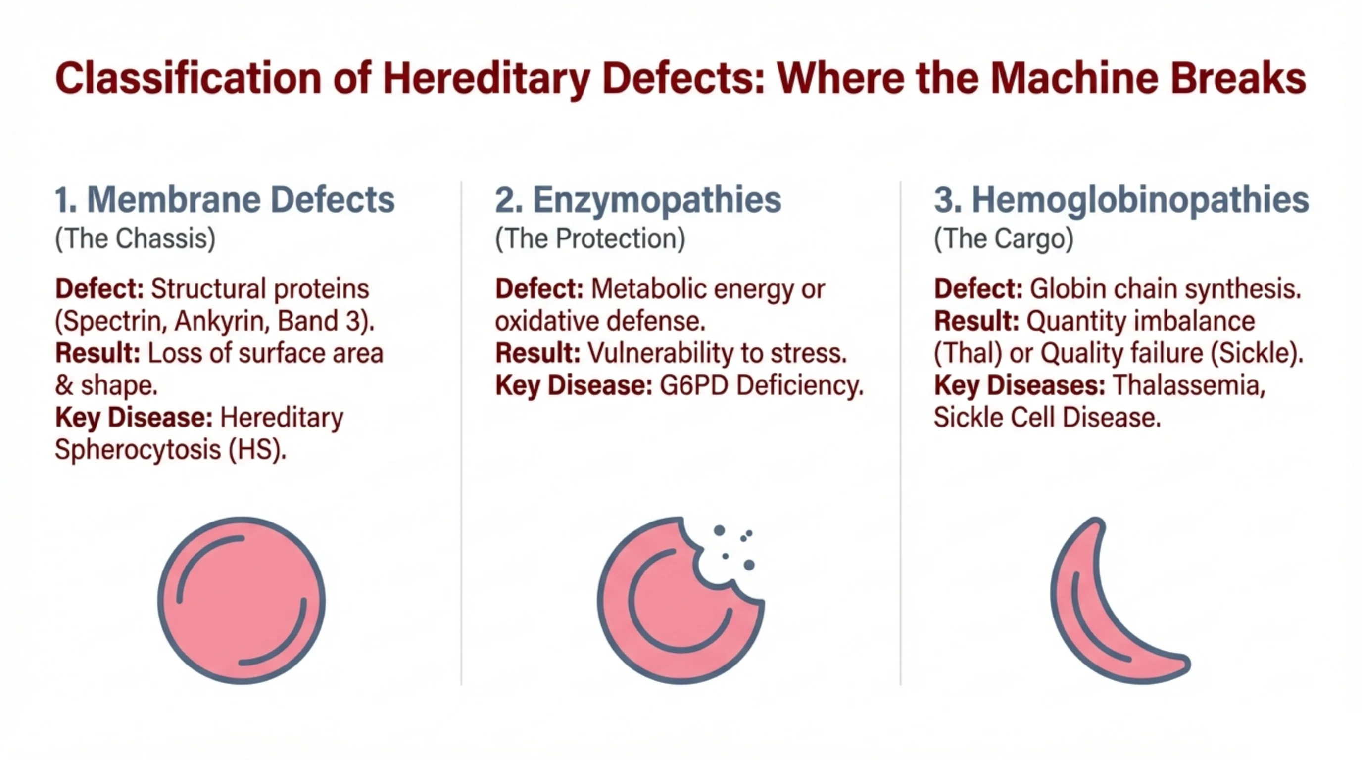
Three families of disease, one for each part
Each of the three parts of the red cell can be defective from birth, and each defect produces a recognisable family of diseases with its own signature cell shape.
Membrane defects. A mutation in spectrin, ankyrin or band 3 weakens the skeleton. The membrane sheds fragments, surface area is lost, and the cell becomes a sphere. The key disease is hereditary spherocytosis, and the signature cell is the spherocyte.
Enzyme defects (enzymopathies). The cell cannot generate enough energy or cannot defend itself against oxidants. The cell looks normal until it is stressed, and then it fails abruptly. The key disease is G6PD deficiency, and the signature findings are Heinz bodies and bite cells during an episode.
Hemoglobin defects (hemoglobinopathies). The cargo itself is wrong. Either there is too little of one globin chain (thalassemia, a quantity problem) or the chain is structurally abnormal (sickle cell disease, a quality problem). The signature cells are the target cell in thalassemia and the sickle cell in sickle cell disease.
If you carry this three-part map in your head, the rest of the lecture is simply filling in the detail for each family, and the last two slides show how the map turns into a diagnostic algorithm.
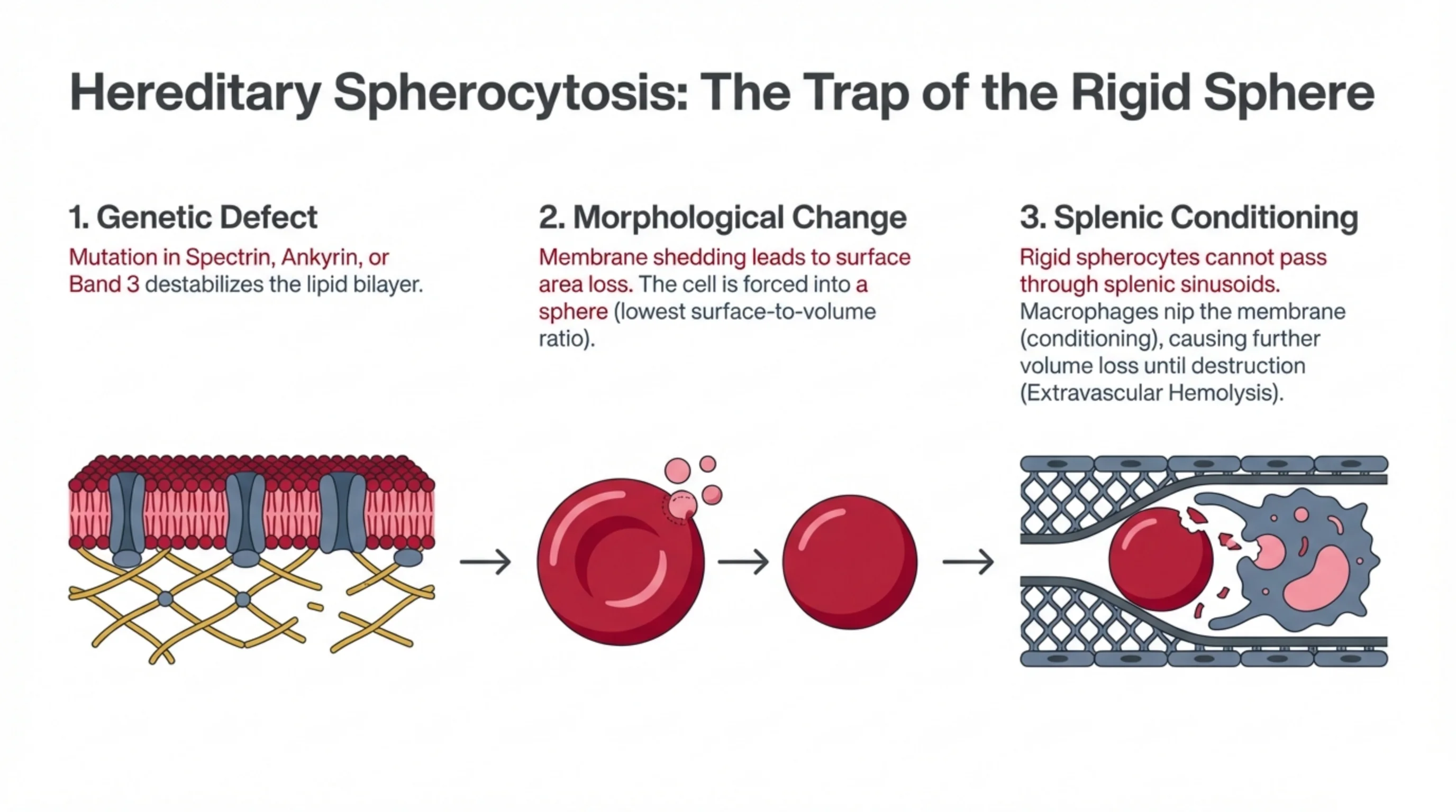
Hereditary spherocytosis: a weak skeleton makes a rigid ball
Hereditary spherocytosis (HS) is the commonest inherited membrane disorder, usually passed down as an autosomal dominant trait, so a family history of anemia, jaundice, gallstones or splenectomy is common. The story unfolds in three steps.
Step 1, the defect. A mutation in one of the skeleton proteins (spectrin, ankyrin or band 3) weakens the link between the protein mesh and the lipid bilayer above it. Parts of the bilayer are no longer held down.
Step 2, the shape change. Unsupported patches of membrane bud off as tiny vesicles. Every time this happens the cell loses surface area but keeps its volume. As in warm AIHA (Part 2, slide 16), geometry forces the cell into the shape with the least surface for its volume: a sphere. The spherocyte is small, dense, has no central pallor, and has lost the slack that let it deform.
Step 3, the spleen finishes it. A rigid sphere cannot squeeze through the narrow slits of the splenic sinusoids. It is held up in the splenic cords, in a low-glucose, acidic environment, where macrophages nip off still more membrane, a process called splenic conditioning. Each passage makes the cell rounder and more fragile until it is finally engulfed. The hemolysis is therefore extravascular, with jaundice, splenomegaly, and a high risk of pigment gallstones from all the bilirubin.
Because the spleen is where the cells die, removing the spleen largely stops the hemolysis, even though the cells remain spherical. That is why splenectomy is the definitive treatment for severe HS, balanced against the lifelong infection risk it brings.
Case: proving hereditary spherocytosis
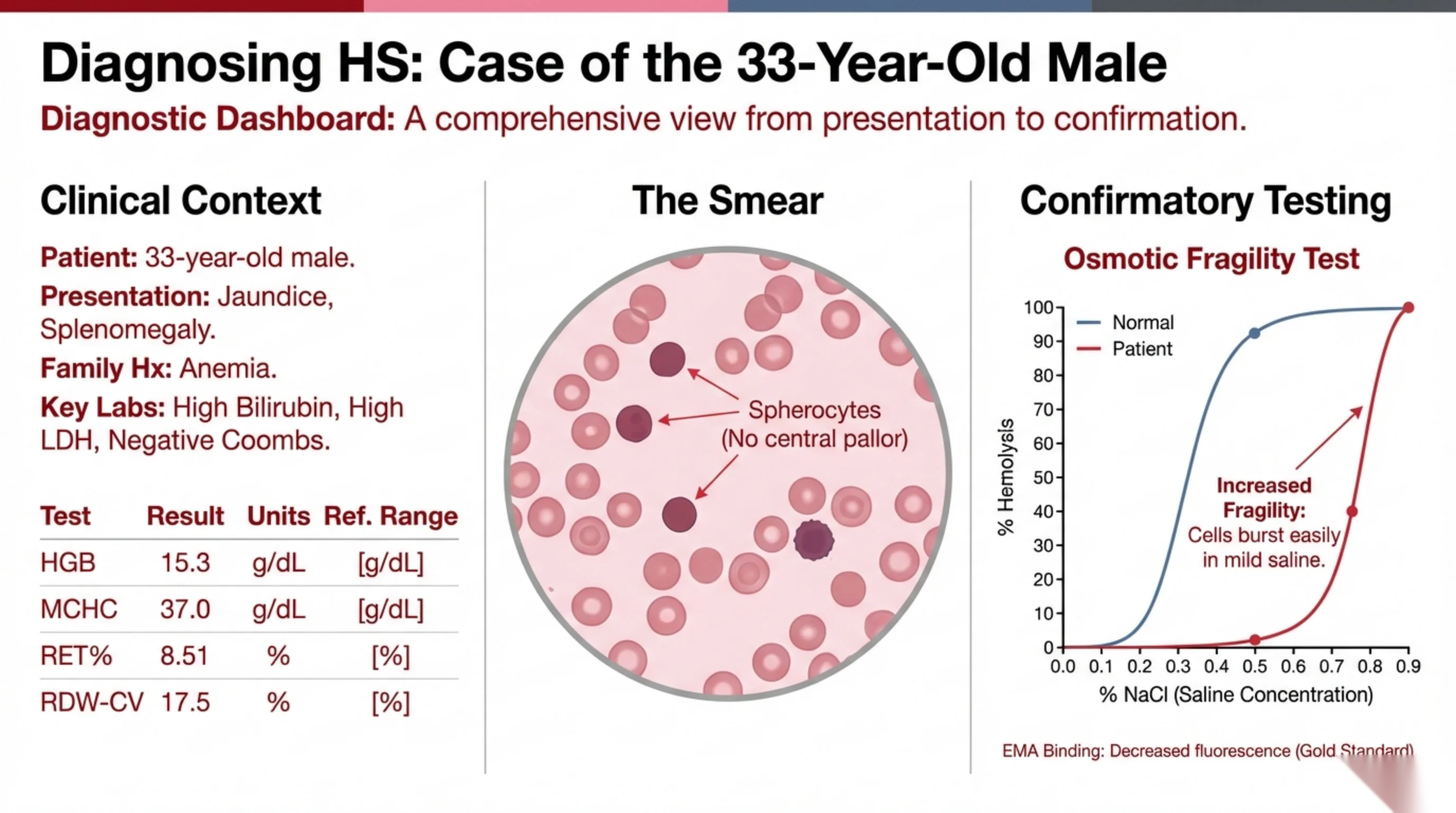
A 33-year-old man presents with jaundice and an enlarged spleen. He mentions that anemia runs in his family. His bilirubin and LDH are high and his Coombs test is negative, so he has hemolysis that is not caused by an antibody. His smear shows spherocytes. Everything points to hereditary spherocytosis, and the laboratory now confirms it.
Look first at the CBC. His hemoglobin is 15.3 g/dL, which is not low: his marrow is fully compensating, a state sometimes called compensated hemolysis, and the reticulocyte count of 8.5% shows how hard it is working. The MCHC, the mean concentration of hemoglobin inside each cell, is 37 g/dL, which is high. This is a very useful clue, because spherocytes have lost membrane but kept their hemoglobin, so they are unusually concentrated. A raised MCHC in a jaundiced patient should make you think of HS. The RDW of 17.5% is raised because spherocytes of many sizes mix with normal cells.
The classic confirmatory test is osmotic fragility. Red cells are placed in a series of salt solutions of decreasing concentration. As the solution becomes more dilute than the cell, water enters and the cell swells. A normal biconcave cell has spare membrane and can swell a great deal before it bursts, so it survives down to quite dilute saline. A spherocyte is already a sphere with no slack; a little extra water bursts it. On the graph, the patient's curve is shifted to the right: his cells burst at higher, milder salt concentrations than normal. That is "increased osmotic fragility".
The modern gold standard is the EMA binding test. Eosin-5-maleimide is a fluorescent dye that binds to band 3 on the red cell surface. Cells that have lost membrane, and with it band 3, bind less dye, so reduced fluorescence on flow cytometry confirms HS quickly and with a small sample.
Further reading. Hereditary Hemolytic Anemias: Membrane and Enzyme Defects compresses this case into three findings worth carrying together: spherocytes, a high MCHC, and a negative direct antiglobulin test — the last of the three being what separates it from the warm AIHA of Part 2. It also gives the management the lecture only gestures at: folate for every patient with a chronic hemolysis, and vaccination against encapsulated organisms before a splenectomy rather than after it. And it returns to the enzyme named on slide 2 and then dropped — pyruvate kinase deficiency, the commonest defect of the glycolytic pathway, which gives a chronic hemolytic anemia with splenomegaly but no spherocytes.
See it online.
- Spherocytes in hereditary spherocytosis ASH Image Bank
G6PD: the only antioxidant defence the red cell has
A red cell is a sack of hemoglobin bathed in oxygen, and oxygen constantly generates reactive by-products that would oxidise the hemoglobin and the membrane. The cell has one line of defence, and it runs through a single enzyme.
The pathway works like this. Glucose-6-phosphate, the first product of glucose entering the cell, can be diverted from glycolysis into the pentose phosphate pathway. The first step of that pathway is catalysed by G6PD (glucose-6-phosphate dehydrogenase), and it generates NADPH. NADPH has one crucial job in the red cell: it recharges glutathione, converting the oxidised form (GSSG) back to the reduced form (GSH). Reduced glutathione is the molecule that actually neutralises the oxidants before they reach hemoglobin. So the chain is G6PD, then NADPH, then GSH, then protection.
Now remove G6PD. Under ordinary conditions the cell may cope, because the demand for NADPH is low. But add an oxidative stress, an infection, fava beans, or one of a list of drugs, and the demand for GSH suddenly rises. With no G6PD there is no NADPH, so no GSH can be regenerated, and the oxidants reach the hemoglobin unopposed. The hemoglobin is oxidised and denatured, and it precipitates into clumps that stick to the inside of the membrane. These clumps are Heinz bodies, and they are the starting point of the hemolysis described on the next slide.
The lesson in one sentence: G6PD deficiency is not a disease of the cell at rest; it is a disease of the cell under attack.
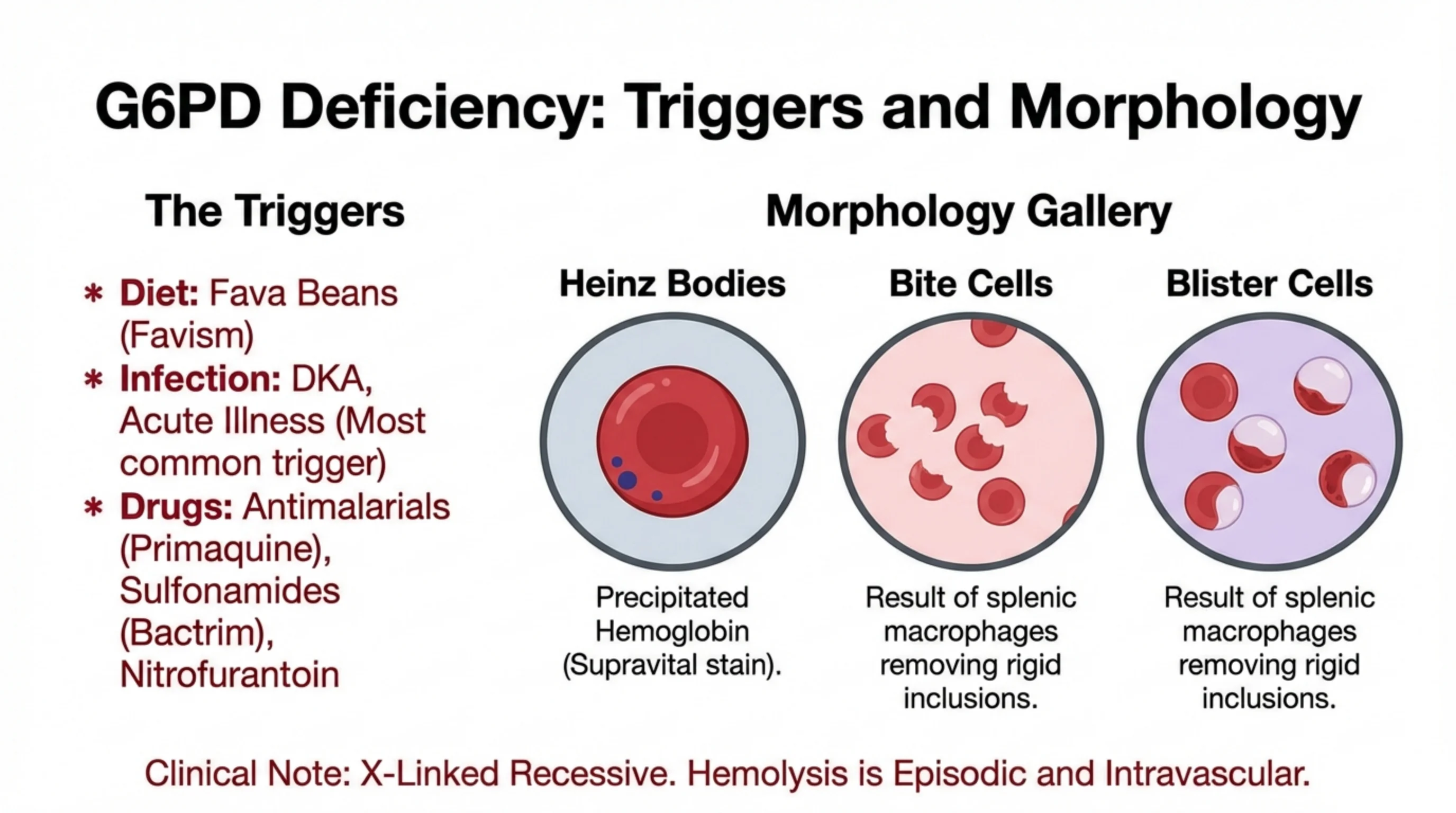
What sets off a G6PD crisis, and what it leaves on the smear
People with G6PD deficiency are usually well. Their cells fail only when an oxidative stress overwhelms the missing defence, so the triggers are the heart of the clinical picture.
- Diet. Fava beans (broad beans) contain compounds that generate oxidants. Hemolysis after eating them, favism, is the classic presentation in children in the Mediterranean and the Middle East.
- Infection. The most common trigger of all. The immune response to infection releases oxidants, and any acute illness, including diabetic ketoacidosis, can precipitate an episode. When a G6PD-deficient patient becomes anemic and jaundiced, look for the infection first.
- Drugs. Antimalarials such as primaquine, sulfonamide antibiotics (co-trimoxazole), and nitrofurantoin are the best-known offenders. Check the G6PD status before prescribing them where the deficiency is common.
The morphology follows from the mechanism. Oxidised hemoglobin precipitates as Heinz bodies, which are invisible on a routine stain but show up as dark dots with a supravital stain. As the cell passes through the spleen, macrophages pluck out the rigid Heinz body along with a piece of membrane, leaving a cell with a semicircular defect in its edge: the bite cell. In some cells the hemoglobin is pushed to one side, leaving a clear, empty-looking area under the membrane: the blister cell. Bite cells and blister cells on a smear in a patient with acute hemolysis are the fingerprints of oxidative damage.
Two final points. The gene is on the X chromosome, so the deficiency is expressed mainly in males, with female carriers usually milder. And the hemolysis is episodic and largely intravascular: dark urine, a sharp fall in hemoglobin, and recovery once the trigger is removed. Between episodes the patient is normal, and management is almost entirely avoidance of the triggers.
Further reading. The casebook runs these triggers through a real workup in the haemolysis section of Case 50 · Macrocytic and Haemolytic Anaemia. Its drug list is longer than the one here — dapsone, methylene blue, rasburicase and some quinolones sit alongside primaquine, the sulphonamides and nitrofurantoin — and it adds a trigger that matters locally: henna applied to newborns has caused severe haemolysis. It also settles the timing of the confirmatory test, which this slide leaves open. The enzyme assay can read falsely normal during the episode itself, because the most deficient cells have already been destroyed and the young cells replacing them are rich in enzyme. Repeat it about three months after the episode before you conclude that a patient is not deficient. Then read Case 42 · Malaria, where the same trigger meets the ward: primaquine causes severe hemolysis in G6PD deficiency, so the enzyme is tested before it is prescribed, always.
See it online.
- Bite cells, blister cells and Heinz bodies in G6PD deficiency ASH Image Bank
- Bite cells ASH Image Bank
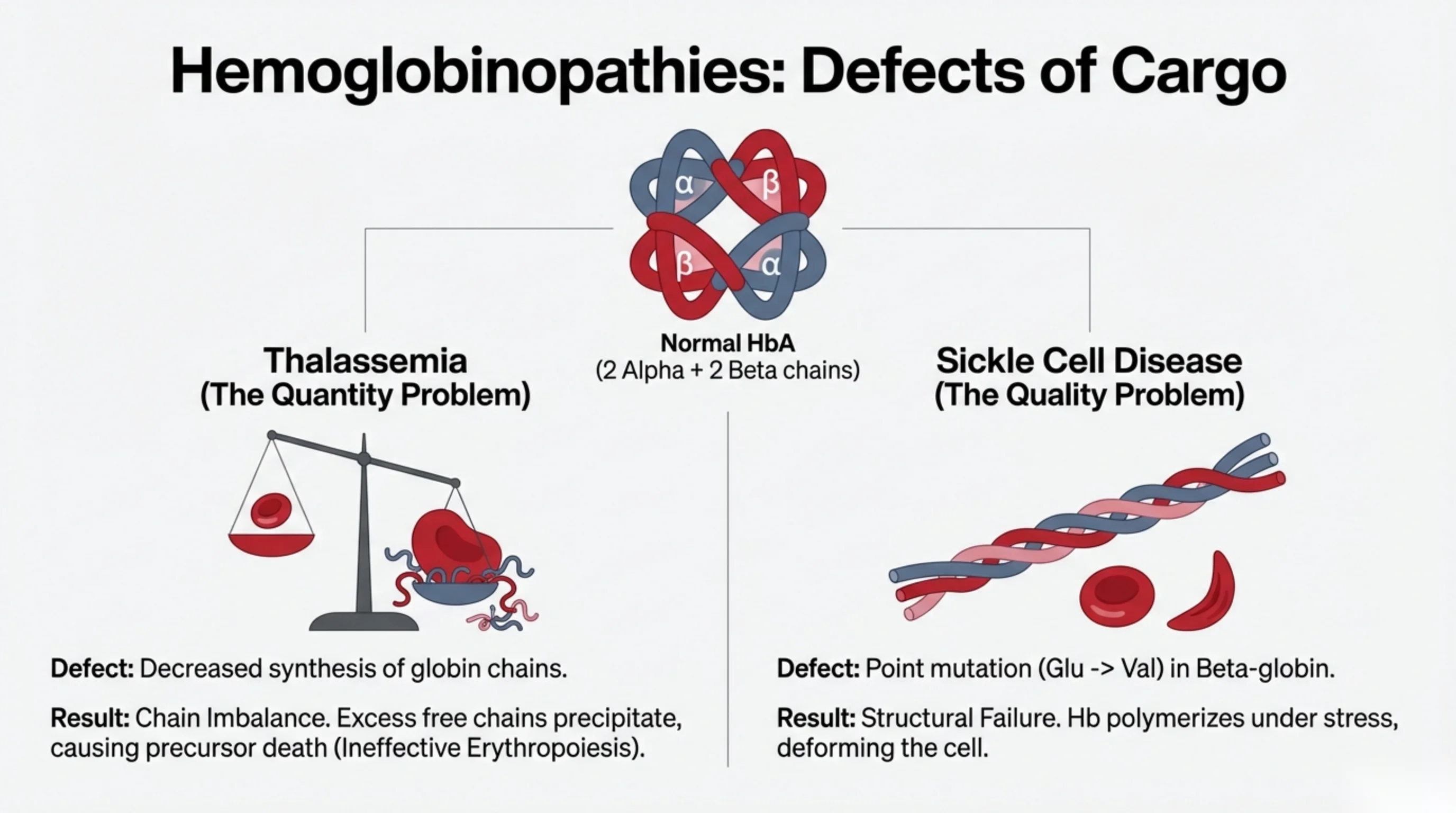
Two ways the hemoglobin itself can be wrong
Normal adult hemoglobin, HbA, is a tetramer of two α-globin chains and two β-globin chains, each carrying a heme. For the molecule to work, the chains must be made in equal amounts and each must fold correctly. A hemoglobinopathy is a failure of one of those two requirements.
Thalassemia is a quantity problem. The globin chains are structurally normal, but one type is produced in reduced amounts or not at all. The chains that are still being made in normal quantity have no partners to pair with. These unpaired chains are unstable; they precipitate inside the developing red cell in the marrow and damage it, so many precursors die before they are ever released. This is ineffective erythropoiesis: a marrow working furiously but producing little. The cells that do escape are small and hemoglobin-poor, which is why thalassemia is a microcytic anemia and lands in the same differential as iron deficiency.
Sickle cell disease is a quality problem. The β-chain is produced in normal amounts, but a single amino-acid substitution (glutamic acid replaced by valine at position 6) changes its surface. The resulting hemoglobin S works normally when oxygenated, but when it gives up its oxygen the altered surface lets molecules stack together into long rigid fibres. The fibres distort the cell into the sickle shape, and the sickled cell is both fragile and prone to jam in small vessels.
Keep the two ideas separate: thalassemia is not enough of a normal chain; sickle cell is enough of an abnormal chain. The next slides take each in turn.
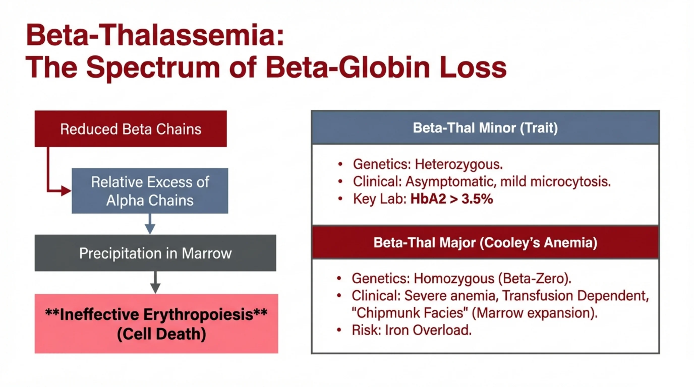
β-thalassemia: from mild trait to transfusion dependence
In β-thalassemia the β-globin genes produce too little β-chain. The α-chains, made in normal amounts, are left without partners. Free α-chains are highly unstable; they precipitate in the developing red cells in the marrow, damage the membrane, and cause the precursors to die before release. This ineffective erythropoiesis is the central pathology, and the severity of the disease depends on how much β-chain is still being made.
β-thalassemia minor (trait) is the heterozygous state: one normal β gene and one defective one. Enough β-chain is made for the patient to be essentially well. The blood count shows mild microcytosis with little or no anemia, and, as you saw in Part 1, the MCV is strikingly low for the degree of anemia and the RDW is normal. The confirmatory test is hemoglobin electrophoresis. Because β-chains are scarce, some δ-chains pair with α instead, and the level of HbA₂ (α₂δ₂) rises above 3.5%. A raised HbA₂ in a microcytic patient with normal iron stores is diagnostic. The main importance of recognising trait is to avoid treating it with iron and to offer genetic counselling, because two carriers have a one-in-four chance of a child with the major form.
β-thalassemia major (Cooley's anemia) is the homozygous state, in the severest form with no β-chain at all (β-zero). Severe anemia appears in the first year of life as fetal hemoglobin declines. The marrow expands enormously in its futile effort to compensate, widening the bones of the skull and face to produce the "chipmunk facies", and the spleen enlarges. These children survive only with regular transfusions. But every unit of blood delivers iron the body cannot excrete, and iron overload, damaging the heart, liver and endocrine glands, becomes the main threat to life. Lifelong iron chelation is therefore as important as the transfusions themselves.
Read next. Case 52 · Sickle Cell Disease and Thalassaemia in the casebook carries the thalassemia detail on from this slide: the transfusion target in the major form, and why cardiac iron overload, not the anemia, is the usual cause of death in a transfused patient.
See it online.
- The blood film in beta-thalassemia major ASH Image Bank
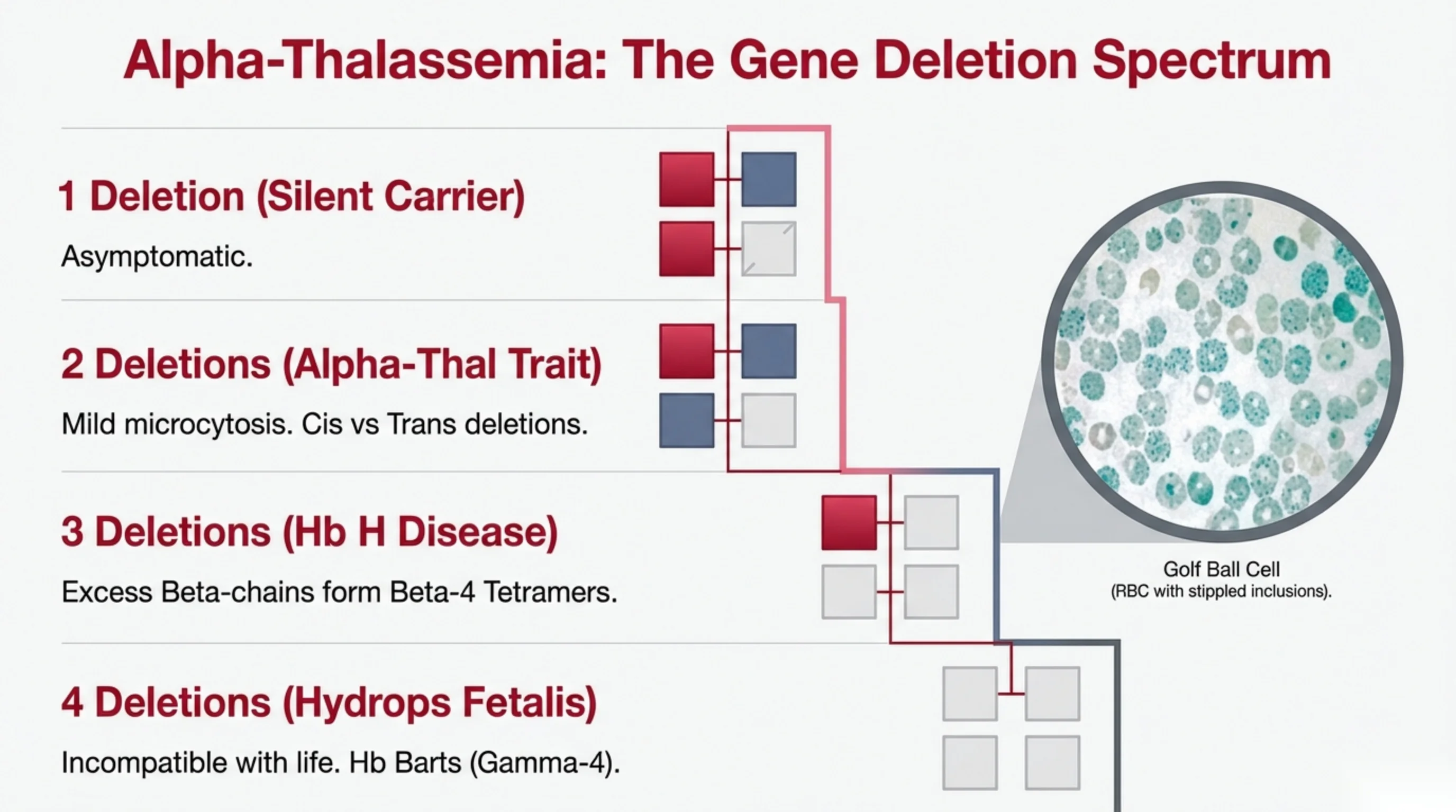
α-thalassemia: severity counts the missing genes
Unlike the β-chain, which is coded by a single gene on each chromosome 11, the α-chain is coded by two genes on each chromosome 16, four in total. α-thalassemia is almost always caused by deletion of these genes, and the clinical picture depends simply on how many of the four are lost.
- One deletion: silent carrier. Three working genes are enough. The patient is entirely well, the blood count is normal or nearly so, and the condition is usually found only by genetic testing or when a child is affected.
- Two deletions: α-thalassemia trait. Mild microcytosis, little or no anemia, normal iron studies, and, importantly, a normal hemoglobin electrophoresis, because the α-chain is shared by HbA, HbA₂ and HbF and all fall together. The two deletions may be on the same chromosome (cis) or one on each (trans). The distinction matters for offspring: a cis carrier can pass both deletions together, which is how Hb H disease and hydrops arise.
- Three deletions: Hb H disease. With only one working gene, α-chains are scarce and the excess β-chains pair with each other to form β₄ tetramers, called hemoglobin H. HbH is unstable and precipitates as the cell ages, causing a moderate chronic hemolytic anemia with splenomegaly. A supravital stain shows the precipitates as fine dots all over the cell, the "golf-ball cell" in the inset.
- Four deletions: hydrops fetalis. No α-chain can be made at all. The fetus makes hemoglobin from γ-chains alone (γ₄, hemoglobin Barts), which binds oxygen so tightly that it releases none to the tissues. The result is severe anemia and heart failure in the womb, and the condition is incompatible with life.
The practical message is the one from Part 1's microcytic algorithm: a microcytic patient with normal ferritin and a normal electrophoresis most likely has α-thalassemia trait, a diagnosis made by excluding the alternatives.
Further reading. The Thalassemias holds the alpha and the beta halves of this story together, and adds two things these slides do not. The first is the clue that separates trait from iron deficiency before any electrophoresis is sent: microcytosis with a high red cell count and only mild anemia points to thalassemia rather than to iron, and the Mentzer index is below 13. The second is beta-thalassemia intermedia, the ground between minor and major, where the anemia is moderate and the patient is not regularly transfusion-dependent. The chapter also states the pitfall that follows from the first point more bluntly than either slide does: do not treat thalassemia trait with long-term iron.
See it online.
- Golf-ball HbH inclusions on a supravital stain ASH Image Bank
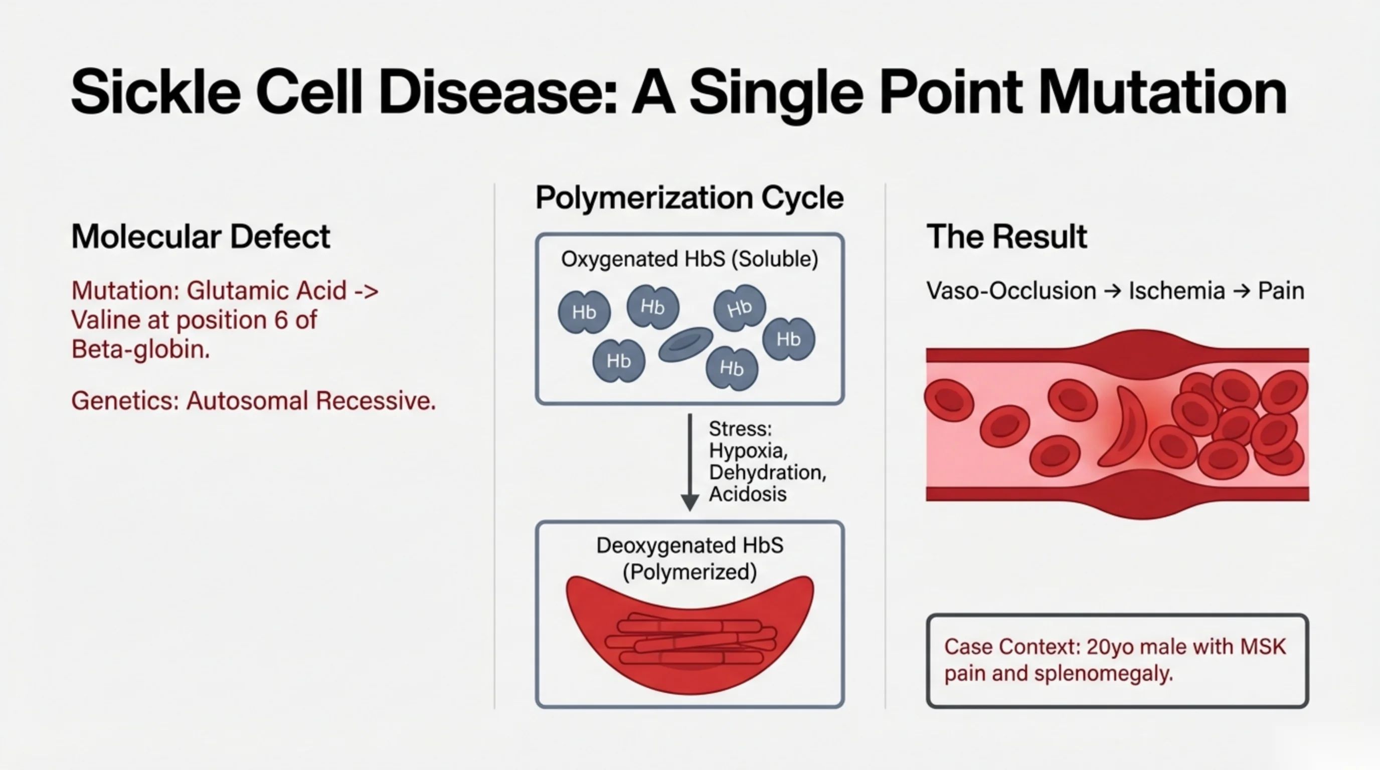
Sickle cell disease: one amino acid changes everything
Sickle cell disease is caused by a single point mutation in the β-globin gene. At position 6 of the β-chain, glutamic acid, which is charged and water-loving, is replaced by valine, which is neutral and water-repelling. The resulting hemoglobin S is fully functional as an oxygen carrier; the problem is what it does when it is not carrying oxygen.
When HbS gives up its oxygen, its shape shifts slightly and the valine at position 6 is exposed on the surface. That hydrophobic patch fits into a pocket on a neighbouring deoxygenated HbS molecule, and molecules begin to stack, one on the next, into long rigid fibres. The fibres grow until they distort the cell from a flexible disc into a stiff crescent: the sickle. This is the polymerisation cycle on the slide. Return oxygen to the cell and the fibres dissolve and the cell recovers its shape, at least for a while; each cycle damages the membrane a little more, and eventually the cell becomes irreversibly sickled.
Anything that lowers oxygen or slows the flow of blood tips the balance toward polymerisation: hypoxia (altitude, lung disease, sleep apnoea), dehydration (which concentrates the hemoglobin), acidosis (which lowers hemoglobin's affinity for oxygen), and cold, infection and stress. This is why a young man from the Southern Region with sickle cell disease may present with a crisis after exertion, a fever, or a journey to altitude.
The consequence is vaso-occlusion. Rigid sickled cells cannot pass through small vessels, they stick to the vessel wall and to each other, blood flow stops, the tissue beyond becomes ischemic, and the patient feels pain. The case on the slide, a 20-year-old man with musculoskeletal pain and an enlarged spleen, is the typical presentation. The disease is autosomal recessive: both parents carry the trait, and a carrier (HbAS) is generally healthy.
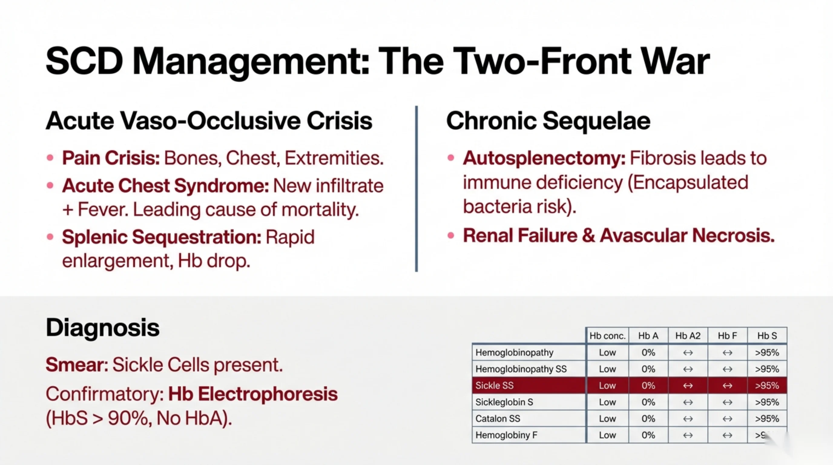
Sickle cell disease: the acute crises and the slow damage
Sickle cell disease is fought on two fronts at once: sudden, dangerous crises, and the slow accumulation of organ damage over a lifetime.
Acute vaso-occlusive events. The pain crisis is the commonest. Sickled cells block the small vessels of bone marrow, chest and limbs, and the resulting ischemia produces severe pain that needs prompt, adequate analgesia, fluids and oxygen. Acute chest syndrome is the most dangerous: a new infiltrate on the chest film with fever, chest pain or falling oxygen, caused by sickling, infection or fat embolism in the lungs. It is the leading cause of death in sickle cell disease and calls for urgent treatment, often including transfusion. Splenic sequestration, seen mainly in children before the spleen has been destroyed, is the sudden trapping of a large volume of blood in the spleen: the spleen enlarges rapidly, the hemoglobin falls, and the child can go into shock.
Chronic sequelae. Repeated infarction of the spleen destroys it by early childhood, an autosplenectomy. Without a spleen the patient cannot clear encapsulated bacteria such as pneumococcus, so vaccination and penicillin prophylaxis are life-saving. Over years the kidneys are scarred, leading to renal failure, and the femoral heads lose their blood supply, leading to avascular necrosis. Other organs suffer too: strokes, leg ulcers, retinal damage and pulmonary hypertension are all part of the picture.
Diagnosis. The smear shows sickled cells, and often target cells and, after autosplenectomy, Howell-Jolly bodies. Confirmation is by hemoglobin electrophoresis, which separates hemoglobins by their electrical charge. A normal adult shows mostly HbA. A patient with sickle cell disease (HbSS) shows HbS above 90%, no HbA at all, and a variable amount of fetal hemoglobin. A carrier shows both HbA and HbS. Because fetal hemoglobin protects against sickling, raising its level with hydroxyurea is one of the main long-term treatments.
Further reading. Sickle Cell Disease carries the crises this slide does not reach: the aplastic crisis, in which parvovirus B19 shuts red cell production down on top of the chronic hemolysis; stroke, with the transcranial Doppler screening that identifies the children at risk; and priapism, which is a urological emergency. It also sets the newer drugs beside hydroxyurea — voxelotor, crizanlizumab and L-glutamine — and the transplantation and gene therapy that can cure selected patients. Then read Case 52 · Sickle Cell Disease and Thalassaemia in the casebook, which is the ward version of the same material: analgesia within thirty minutes of arrival and reassessed every thirty, because under-treatment of sickle cell pain is a documented and repeated failure of care; incentive spirometry every two hours, prescribed as you would prescribe a drug, because it measurably reduces the acute chest syndrome; and every blood count read against the patient's own steady state. It ends on the point that matters most in this country: both diseases are autosomal recessive, consanguineous marriage raises the birth prevalence of both, and premarital screening for the haemoglobinopathies is a national programme whose results you will be asked to explain.
See it online.
- Sickle cells, with Howell-Jolly bodies ASH Image Bank
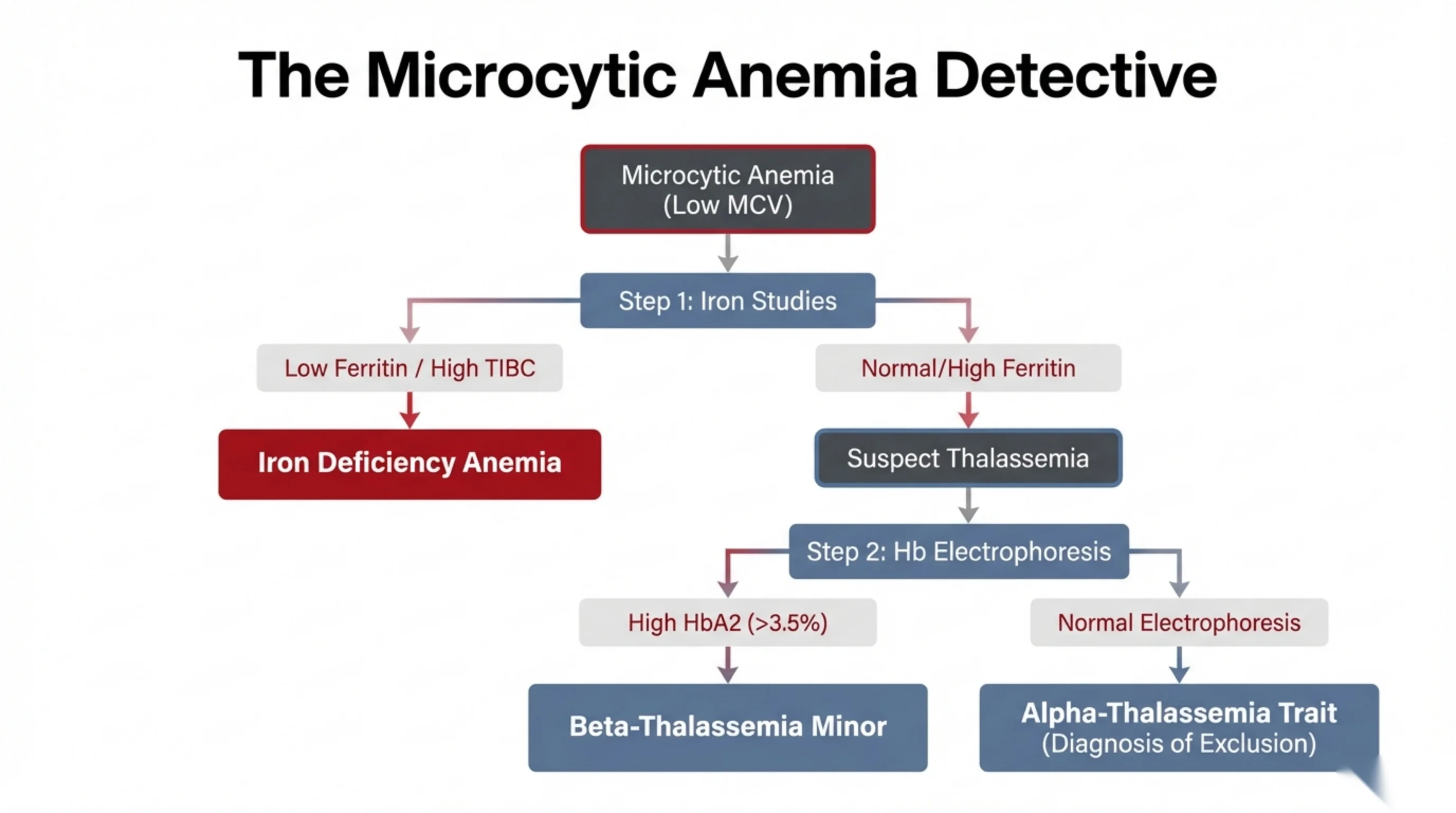
The microcytic algorithm, now complete
Part 1 taught you that a microcytic anemia means a hemoglobin synthesis problem, and that the differential is short: iron deficiency, thalassemia, anemia of inflammation, and rarely sideroblastic anemia. Now that you know the thalassemias, the algorithm can be run to the end.
Step 1: iron studies. Ask first whether the patient is iron deficient, because it is the commonest cause and it is treatable. A low ferritin, together with a high TIBC (total iron-binding capacity, which rises as the body makes more transferrin to capture whatever iron it can find), means iron deficiency anemia. Treat it, and then find the source of the blood loss.
Step 2: hemoglobin electrophoresis. If the ferritin is normal or high, the patient is not short of iron and the small cells need another explanation. Suspect thalassemia, and order electrophoresis. If HbA₂ is raised above 3.5%, the diagnosis is β-thalassemia minor. If the electrophoresis is entirely normal, remember that α-thalassemia trait produces no abnormal band, because the shortage of α-chain lowers every hemoglobin equally; α-thalassemia trait is therefore a diagnosis of exclusion, confirmed if needed by DNA testing.
Two cautions. First, iron deficiency and thalassemia trait often coexist in populations where both are common, and iron deficiency can lower the HbA₂ and mask β-trait, so correct the iron before interpreting electrophoresis. Second, a normal ferritin in an inflamed patient does not exclude iron deficiency (Part 1, case 2); look at the whole picture.
Further reading. Step 2 has a chapter of its own. Hemoglobin Electrophoresis explains what the test separates and what it cannot. The methods sort hemoglobins mostly by electrical charge, so two different variants can travel together — HbS runs with HbD or HbG on an alkaline gel — and an ambiguous band has to be confirmed by a second method: an acid gel, HPLC, or a sickle solubility test. It also states the rule this whole algorithm depends on, that you read the CBC and the film before you interpret an electrophoresis and not after. And it explains the timing you met on the last few slides: beta-chain disorders are hidden at birth by fetal hemoglobin and declare themselves only once the switch to HbA is complete, while alpha-chain disorders affect every hemoglobin and can be found at birth.
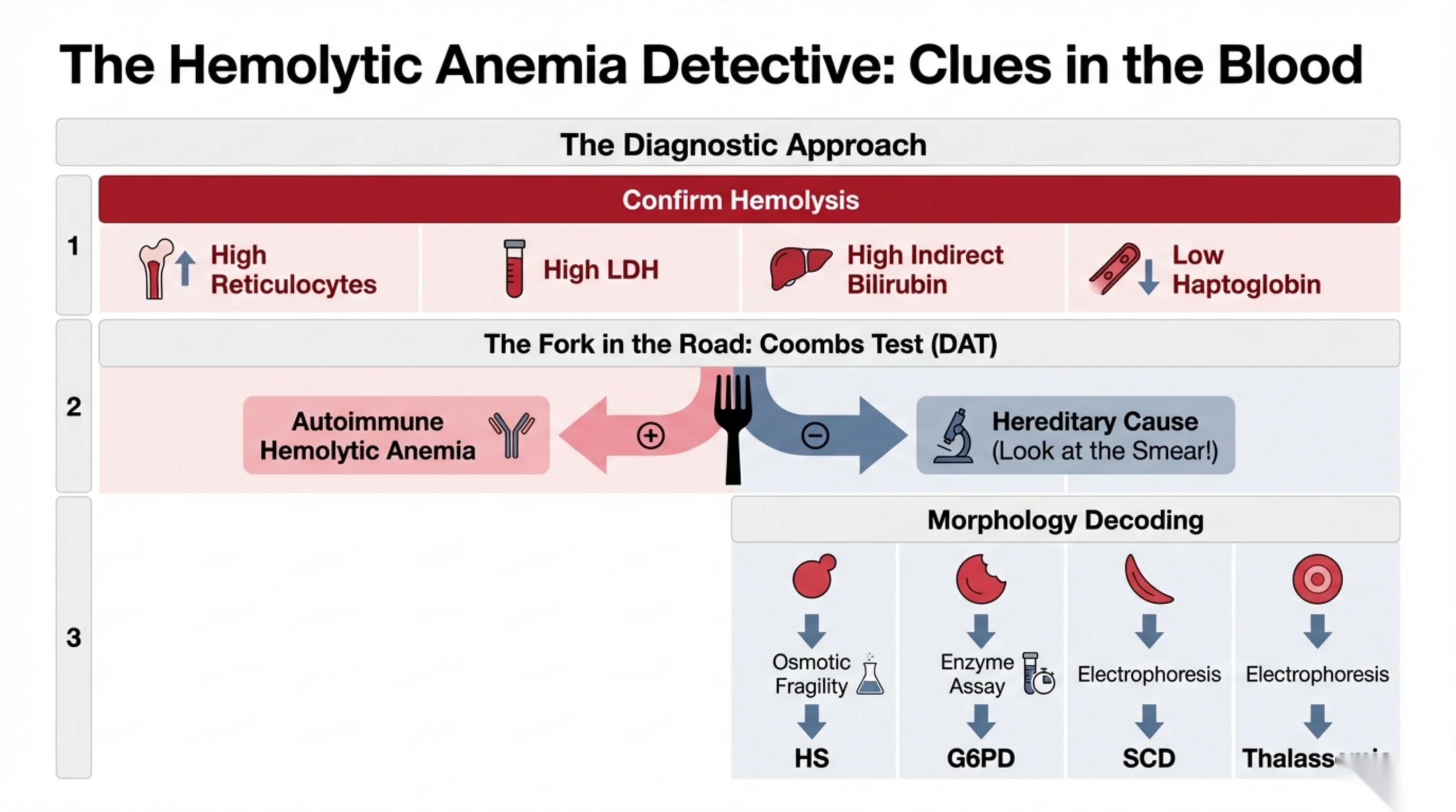
The hemolytic algorithm, now complete
This slide joins Part 2 and Part 3 into one path. It has three steps, and the third is where the hereditary diseases finally appear.
Step 1: confirm hemolysis. High reticulocytes, high LDH, high indirect bilirubin, low haptoglobin. Without these, do not chase a hemolytic diagnosis.
Step 2: the fork in the road, the direct Coombs test. A positive test means antibody on the red cells, and the diagnosis is autoimmune hemolytic anemia; go back to Part 2 for warm versus cold. A negative test means there is no immune attack, and in a patient with lifelong or familial hemolysis the cause is almost certainly hereditary. Now the smear does the work.
Step 3: let the cell shape name the disease.
- Spherocytes mean membrane loss without antibody: hereditary spherocytosis. Confirm with osmotic fragility or the EMA binding test.
- Bite cells (and Heinz bodies on supravital stain) mean oxidative damage: G6PD deficiency. Confirm with an enzyme assay, but remember that during an acute episode the most deficient cells have already been destroyed and the surviving young cells may give a falsely normal result; repeat the assay after recovery.
- Sickle cells mean hemoglobin S: sickle cell disease. Confirm with hemoglobin electrophoresis.
- Target cells with microcytosis mean thalassemia. Confirm with hemoglobin electrophoresis.
Notice how much the peripheral smear carries. In a Coombs-negative hemolytic anemia, a careful look down the microscope tells you which confirmatory test to order, and often what the answer will be before the result comes back.
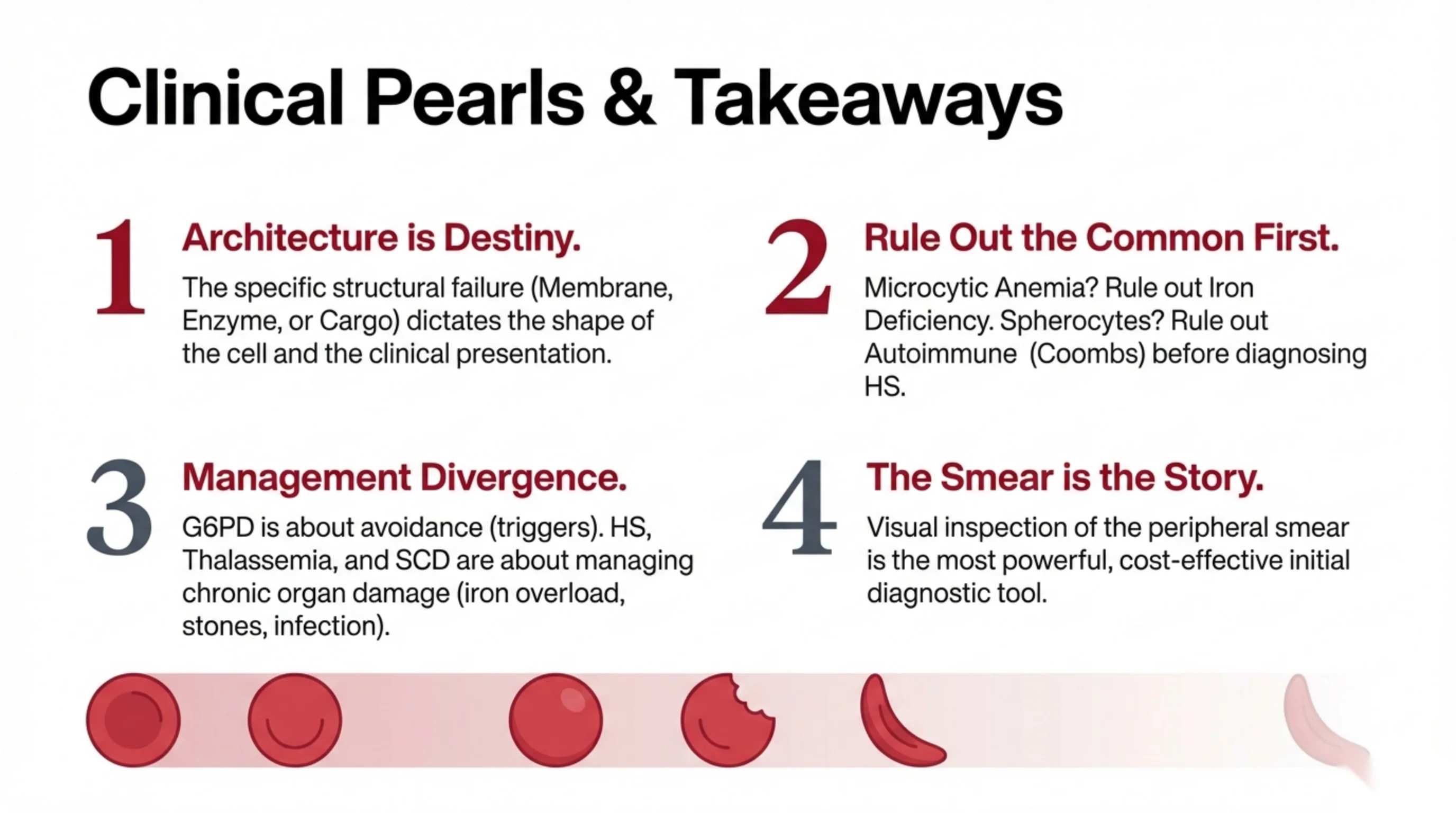
Four ideas to carry out of the series
- Architecture is destiny. Decide which part of the red cell has failed, membrane, enzyme or hemoglobin, and the shape of the cell, the pattern of hemolysis and the clinical presentation all follow. A weak skeleton makes a sphere that dies in the spleen; a missing enzyme makes a cell that survives until an oxidant attacks it; an abnormal hemoglobin makes a sickle that blocks vessels.
- Rule out the common first. A microcytic anemia is iron deficiency until iron studies say otherwise, and only then thalassemia. Spherocytes are autoimmune until the Coombs test is negative, and only then hereditary spherocytosis. Test for the frequent and the treatable before the rare and the inherited.
- Management diverges. G6PD deficiency is managed almost entirely by avoiding the triggers: fava beans, the listed drugs, and prompt treatment of infection. Hereditary spherocytosis, thalassemia and sickle cell disease, by contrast, are lifelong diseases whose care is about preventing and managing chronic organ damage: iron overload from transfusions, pigment gallstones from hemolysis, and infection after loss of the spleen.
- The smear is the story. A drop of blood on a glass slide, examined with care, is the most powerful and least expensive test in hematology. Spherocyte, bite cell, sickle cell, target cell: each one names a disease and tells you which confirmatory test to send.
Look at the shape of the cell, and it will tell you where the machine broke. That sentence is the whole of Part 3, and a good part of clinical hematology.
Read next. In the casebook, Reading the Blood Count in the Interpretation chapter lists the film findings that name a disease on their own, each with the case that works it through.